Digital Posters
Renal Vascular & Parenchymal Pathology
ISMRM & SMRT Annual Meeting • 15-20 May 2021

Digital Posters
Renal Vascular & Parenchymal Pathology
| Concurrent 5 | 19:00 - 20:00 |
2537.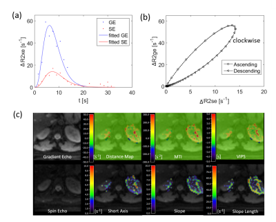 |
Navigator-triggered kidney vessel architecture imaging
Ke Zhang1, Simon M.F. Triphan1, Felix T. Kurz2, Christian H. Ziener3, Heinz-Peter Schlemmer 3, Hans-Ulrich Kauczor1, and Oliver Sedlaczek1,3
1Department of Diagnostic and Interventional Radiology, Heidelberg University Hospital, Heidelberg, Germany, 2Department of Neuroradiology, Heidelberg University Hospital, Heidelberg, Germany, 3Department of Radiology, German Cancer Research Center, Heidelberg, Germany
Vessel architecture imaging (VAI) MRI is a a technique that noninvasively measures parameters who describe the structural heterogeneity of brain microvasculature. To apply VAI in kidney disease respiratory motion artifacts need to be compensated for. In this study, a navigator along the inferior-superior direction was inserted as training data at the beginning of the measurement and interleaved during imaging acquisition. Our preliminary results suggest that respiratory motion can be corrected accurately.
|
|||
2538.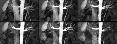 |
Evaluation of Image Quality of Renal Artery based on Balanced Turbo Field Echo Sequence using Compressed Sensing with Acceleration Factors
Haonan Zhang1, Qingwei Song1, Jiazheng Wang2, Zhiwei Shen2, Renwang Pu1, Nan Zhang1, and Ailian Liu1
1Department of Radiology, the First Affiliated Hospital of Dalian Medical University, Dalian, China, 2PHILIPS——Philips Healthcare, beijing, China
Renal artery imaging based on Balanced turbo field echo (B-TFE) sequence need a relatively long scan time and may be disturbed by motion artifacts, such as respiratory movement and other physiological factors. This study aimed to explore the accelerated renal artery imaging based on B-TFE using the compressed-SENSE (CS-SENSE) compared with a parallel imaging technique (SENSE), and to determine an optimal acceleration factor of CS-SENSE to achieved both of the reduction of scan time and a favorable image quality.
|
|||
2539.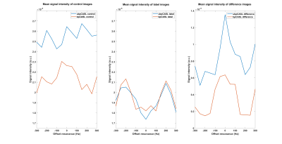 |
Improving the robustness of pseudo-continuous arterial spin labeling for renal perfusion imaging
Limin Zhou1, Yiming Wang1, and Ananth Madhuranthakam1,2
1Department of Radiology, University of Texas Southwestern Medical Center, Dallas, TX, United States, 2Advanced Imaging Research Center, University of Texas Southwestern Medical Center, Dallas, TX, United States
Pseudo-continuous arterial spin labeling (pCASL) has been the recommended ASL method for brain, and one of the two methods for kidneys. However, the reliability of labeling efficiency of pCASL is a major concern, especially in renal imaging due to the increased B0 inhomogeneities. In this study, we implemented and evaluated the optimized unbalanced pCASL gradient scheme generated by numerical simulation with both perfusion phantom and 4 healthy volunteers. The results showed that this optimized unbalanced pCASL gradient scheme was more robust to off-resonance than the corresponding balanced pCASL gradient scheme.
|
|||
2540.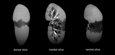 |
Ex-Vivo Renal Vascular Dominant Region Mapping using High Resolution 3D-T2w-MRI and Artery-Selective Contrast Injections in Porcine Kidneys
Nathan Sennesael1,2, Pieter R.E. De Backer3,4,5, Charlotte Debbaut4,6, Karel Decaestecker3,5, Pieter L.J. De Visschere2,7, Marijn M. Speeckaert8,9, Saar Vermijs4,5, Geert Villeirs2,7, and Pim Pullens1,2,7
1GIfMI, Ghent University, Ghent, Belgium, 2Diagnostic Sciences, Ghent University, Ghent, Belgium, 3Urology, Ghent University Hospital, Ghent, Belgium, 4IBiTech-bioMMeda, Ghent University, Ghent, Belgium, 5Human Structure and Repair, Ghent University, Ghent, Belgium, 6Cancer Research Institute Ghent (CRIG), Ghent University, Ghent, Belgium, 7Radiology, Ghent University Hospital, Ghent, Belgium, 8Nephrology, Ghent University Hospital, Ghent, Belgium, 9Internal diseases and Paediatrics, Ghent University, Ghent, Belgium
During partial nephrectomy (PN), a trade-off between clamping arteries to assure optimal conditions for surgery and maximizing oxygenation to tissue unaffected by the tumor, has to be made. Knowledge of the anatomy of the renal vascular dominant regions, can help optimize this trade-off and thus has the potential to significantly improve PN interventions. We found that high resolution 3D-T2w-MRI scans in combination with artery-selective contrast injections is an effective alternative to ex-vivo kidney casting as it allows for 3D mapping of the renal vascular dominant regions and shows potential for segmentation of the renal vascular tree.
|
|||
2541.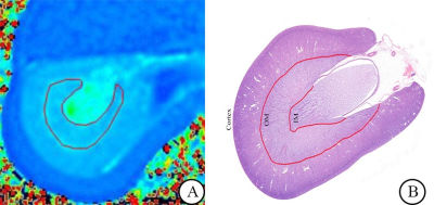 |
T2 mapping in the dynamic evaluation of renal ischemia-reperfusion injury: an animal study
Jing Chen1, Jinggang Zhang1, Weiqiang Dou2, and Jie Chen1
1The Third Affiliated Hospital of Soochow University, Changzhou, Jiangsu, China, 2GE Healthcare, MR Research China, Beijing, Beijing, China
This study explored the relationship between the T2 value of the outer medulla and the pathological characteristics of renal ischemia reperfusion injury (IRI) in an animal model. T2 values of the outer medulla increased at 1 hour after IRI and decreased from 1 hour to 48 hours gradually. T2 values of the outer medulla were significantly correlated with pathological score of renal injury, especially with tubular epithelial edema. Based on the findings, T2 mapping might reflect the dynamic changes of renal IRI and be used to assess the renal IRI in the early stage.
|
|||
2542. |
The value of DKI in assessing the pathologic microstructural changes in the early stages of renal cold ischemia-reperfusion injuries
Yizhong Yuan1, Jipan Xu1, Yan Ren2, Lihua Chen2, Jinxia Zhu3, Robert Grimm4, and Wen Shen2
1First Central Hospital Institute, Tianjin Medical University, Tianjin, China, 2Department of Radiology, Tianjin First Center Hospital, Tianjin, China, 3MR Collaboration, Siemens Healthcare, Beijing, China, 4MR Application development, Siemens Healthcare GmbH, Erlangen, Germany
This study investigated diffusion kurtosis imaging (DKI) values in evaluating the microstructural changes in the early stages of renal cold ischemia-reperfusion injuries (CIRIs). The results showed that both the mean diffusivity (MD) values and mean kurtosis (MK) values were negatively correlated with the pathologic scores, and the MK values showed more detailed microstructural changes than the MD values.
|
|||
2543.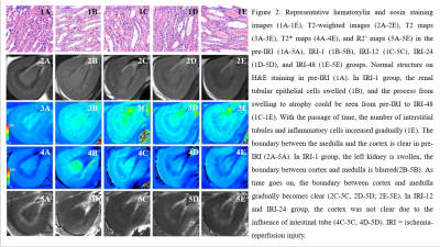 |
Values of R2’ mapping on evaluation of renal ischemia-reperfusion injury:an experimental study
Qin Chen1, JingGang Zhang1, WeiQiang Dou2, Jie Chen1, and Wei Xing1
1The Third Affiliated Hospital of Soochow University, Changzhou, Jiangsu, China, 2GE Healthcare, MR Research, Beijing, China
Ischemia reperfusion injury (IRI) is the main factor that delays the recovery of the renal function and leads to the failure of treatment. To our knowledge, R2’ mapping as a quantitative MRI method may be helpful for assessment of IRI in the early stage. This study compared the R2’ values of outer medulla at different time points in a rabbit model and found significant differences between IRI group and control group. Meanwhile, significant correlation was also found between R2’ values and histopathological features. R2’ mapping can evaluate the dynamic changes of the outer medulla longitudinally.
|
|||
2544.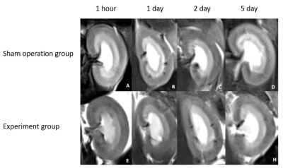 |
Intravoxel Incoherent Motion Imaging of Renal Cold Ischemia Reperfusion Injury in Rats
Yan Ren1, Lihua Chen1, Yizhong Yuan2, Jipan Xu2, Jinxia Zhu3, Robert Grimm4, and Wen Shen1
1Tianjin First Center Hospital, Tianjin, China, 2First Central Hospital Institute, Tianjin Medical University, Tianjin, China, 3MR collaborations, Siemens Healthcare Ltd., Beijing, China, 4Siemens Healthcare GmbH, Erlangen, Germany
The study evaluated the use of Intravoxel Incoherent Motion (IVIM) imaging to detect dynamic changes in renal microvascular characteristics during cold ischemia-reperfusion injuries (CIRIs). As previous studies have investigated warm ischemia-reperfusion injuries, we aimed to assess MR diffusion imaging in a renal CIRI Sprague Dawley rat model. Results showed that IVIM imaging is a sensitive tool to monitor changes in renal functional characteristics.
|
|||
2545.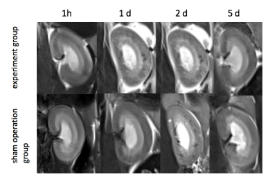 |
Mono-exponential, bi-exponential, and kurtosis diffusion-weighted imaging for renal cold ischemia-reperfusion injury in rats
Lihua Chen1, Yan Ren1, Yizhong Yuan2, Jipan Xu2, Jinxia Zhu3, Xuening Zhang4, Robert Grimm5, and Wen Shen1
1Tianjin First Center Hospital, Tianjin, China, 2First Central Hospital Institute, Tianjin Medical University, Tianjin, China, 3Siemens Healthcare Ltd., Beijing, China, 4Second Hospital of Tianjin Medical University, Tianjin, China, 5Siemens Healthcare GmbH, Erlangen, Germany
Renal dysfunction evaluations after renal cold ischemia-reperfusion injury (CIRI) has important clinical significance for prolonging donor kidney preservation times and improving the survival rates of transplanted kidneys. Diffusion-weighted imaging (DWI) has been accepted for microstructure change evaluations of renal function. In this study, a CIRI rat model was established and compared with a sham-operation group. The value of mono-exponential, bi-exponential, and kurtosis DWI models was compared to evaluate renal changes at different time points after CIRI surgery. The results showed that the bi-exponential and kurtosis models were more sensitive for detecting renal changes compared with the mono-exponential model.
|
|||
2546.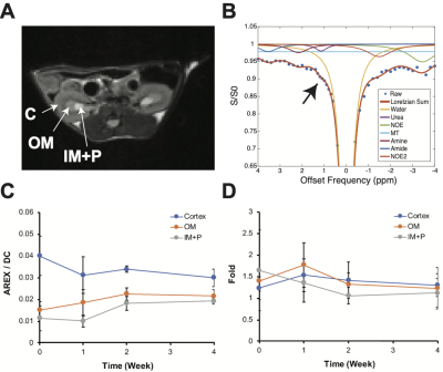 |
Monitoring Subclinical Renal Injury Progression by Saline-responsive CEST and Quantitative MT Imaging
Soo Hyun Shin1, Michael F. Wendland2, and Moriel H. Vandsburger1
1Department of Bioengineering, University of California, Berkeley, Berkeley, CA, United States, 2Berkeley Preclinical Imaging Core (BPIC), University of California, Berkeley, Berkeley, CA, United States
Standardized blood tests of kidney function lack adequate sensitivity to capture chronic progression of renal injuries. We examined whether saline-responsive urea CEST and quantitative MT imaging can be used to detect mild renal injury development. Upon renal injury progression, the urea CEST contrast decreased in the cortex, and the saline-induced fold change of contrast decreased in the inner medulla and papilla. qMT imaging showed the decrease of semi-solid macromolecule pool in the cortex. These results indicate that the saline-responsive CEST and qMT imaging have a potential to detect subclinical renal injury development.
|
|||
2547.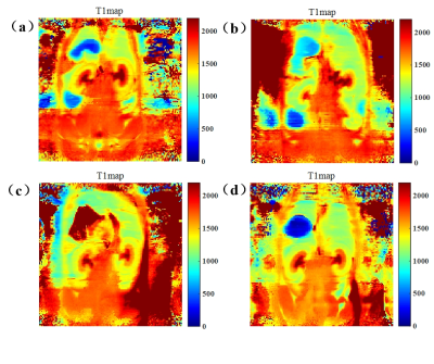 |
MRI enhancement of TPP-TEMPOL during reduces kidney damage
Quan Tao1, Peiwei Yi1, Zimeng Cai1, Yingjie Mei2, Ruiyuan Liu1, and Yanqiu Feng1
1School of Biomedical Engineering, Southern Medical University, Guangzhou, China, 2Philips healthcare, Guangzhou, China
In this study, we synthesis a mitochondria-targeted contrast agent named triphenylphosphine-2,2,6,6-tetramethylpiperidine-1-oxyl (TPP-TEMPOL) to protect the kidney from the damage induced by rhabdomyolysis. At the same time, measured the T1 changes by MRI.
|
|||
2548.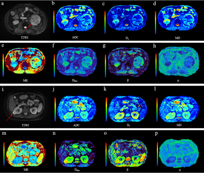 |
Comparison of mono-exponential and three non-Gaussian diffusion models in characterizing low- and high-grade clear cell renal cell carcinoma
Bowen Shi1, Ke Xue2, Yili Yin1, Qing Xu1, Binbin Shi1, Jing Ye1, and Yongming Dai2
1Northern Jiangsu Province Hospital, Yangzhou, China, 2Shanghai United Imaging Healthcare, Shanghai, China
Accurately assessing the tumor grades of ccRCC patients has a significant impact on the selection of the optimal surgical intervention. This study evaluated the performance of FROC, DK, bi- and mono-exponential diffusion models in differentiating low- from high-grade ccRCCs. A spectrum of diffusion metrics from the four models, reflecting cellularity, vascularity and microstructure complexity, were compared between these two groups. As a result, the diffusion parameters from the FROC model outperformed the other three models in characterizing ccRCC grades. This implied the FROC diffusion model held the potential in renal tumor diagnosis, grading, treatment decision and monitoring for treatment response.
|
|||
2549. |
The predictive value of contrast-enhancement MR evaluating the efficacy of preoperative targeted drugs for renal carcinoma with tumor thrombus
Pei Xinlong1 and Yuan Huishu2
1Radiology Department, Peking University Third Hospital, Beijing, China, 2Peking University Third Hospital, Beijing, China
The target drug is designed to shrink renal carcinoma and tumor thrombi. MR is promising about predicting the efficacy of targeted drugs before operation. We attempts to summarize and analyze this aspect.
|
|||
2550.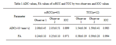 |
Diffusion Tensor Imaging in the Differential Diagnosis of Clear Cell Renal Cell Carcinoma invasion pelvis and Transitional Cell Carcinoma
Jinghong Liu1, Mingzhe Xu2, Ailian Liu1, Qingwei Song1, Lihua Chen1, Ru Cao1, Weilin Li1, and Juan Ruan1
1Radiology, First Affiliated Hospital of Dalian Medical University, Dalian, China, 2Henan Cancer Hospital, Henan, China
Centripetal infiltration into the renal parenchyma often accompanies with the growth of infiltrative RTCC.
|
|||
2551.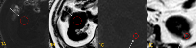 |
Differential diagnosis of clear renal cell carcinoma and renal angiomyolipoma without visible fat by IDEAL-IQ sequence
Xinmiao Bu1, Ailian Liu1, Jinghong Liu1, Qingwei Song1, Juan Ruan2, Weilin Li2, and Ru Cao2
1The First Affiliated Hospital of Dalian Medical University, Dalian, China, 2Dalian Medical University, Dalian, China
IDEAL-IQ is a new scanning sequence based on the principle of three-point Dixon asymmetric echo technology. The images of water phase, lipid phase, in-phase, reverse phase, fat fraction and R2* relaxation images can be generated at the same time in one scan to realize the quantitative measurement of fat fraction. The R2* value of the ccRCC group was greater than that of the RAMLwvf group, and the FF value was lower than RAMLwvf group, the difference was statistically significant (P <0.05). The AUC value of R2* value and FF value for identifying ccRCC with RAMLwvf groups are 0.893 and 0.905.
|
|||
2552.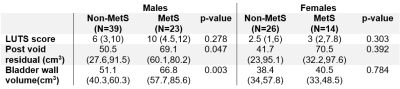 |
Effect of metabolic syndrome on anatomy and function of the lower urinary tract
Cody Johnson1, Alex Tannenbaum1, Samuel Koebe1, Lucille Anzia1, Lu Mao2, Matthew Grimes3, Diego Hernando1, Alejandro Roldán-Alzate1,4,5, and Shane Wells1
1Radiology, University of Wisconsin-Madison, MADISON, WI, United States, 2Biostatics and Medical Informatics, University of Wisconsin-Madison, Madison, WI, United States, 3Urology, University of Wisconsin-Madison, Madison, WI, United States, 4Biomedical Engineering, University of Wisconsin-Madison, MADISON, WI, United States, 5Mechanical Engineering, University of Wisconsin-Madison, Madison, WI, United States
Metabolic syndrome (MetS) contributes to lower urinary tract symptoms (LUTS). However, non-invasive methods for studying this association are limited. This study investigated the relationship of MetS, LUTS, anatomy, and function of the bladder and prostate in men and women. Manual segmentation with 3D rendering of the bladder and prostate were performed from MRI. We found that MetS is associated with increased bladder wall volume and postvoid residual in men but not women, suggesting that the effect of MetS on the prostate contributes to anatomic and functional changes of the bladder in men.
|
|||
2553.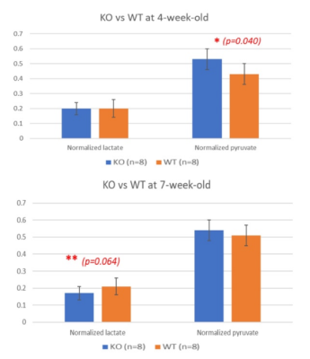 |
In Vivo Assessment Of Metabolic Abnormality In Alport Syndrome Using Hyperpolarized [1-13C]Pyruvate MR Spectroscopic Imaging
Nguyen Trong Nguyen1, Ilwoo Park2,3, Ngoc Luu Do2, Tien Anh Nguyen2, Sang Heon Suh 4, Eun Hui Bae4, and Sang Soo Shin2
1Department of Biomedical Science, Chonnam National University, Gwangju, Korea, Republic of, 2Department of Radiology, Chonnam National University Medical School and Hospital, Gwangju, Korea, Republic of, 3Department of Artificial Intelligence Convergence, Chonnam National University, Gwangju, Korea, Republic of, 4Department of Internal Medicine, Chonnam National University Hospital, Gwangju, Korea, Republic of
Alport Syndrome (AS) is an inherited kidney disease with progressive kidney failure. We investigate the potential of hyperpolarized 13C MRI to be used as an non-invasive technique for detecting early abnormal changes in renal metabolism. Hyperpolarized [1-13C]pyruvate imaging was applied to AS and wild-type mice at 4- and 7-week-old. Normalized Lactate in AS model was significantly lower at 7-week compared to 4-week. This abnormal renal metabolism in AS mice was consistent with PEPCK analysis, which showed increased expression level as the disease progressed. We demonstrated the feasibility of this technique for assessing early renal function in mouse model of AS.
|
|||
2554.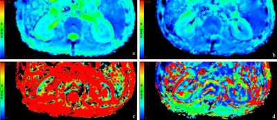 |
Intravoxel Incoherent Motion Diffusion-weighted MRI in Kidneys of Acute Leukemia and Its Clinical Significance: A Pilot Study
Jianting Li1, Jinliang Niu1, and Rong Zheng2
1The second hospital of Shanxi medical university, Taiyuan, China, 2The first hospital of Shanxi medical university, Taiyuan, China
36% of high-grade hematological malignancies patients suffer acute kidney injury (KI), including leukemia. Autopsy data suggested that kidney involvement represent a significant proportion of patients with acute leukemia (AL), ranging from 50% to 100%. Therefore, it is crucial to diagnose KI early and accurately. Clinical symptoms and renal biopsy are limited in diagnosing KI. Intravoxel incoherent motion MRI (IVIM) can reflect the diffusion and perfusion of kindeys. It is reported that some parameters are sensitive to renal pathological processes of focal and diffuse lesions. Thus, IVIM may offer an opportunity to identify early changes in renal function in AL.
|
|||
2555.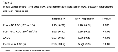 |
Can Diffusion Weighted MRI Predict the Response Prior to Neoadjuvant Chemotherapy in Patients with Muscular Invasive Bladder Cancer?
Xinxin Zhang1 and Yan Chen1
1Department of Imaging Diagnosis, National Cancer Center/National Clinical Research Center for Cancer/Cancer Hospital,Chinese Academy of Medical Sciences and Peking Union Medical College, BeiJing, China
Neoadjuvant chemotherapy(NAC) becomes the standard treatment for muscular invasive bladder cancer (MIBC), some patients do not respond to NAC. So the purpose of this study was to evaluate the potential of diffusion-weighted (DW) magnetic resonance imaging (MRI) with an ADC map in the prediction of response to NAC in patients with MIBC. In our study, all the sixty-two MIBC patients have underwent DW-MRI before and after NAC. After NAC, patients were classified into responders or non-responders by the Response Evaluation Criteria in Solid Tumors. However, there was no statistically significant difference in ADC between responders and non-responders before NAC.
|
The International Society for Magnetic Resonance in Medicine is accredited by the Accreditation Council for Continuing Medical Education to provide continuing medical education for physicians.