Digital Posters
Liver Function & Pancreas
ISMRM & SMRT Annual Meeting • 15-20 May 2021

| Concurrent 5 | 13:00 - 14:00 |
2753.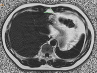 |
The relationship between preperitoneal fat and cardiometabolic risk factors
Qin-He Zhang1, Li-Zhi Xie2, and Ai-Lian Liu1
1The First Affiliated Hospital of Dalian Medical University, Dalian, China, 2MR Research, Beijing, China
This study assessed the correlation between preperitoneal fat and cardiometabolic risk factors. The results showed that preperitoneal FF and area were associated with specific risk factors, and the correlation coefficient varied by sex. This plays an important role in our further understanding about the association between ectopic fat deposition and cardiometabolic risk factors.
|
|||
2754.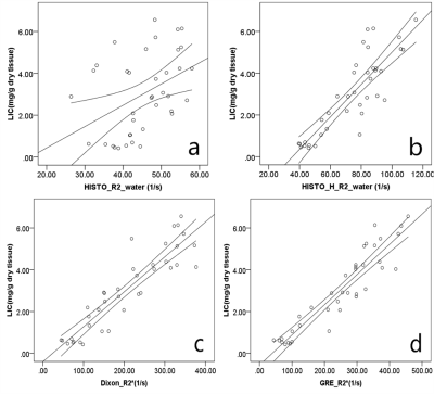 |
Comparison of multi-echo Dixon andmulti-echo MRS sequences to quantify hepatic iron overload in rabbits with or without fatty liver
Zhao Fan yu1, Chen Yidi1, Zhang Huiting2, Stephan Kannengiesser3, and Long Liling1
1Radiology Department, The First Affiliated Hospital of Guangxi Medical University, Nanning, China, 2Siemens Healthcare Ltd, MR Scientific Marketing, Wuhan, China, 3Siemens Healthcare GmbH, MR Application Development, Erlangen, Germany
Feasibility and accuracy of confounder-corrected 3D multi-echo-Dixon (ME-Dixon) imaging and T2-corrected multi-echo single-voxel (HISTO) spectroscopy for the quantification of liver iron content at 3T MRI were investigated in rabbits with or without fatty liver, compared with conventional 2D multi-gradient-echo (2D GRE) imaging, using histopathology as a reference. The results showed that compared with HISTO (R2_water) method, the multi-echo Dixon (R2*) method was found to be superior for liver iron and fat evaluations. AUC calculations showed that both the ME-Dixon and 2D GRE values had high accuracy for diagnosing liver iron overload in the rabbit model for LICs up to 7mg/g.
|
|||
2755.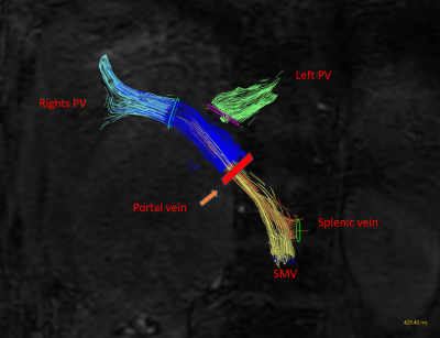 |
Portal Venous Hemodynamics with an abdominal 4D-flow MR imaging in patients with and without chronic liver disease
Atsushi Higaki1, Tsutomu Tamada1, Yu Ueda2, Akihiko Kanki3, and Akira Yamamoto3
1Radiology, Kawasaki Medical School, Okayama, Japan, 2Phillips Japan, Tokyo, Japan, 3Kawasaki Medical School, Okayama, Japan
For the early diagnosis of portal hypertension, the relationship between portal venous hemodynamic changes and degree of liver fibrosis was evaluated using abdominal 4D-flow MR imaging. Flow quantification was performed in the middle part of the portal trunk. The forward flow was significantly correlated with Fib-4 index. In the comparison between the three groups divided by the degree of fibrosis, only the average velocity was significantly different. As the liver fibrosis progressed, the forward flow of the portal vein increased. The average velocity of the portal vein may be useful for early diagnosis of liver fibrosis.
|
|||
2756.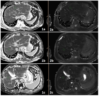 |
Correlation between liver function indicators in patients with cirrhosis and quantitative parameters of MRI---4D flow and T1/T2 relaxation time
Wenjun Zhang1, Nan Wang1, Ailian Liu1, Jiazheng Wang2, Qingwei Song1, Renwang Pu1, and Lihua Chen1
1the First Affiliated Hospital of Dalian Medical University, Dalian, Dalian, China, 2Philips Healthcare, Dalian, China
Liver function information plays an important role in the decision-making of treatment strategy in liver cirrhosis and in the prevention of liver failure after treatment. In recent years, more and more evidence of Gd-EOB-DTPA MRI T1mapping can be used to the liver and partial liver function. As cirrhosis often leads to changes in liver-related blood flow, we demonstrate the feasibility of 4D Flow MRI on quantitative assessment of the disease.
|
|||
2757.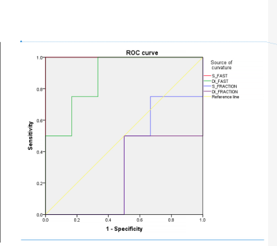 |
A quantitative perfusion value is a good discriminator of liver fibrosis level in patients with chronic hepatitis B
Lesheng Huang1, Weiyin Vivian Liu 2, Tianzhu Liu 1, Hongyi Li1, Jinghua Jiang1, Wanchun Zhang1, Jiahui Tang1, Meng Hu1, Dong Zhang1, Guangjun Tian1, Jun Chen1, Tao He1, Kaili Cai1, and Yifeng Wang1
1Guangdong Hospital of Traditional Chinese Medicine, Zhuhai, China, 2MR Research, GE Healthcare, Beijing, China 100176, Beijing, China
IVIM is reckoned as a valuable tool for non-invasive detection and evaluation of liver fibrosis staging. However, only more small b values used in one scan improve the diagnosis of liver fibrosis. In clinics, the different fibrosis stages overlap in the same inflammation level. However, both physiological expressions were different. To distinguish fibrosis from inflammation on images may help clinical diagnosis and provide more effective treatments for patients. Our study demonstrated that Dfast indeed had excellent discrimination ability of early liver fibrosis S1 and S2 in CHB patients with the same inflammation grade.
|
|||
2758.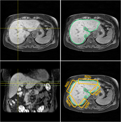 |
Regional variation of liver surface nodularity scores for evaluating hepatic fibrosis on a single axial MR image
Tae-Hoon Kim1, Youe Ree Kim2,3, Chang-Won Jeong1, Chungsub Lee1, SiHyeong Noh1, Ji Eon Kim1, Young Hwan Lee2,3, and Kwon-Ha Yoon2,3
1Medical Convergence Research Center, Wonkwang University, Iksan, Korea, Republic of, 2Radiology, Wonkwang University School of Medicine, Iksan, Korea, Republic of, 3Radiology, Wonkwang University Hospital, Iksan, Korea, Republic of
The assessment of liver surface nodularity (LSN) is emerging importance to diagnose hepatic fibrotic changes in clinical. The imaging techniques MRI and CT are gold-standard methods to estimate LSN scores. However, in clinical practice, the manual LSN assessment of whole liver is time-consuming. Therefore, it is powerful for assessing LSN score from a single slice image instead of whole liver images. This study compared the regional variation of LSN score for assessing fibrotic changes on a single liver MR image in chronic liver disease (CLD).
|
|||
2759.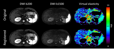 |
Diffusion-weighted MRI-based Virtual Elastography in liver: Enhancement prospect through diffusion image registration
Valentin H. Prevost1, Julien Rouyer2, Wolter de Graaf3, and Bruno Triaire1
1Canon Medical Systems Corporation, Tochigi, Japan, 2Department of Research & Innovation, Olea Medical, La Ciotat, France, 3Canon Medical Systems Europe, Zoetermeer, Netherlands
Diffusion-weighted based virtual elastography is a new approach proposing for liver elasticity assessment without the use of additional equipment. As abdominal imaging is challenging in a clinical context, this work investigated the potential of a respiratory triggered implementation with a dedicated image registration pipeline. Results analysis concluded to a signal-to-noise ratio increase, an organ delineation improvement, an overall signal dispersion reduction in liver and equal results for registered images using only two averages compared to six averages without registration. This dedicated postprocessing appears to enhance the liver elasticity assessment accuracy, with scan times that are feasible in a clinical context.
|
|||
2760.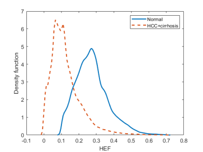 |
Voxel-wise hepatocellular function prediction using population-based probability density function
Monchai Phonlakrai1, Behzad Asadi2, Neda Gholizadeh3, Kate Skehan2, Liam Hilleary2, Jameen Arms4, Saadallah Ramadan5,6, John Simpson2,3, Jonathan Goodwin2,3, Jarad Martin2,7, Yuvnik Trada2, Swetha Sridharan2,7, and Peter Greer2,3
1School of Health Sciences, The University of Newcastle, Newcastle, Australia, 2Radiation Oncology, Calvary Mater Newcastle Hospital, Newcastle, Australia, 3School of Mathematical and Physical Sciences, The University of Newcastle, Newcastle, Australia, 4Diagnostic Radiology, Calvary Mater Newcastle Hospital, Newcastle, Australia, 5Faculty of Health and Medicine, The University of Newcastle, Newcastle, Australia, 6HMRI Imaging Centre, John Hunter Hospital, Newcastle, Australia, 7School of Medicine and Public Health, The University of Newcastle, Newcastle, Australia
Dynamic gadoxetate contrast-enhanced MRI yields spatial hepatocellular function through hepatic extraction fraction map. This allows well-functioning hepatocyte sparing in radiotherapy to avoid radiation-induced liver toxicity. However, the major challenge of using this parametric map in a clinical practice for normal function sparing is the lack of standard method to determine liver function at a voxel level within the same patient. As such, population-based kernel density function was proposed to deal with this problem to predict voxel-based probability of liver function. This novel approach also allows derivation of functional probability map that could be used for radiation beam guidance in function-based radiation treatment planning.
|
|||
2761.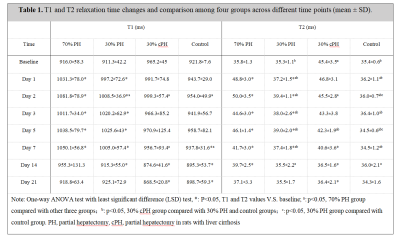 |
T1 and T2 mapping for Monitoring Liver Regeneration in Normal and Cirrhotic Liver Rats after Partial Hepatectomy
Qiu caixin1, Xie shuangshuang1, Sun yajie1, Zhu jinxia2, Xu yahui1, and Shen wen1
1Radiology Department, Tianjin First Center Hospital, TIANJIN, China, 2MR Collaboration, Siemens Healthcare Ltd., BEIJING, China
In this study, we used T1 and T2 mapping to quantify the microscopic changes of residual liver in normal and cirrhotic liver rats after partial hepatectomy (PH). The results showed that liver regeneration activity was more active in the rats with 70% PH than 30% PH, consistent with changes in T1 and T2 values. Furthermore, regeneration in cirrhotic liver was slower than in normal liver. However, in the process of regeneration, liver cirrhosis may be reversed to different degrees. The results suggested that T1 and T2 mapping can be used to assess liver regeneration after PH.
|
|||
2762.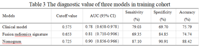 |
Diagnostic value of radiomics analysis based on multimodal MRI for advanced liver fibrosis in patients with hepatitis B
Shuangshuang Lu1, Jian Lu1, Aina Huang1, Xueqin Zhang1, Tao Zhang1, and Weibo Chen2
1Nantong University Affiliated Nantong Third People's Hospital, Nantong, China, 2Philips Healthcare Shanghai, China, Shanghai, China
The purpose of this study was to develop and validate an optimal radiomics-based model for diagnosis of advanced liver fibrosis. Firstly, multimodal MRI was performed before and 20 minutes after Gd-EOB-DTPA administration. Secondly, the clinical diagnosis model, fusion radiomics signature and radiomics nomogram model were established in the training cohort. Finally, the diagnostic value of three models was confirmed in the validation cohort.
|
|||
2763.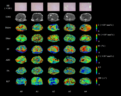 |
Multiparametric magnetic resonance imaging of liver regeneration in rabbits with warm ischemia-reperfusion injury
Zhengzheng Tao1,2, Zhiqiang Chu3, Jiabing Jiang1,2, Qing He1,2, Jinxia Zhu4, Robert Grimm5, and Qian Ji2
1First Central Clinical College of Tianjin Medical University, Tianjin, China, 2Department of Radiology, Tianjin First Central Hospital, Tianjin, China, 3Department of Transplantation, Tianjin First Central Hospital, Tianjin, China, 4MR Collaboration, Siemens Healthcare, Beijing, China, 5Siemens Healthcare GmbH, Erlangen, Germany
Multiparametric magnetic resonance imaging (MRI) provides various noninvasive and quantitative diagnostic information. This study assessed the value of multiparametric MRI in liver regeneration after warm ischemic-reperfusion injury (WIRI) in a rabbit model with different warm ischemia and reperfusion times. The results showed that the tissue diffusivity (Dslow), pseudo-diffusion coefficient (Dfast), perfusion fraction (PF), apparent diffusion coefficient (ADC), fractional anisotropy (FA), and R2* maps reflected changes in hepatic microcirculation perfusion, micro-dispersion, and oxygenation of hepatic WIRI after partial hepatectomy. Liver regeneration capacities were enhanced when warm ischemia times were 30 minutes or less; however, they were diminished after 30 minutes.
|
|||
2764.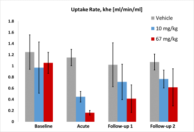 |
Acute and chronic rifampicin effect on gadoxetate uptake in rats using gadoxetate DCE-MRI
Mikael Montelius1, Steven Sourbron2, Nicola Melillo3, Daniel Scotcher3, Aleksandra Galetin3, Gunnar Schuetz4, Claudia Green4, Edvin Johansson1, John C. Waterton3,5, and Paul Hockings1
1Antaros Medical, BioVenture Hub, Mölndal, Sweden, 2University of Sheffield, Sheffield, United Kingdom, 3University of Manchester, MANCHESTER, United Kingdom, 4Bayer Pharma AG, BERLIN, Germany, 5Bioxydyn, Manchester Science Park, MANCHESTER, United Kingdom
Drug Induced Liver Injury causes liver failure and impedes drug development, and Drug-Drug Interactions affect the pharmacokinetics of drug metabolism and excretion. Non-invasive biomarkers are needed to monitor these processes. We used gadoxetate DCE-MRI to measure clinical and high dose rifampicin effects on hepatocellular uptake in acute and chronic dosing regimens in rats. High dose rifampicin caused significantly reduced gadoxetate uptake acutely, whereas uptake rates returned to baseline values after chronic dosing. Similar but non-significant effects were seen at clinical dose levels. We demonstrated the potential of gadoxetate DCE-MRI to non-invasively assess drug-induced inhibition of hepatocellular transport and DDIs.
|
|||
2765.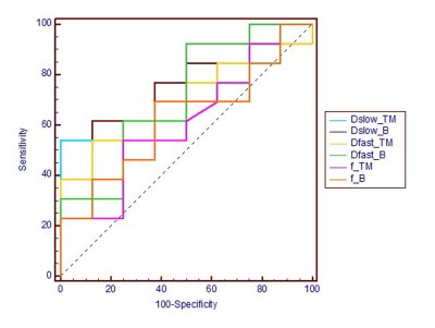 |
Distinction of early fibrosis stages in patients with chronic hepatitis B using intra voxel incoherent motion MR imaging
Hongyi Li1, Weiyin Vivian Liu2, Lesheng Huang1, Tianzhu Liu1, Jinghua Jiang1, Wanchun Zhang1, Jiahui Tang1, Tao He1, and Jun Chen1
1Guangdong Hospital of Traditional Chinese Medicine, Zhuhai, China, 2MR Research, GE Healthcare, Beijing, China, Beijing, China
The purpose was to evaluate the feasibility of IVIM variables in distinction of patients with early liver fibrosis stages(F1-2). Eight volunteers and 21 patients suspected chronic hepatitis B with fibrosis stage F1 or F2 were recruited. Correlations between all IVIM and DWI variables and ALT, AST, GGT values were analyzed. Two-segment mono-exponential model derived Dslow,TM was statistically different between volunteers and F1 patients(p< 0.05) with AUC of 0.740. The correlation between bi-exponential model derived Dslow,B and GGT was fair(r =0.513, p < 0.05).
|
|||
| 2766. | ROI-based intravoxel incoherent motion (IVIM) shows good diagnostic performance on fibrosis severity of patients with chronic hepatitis B
Jiang jinghua1, Weiyin Vivian LIu2, Huang Lesheng1, Liu Tianzhu1, Li Hongyi 1, Chen Jun 1, Zhang Wanchun 1, He Tao 1, and Tang Jiahui 1
1Department of Radiology, Guangdong Hospital of Traditional Chinese Medicine, Zhuhai,519000, China, zhuhai, China, 2MR Reaearch,GE healthcare, Beijing,China, China
IVIM, as a noninvasive tool, has good diagnostic performance in the detection and staging grading of liver fibrosis
|
|||
2767.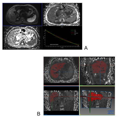 |
Whole-Liver Histogram Analysis of Diffusion Kurtosis Imaging Metrics in Predicting Acute Rejection After Orthotopic Liver Transplantation
Chang Li1, Mengzhu Wang2, and Canhui Sun1
1The First Affiliated Hospital, Sun Yat-Sen University, Guangzhou, China, 2MR Scientific Marketing, Siemens Healthcare, Guangzhou, China
Acute cellular rejection (ACR) occurs in 10-40% patients following liver transplantation (LT). Diffusion kurtosis imaging (DKI) provides more information about microstructure than standard monoexponential diffusion method. We analyzed the whole-liver volume histogram metrics of D map, K map, and ADC map with comparison those of the ACR and non-ACR group. Statistical analysis shows that most metrics of D maps were significantly lower in the ACR group than those in the non-ACR group, while most metrics of K maps were significantly higher in the ACR group those in the non-ACR group. None of the metrics of ADC maps were significantly different.
|
|||
2768.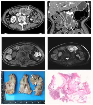 |
Colloid carcinoma arising from IPMN of pancreas: imaging features and differentiation with ductal adenocarcinoma from IPMN
Xu Fang1, Yun Bian1, Kai Cao1, Chengwei Shao1, Li Wang1, and Jianping Lu1
1Changhai Hospital of Shanghai, Shanghai, China
N/A
|
|||
2769.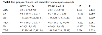 |
Value of diffusion tensor imaging quantitative parameter in differentiating cystic pancreatic cancer from solid pseudopapillary neoplasm
Xinqi Wang1, Yi Wang2, Ailian Liu3, and Qinhe Zhang3
1School of Medical Imaging, Dalian Medical University, Dalian, China, 2Department of Radiology, Dalian Friendship Hospital, Dalian, China, 3Department of Radiology, the First Affiliated Hospital of Dalian Medical University, Dalian, China
It is difficult to differentiate the pancreatic ductail adenocinama(PDAC) from solidpseudopapillary tumer of the pancreas(SPTP),due to their similar imaging characteristics,especially in pancreatic cancer sac change occurs.Diffusion tensor imaging (DTI) is a kind of magnetic resonance technology which can image the diffusion of water molecules in living tissues. There was significant difference in DTI quantitative parameters Fractional Aniso (FA) ,Volume ratio Aniso (VRA), Isotropic image(Iso) and T2-weightial trace(T2-T) between PDAC and SPTP. Therefore, DWI derivative sequences may be an effective method to identify PDAC and SPTP.
|
The International Society for Magnetic Resonance in Medicine is accredited by the Accreditation Council for Continuing Medical Education to provide continuing medical education for physicians.