Digital Posters
Body Imaging Quality Improvement
ISMRM & SMRT Annual Meeting • 15-20 May 2021

Digital Posters
Body Imaging Quality Improvement
| Concurrent 5 | 13:00 - 14:00 |
2770.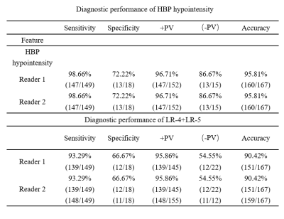 |
Hepatobiliary phase hypointensity improves the diagnostic performance of version 2018 LI⁃RADS on hepatocellular carcinoma
Bin Lin1, WenHai Dai1, JiaYan Chen1, YangDong Zeng1, ZhiPeng Zhou1, Long Qian2, and Weiyin Vivian Liu2
1Department of Radiology,Affiliated Hospital of Guilin Medical University, Guilin, China, 2MR Research, GE Healthcare, Beijing, China
Hypointensity in the hepatobiliary phase is an important ancillary feature in the diagnosis of liver malignancies in the version 2018 of LI-RADS. In this study, Hypointensity in the hepatobiliary phase was used as the major features to explore the effect of hepatobiliary hypointensity on the diagnosis performance of LI-RADS in small hepatocellular carcinoma (sHCC). Our results indicated that the sensitivity of diagnosing sHCC can be significantly improved without affecting the specificity of diagnosing sHCC when Hypointensity in the hepatobiliary phase was used as the major features for classification of LR-3,LR-4.
|
|||
2771.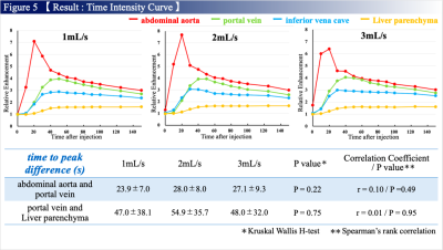 |
Effect of Injection Rates in Free-Breathing Gadoxetic Acid-Enhanced MRI with K-Space Weighted Image Contrast Reconstruction.
RYUJI SHIMADA1, Keitaro Sofue2, Yuichiro Somiya1, Tomohiro Noda1, Shintaro Horii1, Yoshiko Ueno2, Naoki Yoshida1, Wakiko Tani1, Yu Ueda3, Akiko Kusaka1, and Takamichi Murakami2
1Center of Radiology and Radiation Oncology, Kobe University Hospital, Kobe, Japan, 2Department of Radiology, Kobe University Graduate School of Medicine, Kobe, Japan, 3MR Clinical Science, Philips Japan, Tokyo, Japan
We prospectively evaluated the effect of injection rates on TIC analysis in free-breathing gadoxetic acid-enhanced MRI using radial sampling with KWIC reconstruction with different injection rate of 1, 2, and 3 mL/s in 48 patients. Time intensity curve analysis was performed and compared among the three injection rate groups. Time to peak in abdominal aorta was significantly shorter with higher injection rates, and the other parameters regarding portal vein, inferior vena cava, and liver parenchyma were not affected by the injection rates. Higher injection rate is recommended for the better discrimination on arterial input data.
|
|||
2772. |
Assessment of hepatic signal change in free breathing continuous multiphasic dynamic EOB-MRI with compressed sensing and self-gating technique
Masaya Tanabe1, Masahiro Tanabe1, and Katsuyoshi Ito1
1Yamaguchi University, Ube City, Japan
In the analysis of the hepatic contrast enhancement parameters based on continuous data of the signal changes over time obtained by free-breathing continuous multiphasic dynamic EOB-MR imaging using compressed sensing and self-gating technique, contrast enhancement ratio (CER) in the portal phase and 5min early hepatocyte phase had significant correlation with hepatic contrast enhancement effects in the 20min hepatobiliary phases and showed significant difference between sufficient and insufficient hepatobiliary phases enhancement groups, suggesting that sufficient 20min hepatobiliary phases enhancement may be estimated by the CER in the portal phase and 5min early hepatocyte phase.
|
|||
2773. |
The application value of CAIPIRINHA-VIBE sequence with non-rigid 3D-registration motion correction in liver
Junjiao Hu1, Weijun Situ1, and Huiting Zhang2
1Department of Radiology, the Second Xiangya Hospital of Central South University, Changsha, China, 2MR Scientific Marketing, Siemens Healthcare Ltd., Wuhan, China This prospective study aimed to compare the image quality of T1w-VIBE sequence using generalized autocalibrating partially parallel acquisitions (GRPPA-VIBE) and controlled aliasing in parallel imaging results in higher acceleration (CAIPIRINHA-VIBE) and the spatial position consistency of CAIPIRINHA with/without non-rigid 3D-registration motion correction (MOCO) in liver MRI. The results showed that the CAIPIRINHA had better image quality than the GRPPA with respect to respiratory motion artifact suppression, liver edge sharpness and intrahepatic vascular sharpness, and the spatial position consistency of the liver using CAIPIRINHA with MOCO was significantly better than that of images without MOCO. |
|||
2774.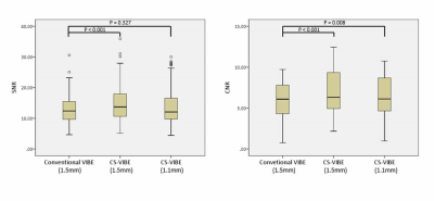 |
Utility of compressed sensing VIBE for hepatobiliary phase of Gd-EOB-DTPA-enhanced MRI: comparison with conventional VIBE
Hirokazu Otsuka1, Yoshihiko Fukukura2, Takashi Iwanaga1, Yuichi Kumagae2, Yasumasa Saigo1, Hiroshi Imai3, and Takashi Yoshiura2
1Kagoshima University Hospital, Kagoshima, Japan, 2Kagoshima University Graduate School of Medical and Dental Sciences, Kagoshima, Japan, 3Siemens Healthcare K.K., Tokyo, Japan
This study focused on the feasibility of volumetric-interpolated breath-hold examination (VIBE) using compressed sensing (CS) acceleration (CS-VIBE) for the hepatobiliary phase of Gd-EOB-DTPA-enhanced MRI in comparison with conventional VIBE. Our results showed a significantly higher signal-to-noise ratio and contrast-to-noise ratio (CNR) in CS-VIBE than in conventional VIBE. CS-VIBE showed a significantly higher CNR than conventional VIBE, even when decreasing a slice thickness from 1.5 mm to 1.1 mm of CS-VIBE. These results suggested CS-VIBE could replace conventional VIBE with superior CNR and spatial resolution.
|
|||
2775.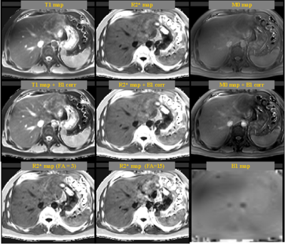 |
A Rapid Simultaneously 3D T1/T2* Quantitative Imaging (TXI) in Body within 3 Breath-holds
Minxiong Zhou1, Zhongshuai Zhang2, Huiting Zhang2, Guang Yang3, Jinrong Qu4, and Xu Yan2
1Shanghai University of Medicine & Health Sciences, Shanghai, China, 2MR Scientific Marketing, Siemens Healthcare, Shanghai, China, 33. Shanghai Key Laboratory of Magnetic Resonance, East China Normal University, Shanghai, China, 4Department of Radiology, the Affiliated Cancer Hospital of Zhengzhou University & Henan Cancer Hospital, Zhengzhou, China Simultaneously multi-parameter quantification is recently a hot research topic for MRI. A series of techniques are available, such as MR fingerprinting, MAGIC and STAGE. This study proposed a rapid and high-quality T1/T2* quantification method, called TXI. The results showed that it can generate high SNR T1 and R2* maps with less motion artifacts, which is superior to the conventional T1 and R2* mapping method. The T1 map is significantly improved with higher measurement consistency among different liver regions than conventional method. |
|||
2776.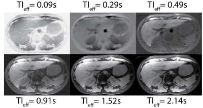 |
Free-Breathing, 3D Phase Sensitive Inversion Recovery MRI with Stack-of-Stars SGRE and Locally Low Rank Reconstruction
Yavuz Muslu1,2, Ty A. Cashen3, Sagar Mandava4, and Scott B. Reeder1,2,5,6,7
1Department of Biomedical Engineering, University of Wisconsin-Madison, Madison, WI, United States, 2Department of Radiology, University of Wisconsin-Madison, Madison, WI, United States, 3Global MR Applications and Workflow, GE Healthcare, Madison, WI, United States, 4Global MR Applications and Workflow, GE Healthcare, Atlanta, GA, United States, 5Department of Medical Physics, University of Wisconsin-Madison, Madison, WI, United States, 6Department of Medicine, University of Wisconsin-Madison, Madison, WI, United States, 7Department of Emergency Medicine, University of Wisconsin-Madison, Madison, WI, United States
Gadoxetic acid (GA)-enhanced MRI is an important tool for the detection of liver metastases. Through GA dose optimization and novel pulse sequence strategies, small lesions are often resolved using only high resolution GA-enhanced hepatobiliary phase T1w-MRI. However, such small lesions are often occult in T2w, diffusion weighted imaging (DWI) and dynamic contrast enhanced (DCE) T1w-MRI, which hinders the ability of MRI to characterize these lesions. In this work, we propose the development of a phase sensitive inversion recovery (PSIR) T1w-MRI in combination with 3D radial stack-of-stars imaging for detection and characterization of small focal liver lesions.
|
|||
2777.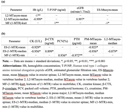 |
Feasibility of amide proton transfer imaging on distinction of the vertebral body and paravertebral muscles in patients with CKD
Yan Xiong1, Weiyin Vivian Liu2, Tongxiang He1, Donglin Wen 1, Fan He1, and Xiaoming Li3
1Radiology, Tongji Hospital, Tongji Medical College, Huazhong University of Science and Technology, Wuhan, China, 2MR Research, GE Healthcare, Beijing, China, 31Radiology, Tongji Hospital, Tongji Medical College, Huazhong University of Science and Technology, Wuhan, China
Amide proton transfer-weighted (APTw) imaging is a molecular MRI technique that generates image contrast based predominantly on the amide protons in mobile cellular proteins and peptides that are endogenous in tissue. Due to decreased vascular perfusion and increased glycolytic metabolism, tissue hypoxia is usually accompanied by acidosis; however, osteoclasts show significant functional enhancement in hypoxia and acidosis. The present study demonstrates that APT had higher diagnostic performance on identifying the differences of both bone and muscles between patients with different levels of CKD. It may have potential in distinction patients with CKD in accompanied with Mineral bone disease (MBD).
|
|||
2778.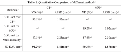 |
Using deep domain adaptation method to improve liver segmentation performance in Limited MRI images
Meng Dou1,2, Ailian Liu3, Yu Yao1,2, ZheBin Chen1,2, Han Wen1,2, Xu Luo1,2, and Ying Zhao3
1Chengdu Institute of Computer Application, Chinese Academy of Sciences, Chengdu, China, 2University of Chinese Academy of Sciences, Beijing, China, 3Department of Radiology, the First Affiliated Hospital of Dalian Medical University, Dalian, China
In clinical practice, it is too expensive to collect large-scale labeled Magnetic Resonance Imaging liver scans which constrains the segmentation performance. However, we noted that plenty of labeled Computed Tomography datasets aimed at liver have been published. Inspired by this, we proposed a deep domain adaptation method which can exploit the published CT datasets to improve the segmentation performance on MRI images. Our experiments showed that the liver segmentation performance is boosted on limited labeled MRI images (20 cases). Lastly, our method achieved competitive performance on both modality images. This work will be benefit to computer-aided diagnosis and treatment planning.
|
|||
2779.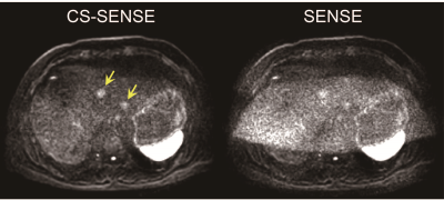 |
Improved image quality of liver diffusion-weighted imaging by combining compressed sensing and sensitivity encoding
Naoki Ohno1, Satoshi Kobayashi1, Tosiaki Miyati1, Yu Ueda2, Masami Yoneyama2, and Toshifumi Gabata1
1Kanazawa University, Kanazawa, Japan, 2Philips Japan, Tokyo, Japan
Although sensitivity encoding (SENSE) technique is commonly used to reduce distortion in diffusion-weighted imaging (DWI), highly accelerated SENSE results in significantly increased noise, leading to systematic errors to the quantification of apparent diffusion coefficient. In this study, we proposed a novel method using compressed sensing combined with a highly accelerated sensitivity encoding (CS-SENSE) to reduce both the distortion and noise in liver DWI. The CS-SENSE demonstrated the higher SNR and reduced geometric distortion compared with conventional SENSE reconstruction. Liver DWI with the proposed method can improve the image quality with better lesion conspicuity.
|
|||
2780.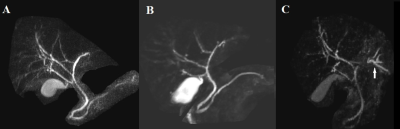 |
3D breath-hold magnetic resonance cholangiography using compressed sensing acceleration in living donor liver transplantation donors
shuangshuang xie1, yajie sun1, caixin qiu1, jinxia zhu2, Elisabeth Weiland3, Bernd Kühn3, and wen shen1
1Tianjin First Central Hospital, Tianjin, China, 2MR Collaboration, Siemens Healthcare Ltd., Beijing, China, 3MR Application Development, Siemens Healthcare GmbH, Erlangen, Germany
This study investigated the feasibility of optimized breath-hold compressed-sensing accelerated magnetic resonance cholangiography (BH-CS-MRCP) in visualizing the biliary system in living donor liver transplantation (LDLT) donors. Conventional navigator-triggered (NT) MRCP was performed preoperatively, and optimized BH-CS-MRCP was performed both preoperatively and one month after surgery. The optimized BH-CS-MRCP protocol showed similar motion or blurring artifacts, overall image quality, background suppression, and biliary duct depiction compared with conventional NT-MRCP protocols. This suggests that optimized BH-CS-MRCP with a short acquisition time (17 s) can be used for preoperative and postoperative evaluation of biliary ducts in LDLT donors.
|
|||
2781.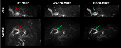 |
Free-breathing MRCP using motion compensated 3D Cartesian turbo spin-echo imaging
Tom Bruijnen1,2, Tim Schakel1, Jan Lagendijk1, Liesbeth Geerts3, Johannes Peeters3, Niels Blanken4, Wouter Veldhuis4, and Cornelis van den Berg1,2
1Department of Radiotherapy, University Medical Center Utrecht, Utrecht, Netherlands, 2Computational Imaging Group for MRI diagnostics and therapy, Centre for Image Science, Utrecht, Netherlands, 3BIU MR, Philips Healthcare, Best, Netherlands, 4Department of Radiology, University Medical Center Utrecht, Utrecht, Netherlands
Magnetic resonance cholangiopancreatography (MRCP) is crucial for the diagnosis of biliary/pancreatic duct disease. However, conventional respiratory triggered (RT-MRCP) scans require long acquisition times and ultimately fail in patients with irregular breathing. In this work we propose a free-breathing motion corrected MRCP scan (MOCO-MRCP) based on a rewinded Cartesian Acquisition with Spiral Profile ordering (rCASPR). MOCO-MRCP eliminates idle time associated with respiratory triggering, thereby increasing the robustness to motion artefacts while reducing the overall scan time compared to RT-MRCP. We show preliminary results of MOCO-MRCP in three patients with suspected biliary/pancreatic duct disease.
|
|||
2782.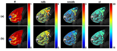 |
The Effect of the Scanner's Frequency Variation on Estimating the Fatty Acid Composition in Adipose Tissue
Mehran Baboli1, Pippa Storey2, Terlika Pandit Sood2, Justin Fogarty2, Melanie Moccaldi2, Alana Lewin2, Linda Moy2, and Sungheon Gene Kim1
1Radiology, Weill Cornell Medicine, New York, NY, United States, 2Radiology, NYU Langone Health, New York, NY, United States
We assessed the influence of frequency variation on measurements of fatty acid composition in adipose tissue. A 3D bilateral gradient spectroscopic imaging sequence with a simultaneous dual-slab excitation followed by 128 monopolar echoes was used. The sequence included a short train of 12 navigator echoes without phase encoding at the beginning of each TR period to correct frequency variations due to hardware heating and patient respiration. The proposed method was tested in oil phantoms and ten postmenopausal women. Phase correction reduced measurement error in the phantom and spatial variation in estimates of fatty acid composition in vivo.
|
|||
2783.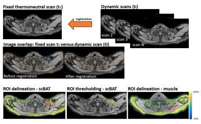 |
High temporal-resolution dynamic MRI for the assessment of brown adipose tissue during mild-cold exposure in young healthy adults.
Aashley S.D. Sardjoe Mishre1, Maaike E. Straat2, Borja M. Martinez-Tellez2, Oleh Dzyubachyk3, Mariette R. Boon2, Patrick C.N. Rensen2, Andrew G. Webb1, and Hermien E. Kan1
1Department of Radiology, C.J. Gorter Center for High Field MRI, Leiden University Medical Center (LUMC), Leiden, Netherlands, 2Department of Medicine, Division of Endocrinology and Einthoven Laboratory for Experimental Vascular Medicine, LUMC, Leiden, Netherlands, 3Department of Radiology, Division of Image Processing (LKEB) and Department of Cell and Chemical Biology, Electron Microscopy section, LUMC, Leiden, Netherlands
Brown adipose tissue (BAT) is considered as a potential therapeutic target against cardiometabolic diseases. Activated BAT combusts fatty acids leading to a reduction in fat fraction (FF). Both cold exposure and pharmacological stimuli can activate BAT, but the short-term dynamics of BAT activation are still unknown. To assess supraclavicular BAT (scBAT) FF dynamics during cold-exposure, we developed a 1-minute time resolution MRI protocol using breath-holds and co-registration to minimize motion-artefacts, and tested the protocol in five individuals. Co-registration resulted in low variation (<0.5%) and dynamic FF changes during cooling differed between subjects, possibly due to variable responses to thermoneutral conditions.
|
|||
2784.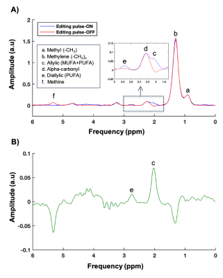 |
Application of MEGA-sLASER for detection of lipid composition in the human liver
Pandichelvam Veeraiah1, Lucas Lindeboom1,2, Kim Brouwers1, Joachim E Wildberger1, and Vera B Schrauwen-Hinderling1,2
1Department of Radiology and Nuclear Medicine, NUTRIM School for Nutrition and Translational Research in Metabolism, Maastricht University Medical Center, Maastricht, Netherlands, 2Nutrition and Movement Sciences, NUTRIM School for Nutrition and Translational Research in Metabolism, Maastricht University, Maastricht, Netherlands
1H-MRS has been widely used to measure total intrahepatic lipid content, but measuring lipid composition with specifically differentiating saturated, mono- and poly-unsaturated fatty acids in the liver, is challenging. At clinical field strength (3T), the allylic peak is overlapping with the alpha-carbonyl methylene resonance, which we recently addressed with a sophisticated fitting routine. However, this approach is difficult when shimming is suboptimal or in volunteers with low liver fat content (1-2%). Here, we evaluated the in vivo feasibility of J-difference editing, using MEGA-sLASER in the liver, to separate allylic from the alpha-carbonyl resonance for estimation of hepatic lipid composition.
|
|||
2785.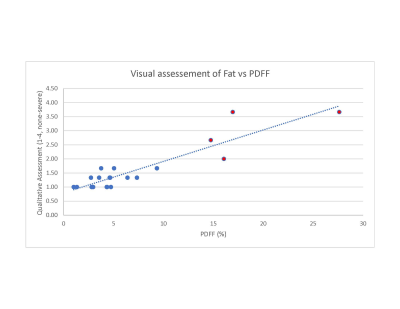 |
Qualitative Assessment of Hepatic Fat and Iron as a Screening Method
Kathan A Amin1, Achille Mileto1, and Orpheus Kolokythas1
1Radiology, University of Washington, Seattle, WA, United States
Qualitative assessment of hepatic fat and iron from three radiologists were compared to the quantitative measurements from the mDIXON-quant sequences. There was a robust correlation providing support for the ability to use qualitative assessment to screen patients on routine abdominal MRI exams and recommend further evaluation with specialized quantitative sequences.
|
|||
2786.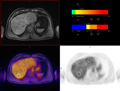 |
Increased Hepatic FDG Uptake in Patients with Metabolic Associated Fatty Liver Disease on PET/MR
Zhaoting Meng1, Gang Feng1, Mingxiang Sun1, Liling Peng1, Min Zhu1, Mu Lin2, and Xin Gao1
1Shanghai Universal Medical Imaging Diagnostic Center, Shanghai, China, 2MR Collaboration, Diagnostic Imaging, Siemens Healthineers Ltd., Shanghai, China
Globally, nonalcoholic associated fatty liver (NAFLD) is the predominant chronic liver disease, and it correlates to metabolic syndrome and type 2 diabetes, leading to cardiac and cerebral arteriosclerosis. In 2020, metabolic associated fatty liver disease (MAFLD) was newly defined to clarify the pathogenic mechanisms of this disease and to better guide clinical treatment. We hypothesized that with MAFLD, previously contradictory imaging results could be better explained. Liver MRS and FDG PET imaging were conducted simultaneously on an integrated PET/MR platform, which improved data accuracy. The study also analyzed whether the liver could be used as a reference tissue for PET imaging.
|
|||
2787.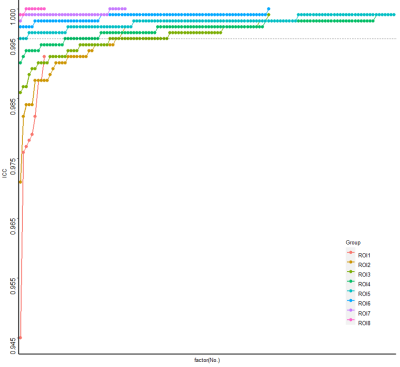 |
Optimization of ROI sampling strategies for proton density fat-fraction MRI of hepatic steatosis before liver transplantation in ex vivo
Gen Chen1, Weiyin Vivian Liu2, Hao Tang1, Lifen Zhou1, Daoyu Hu1, and Zhen Li1
1Tongji Hospital, Tongji Medical College, Huazhong University of Science and Technology, Wuhan, China, 2MR Research, GE Healthcare, Beijing, China, Beijing, China
This study aims to determine sampling strategy (SS) with the fewest regions of interest (ROIs) that could diagnose hepatic steatosis (HS) as reliably as the 9-ROI strategy before liver transplantation in ex vivo. All strategies with ≥5 ROIs had an intraclass correlation coefficients(ICC) ≥ 0.995 and limit of agreement (|LOA|)≤ 1.5%. The average proton density fat-fraction (PDFF) was moderately correlated with the histological diagnosis. The 5-ROI SS obtains similar PDFF results as the 9-ROI strategy before liver transplantation in ex vivo. After further verification, this method may become an available standard to assess HS before liver transplantation in ex vivo.
|
|||
 |
2788.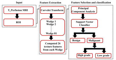 |
Computer-aided diagnosis of human breast lesion in T1 perfusion MRI using curvelet based features.
Snekha Thakran1,2
1University of Pennsylvania, Philadelphia, PA, United States, 2, Indian Institute of Technology Delhi, New Delhi, India, Delhi, India
Curvelet transform is used as a multi-scale level decomposition to represent images. It was hypothesized that curvelet based texture features extraction can improve accuracy of tumor classification. The objective of this study was to differentiate the breast tumor using curvelet based features extraction followed by principal component analysis(PCA) for feature reduction and support vector machine(SVM) classifier. The study included T1 perfusion MRI data of 40 patients with breast cancer. The curvelet based texture feature using PCA with SVM classifier provided high average accuracy(0.93±0.04) in classification of malignant vs. benign and average accuracy (0.86±0.06) in characterization of high- vs. low-grade.
|
||
2789.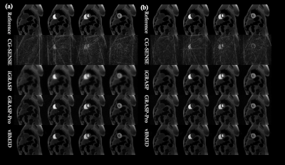 |
A Video Block Matching 3D Transform-domain Collaborative Filtering Approach for High Spatiotemporal Resolution DCE-MRI
Zhongbiao Xu1, Zhenguo Yuan2, Yaohui Wang3, Junying Cheng4, Rongli Zhang5, Ling Xia6, Yanqiu Feng7, Feng Liu8, and Zhifeng Chen7
1Guangdong Provincial People's Hospital, guangzhou, China, 2shandong medical imaging research institute ,shandong provincial hospital ,afflliated shangdong first medical uinversity, jinan, China, 3Institute of Electrical Engineering, Chinese Academy of Sciences, Beijing, China, 4First Affiliated Hospital of Zhengzhou University, zhengzhou, China, 5Department of Imaging and Interventional Radiology, The Chinese University of Hong Kong, Hongkong, China, 6Department of Biomedical Engineering, Zhejiang University, HangZhou, China, 7Southern Medical Southern, guangzhou, China, 8School of Information Technology and Electrical Engineering, The University of Queensland, Brisbane, Australia
High spatiotemporal resolution DCE-MRI has great clinical value in disease diagnosis and treatment. In this study, we propose to use a video block matching 3-D filtering approach to improve high spatiotemporal resolution DCE-MRI. Both phantom and in vivo experiments were performed in this work. The phantom experiment indicated that the proposed approach outperforms iGRASP and GRASP-Pro methods with lower reconstruction errors, especially in cases involving super high reduction factors. In vivo experiments draw similar conclusion. This new technique can provide a potential solution for real-time imaging and image guided radiation therapy.
|
The International Society for Magnetic Resonance in Medicine is accredited by the Accreditation Council for Continuing Medical Education to provide continuing medical education for physicians.