Digital Posters
Liver & Other Body Cancers
ISMRM & SMRT Annual Meeting • 15-20 May 2021

| Concurrent 5 | 17:00 - 18:00 |
2338.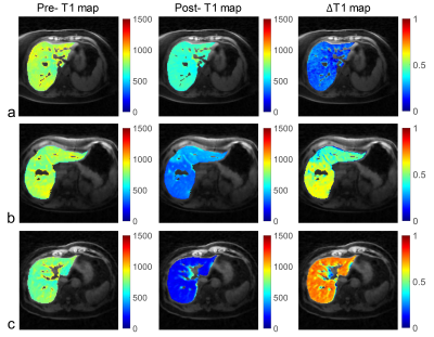 |
Free-breathing T1 Mapping of the Whole Liver Using GOAL-SNAP Sequence in Patients with Hilar Cholangiocarcinoma
Yajie Wang1, Ming Xiao2, Canhong Xiang2, Yuewei Zhang2, Haikun Qi3, Yishi Wang4, Jiahong Dong2, and Huijun Chen1
1Center for Biomedical Imaging Research, Department of Biomedical Engineering, School of Medicine, Tsinghua University, Beijing, China, 2Hepato-pancreato-biliary Center, Beijing Tsinghua Changgung Hospital, School of Medicine, Tsinghua University, Beijing, China, 3School of Biomedical Engineering and Imaging Sciences, King's College London, London, United Kingdom, 4Philips Healthcare, Beijing, China
T1 mapping combined with MR contrast agent administration has been applied to evaluate the liver function. However, conventional liver T1 mapping techniques have some limitations, such as the need of breath-holding, limited slice coverage or the need of multiple acquisitions. In this study, a free-breathing whole liver T1 mapping technique was proposed using a single scan. Preliminary results in patients with HCCA demonstrated its great potential for T1 quantification of the liver and liver function estimation.
|
|||
2339.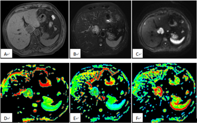 |
Diagnosis of Intrahepatic Cholangiocarcinoma and Hepatocellular Carcinoma Using Diffusion-tensor Imaging
Lihua Chen1, Ailian Liu1, Qingwei Song1, and Lizhi Xie2
1The First Affiliated Hospital of DaLian Medical University, DaLian , China., Dalian, China, 2GE Healthcare China, Beijing, China, Beijing, China
Treatment options differ significantly between ICC and HCC; the only curative treatment option for ICC is surgical resection, whereas for HCC, liver transplantation, percutaneous ethanol injection, and radiofrequency ablation are available. So, it is very important to accurately distinguish between these two tumors before therapy is planned. The purpose of this study is to evaluate and compare the FA value of DTI and the ADC value of DWI in ICC and HCC.
|
|||
2340.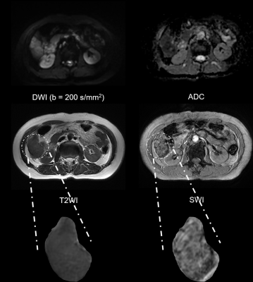 |
Radiomics analysis on SWI in hepatocellular carcinoma: exploring the correlation between histopathology and radiomics features
Zhijun Geng1, Yunfei Zhang2, Chuanmiao Xie1, and Yongming Dai2
1Sun Yat-sen University Cancer Center, Guangzhou, China, 2Central Research Institute, United Imaging Healthcare, Shanghai, China
Susceptibility weighted imaging (SWI) has shown tremendous clinical potential in visualizing the micro-haemorrhage, calcification, neovascularization and so forth, which serve as important micro-structural manifestations during the carcinogenesis. Radiomics is of great significance for providing valuable quantitative image markers via high throughput feature extraction. However, hardly have the radiomics been applied for extracting the quantitative markers from SWI images. This research, hence, aims to extract the diagnostic markers from the SWI images for evaluating several important prognostic markers of hepatocellular carcinoma including histopathologic grade, microvascular invasion (MVI) as well as the expression status of cytokeratin 7, cytokeratin 19 and Glypican-3.
|
|||
2341.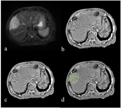 |
Value of intratumoral susceptibility signal intensities in quantitatively and automatically evaluating histological grade of HCC using ESWAN
Dahua Cui1, Ailian Liu1, Hongkai Wang2, Mingrui Zhuang2, Ying Zhao1, and Qingwei Song1
1The First Affiliated Hospital of Dalian Medical University, Dalian, China, 2Dalian University of Technology, Dalian, China
The aim of this study was to explore the value of intratumoral susceptibility signal intensities (ITSS) in quantitatively and automatically evaluating histological grading of hepatocellular carcinoma (HCC) by using enhanced T2 star-weighted angiography (ESWAN). The results showed that quantitative ITSS providied a promising differential performance (AUC = 0.812, P< 0.0001, sensitivity of 83.33%, specificity of 66.67%) in automatically evaluating histological grading of HCC.
|
|||
2342.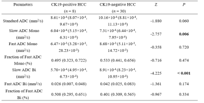 |
Intravoxel incoherent motion diffusion-weighted MR imaging for preoperatively identifying CK19-positive hepatocellular carcinoma
Ying Zhao1, Ailian Liu1, Wenjing Qi2, Xue Ren1, Tao Lin1, and Qingwei Song1
1The First Affiliated Hospital of Dalian Medical University, Dalian, China, 2Department of Pathology, The First Affiliated Hospital, Dalian Medical University, Dalian, China
Cytokeratin 19 (CK19) is well acknowledged as a biliary/progenitor cell marker and a marker of tumor stem cell. CK19-positive HCCs demonstrate more aggressive behaviors and poorer outcomes. This worked aimed at exploring the value of intravoxel incoherent motion (IVIM) diffusion-weighted (DW) magnetic resonance (MR) imaging in the diagnosis of CK19-positive HCC. The results showed that Slow ADC Mono and Slow ADC Bi were significantly different between the CK19-positive and CK19-negative HCC groups. The area under the curves (AUCs) of Slow ADC Mono, Slow ADC Bi, and combine (Slow ADC Mono & Slow ADC Bi) were 0.817, 0.988, and 0.992, respectively.
|
|||
2343.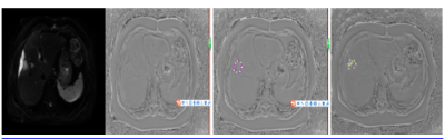 |
The value of intratumoral susceptibility signal intensities in quantitatively and automatically differentiating ICC from HCC
Changjun Ma1, Ailian Liu1, Dahua Cui1, Ying Zhao1, Hongkai Wang2, and Mingrui Zhuang2
1Radiology Department, The First Affiliated Hospital of Dalian Medical University, Dalian, China, Dalian,China, China, 2the School of Biomedical Engineering, Dalian University of Technology, Dalian,China, China
The aim of this study was to explore the value of intratumoral susceptibility signal intensities (ITSS) in quantitatively and automatically differentiating intrahepatic cholangiocarcinoma (ICC) from hepatocellular carcinoma (HCC) using enhanced T2 star-weighted angiography (ESWAN). The results showed that quantitative ITSS could provide a promising differential performance (AUC = 0.927, sensitivity = 85%, specificity = 92.3%) in quantitatively differentiating ICC from HCC.
|
|||
2344.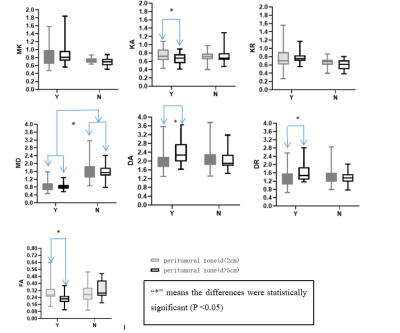 |
DKI for Assessing the Therapeutic Response of TACE in Hepatocellular Carcinoma and Invasion of Peritumoral Zone
Junying wang1, Weiqiang Dou2, Xiaoyi He3, and Hao Shi4
1Medical Imaging, Shandong Provincial Qianfoshan Hospital, the First Hospital Affiliated with Shandong First Medical University, Jinan, China, 2GE Healthcare, MR Research China, Bejing,, Beijing, China, 3Medical Imaging, Shandong Provincial Qianfoshan Hospital, the First Hospital Affiliated with Shandong First Medical University, Jinan, Shandong, China, 4Shandong Provincial Qianfoshan Hospital, the First Hospital Affiliated with Shandong First Medical University, Jinan, Shandong, China
Diffusion kurtosis imaging (DKI) is able to describe the deviations of water molecules diffusion from Gaussian form and depict the microstructural environment precisely. Previous study applied DKI in assessing the therapeutic response of Transcatheter Arterial Chemoembolization (TACE) in Hepatocellular Carcinoma (HCC), and showed promising results. On this basis, this study further explored the clinical value of DKI in assessing liver cancer and tumoral cell invasion of peritumoral zone between HCC progressive group and pseudo-progressive group after TACE treatment, in order to provide new ideas for clinical follow-up.
|
|||
| 2345. | Synthetic Diffusion-Weighted MRI in Patients with Hepatocellular Carcinoma: Feasibility and Performance Versus the Conventional Acquired
Shan Yao1, Yi Wei1, Zheng Ye1, and Bin Song1
1Department of Radiology, West China Hospital, Sichuan University, Chengdu, China
Hepatocellular carcinoma (HCC) is the most common malignancy cancer and the second major contributor to cancer-related mortality worldwide. Diffusion-weighted imaging (DWI) has been widely used in HCC evaluation and showed great potential, while with some limitations like low SNRs and artifacts causing by high b values, and prolonged acquisition time. Synthetic DWI is mathematically derived from directly acquired DWI with at least two different b-values, which may tackle the limitations of conventional acquired DWI. This study aims to investigate the clinical feasibility and diagnosis performance of synthetic DWI in patients with HCC compared with conventional acquired DWI qualitatively and quantitatively.
|
|||
2346.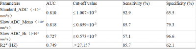 |
The value of IVIM and enhanced T2 star weighted angiography (ESWAN) for prediction of microvascular invasion in hepatocellular carcinoma
Huanhuan Chen1, Ailian Liu1, Ying Zhao1, Qingwei Song1, Xin Li2, Yan Guo2, and Tingfan Wu2
1The First Affiliated Hospital of Dalian Medical University, Dalian, China, 2GE Healthcare, Shanghai, China
Hepatocellular carcinoma (HCC) is the most common primary liver cancer, and surgical resection has been regarded as the most effective treatment of HCC; however, the high postoperative recurrence rate leads to poor prognosis. Microvascular invasion (MVI) is a risk factor for postoperative recurrence of HCC [1]. This study aimed to investigate the value of intravoxel incoherent motion (IVIM) and enhanced T2 star weighted angiography (ESWAN) for prediction of MVI in HCC.
|
|||
2347.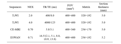 |
Susceptibility weighted imaging for quantitatively evaluation of dual‑phenotype hepatocellular carcinoma
Ying Zhao1, Ailian Liu1, Wenjing Qi2, Xue Ren1, Tao Lin1, and Qingwei Song1
1The First Affiliated Hospital of Dalian Medical University, Dalian, China, 2Department of Pathology, The First Affiliated Hospital, Dalian Medical University, Dalian, China
Dual-phenotype hepatocellular carcinoma (DPHCC) is associated with high rate of post-operative recurrence and low rate of survival. This worked aimed at exploring the value of susceptibility weighted imaging (SWI) quantitative parameters for diagnosis of DPHCC. The results showed that R2* provided a promising performance (AUC = 0.830, sensitivity = 85.0%, specificity = 77.3%) in quantitatively evaluating DPHCC.
|
|||
2348.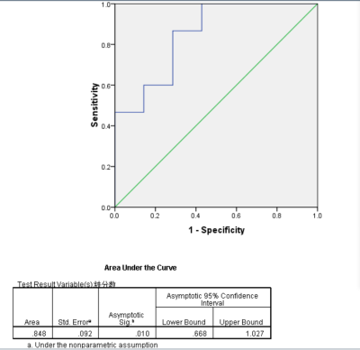 |
The valuation of MRI based on IDEAL-IQ in differential diagnosis between HCC with Negative Alpha Fetal Protein and FNH
LI SHAO PENG1, DENG KE XUE1, and WANG PENG2
1Department of Radiology, The First Affiliated Hospital of USTC, Southern District of Anhui Provincial Hospital, Hefei, China, 2The First Affiliated Hospital of USTC, Southern District of Anhui Provincial Hospital, hefei, China
To investigate the MRI technique based on the sequence of IDEAL-IQ (iterative decomposition of water and fat with echo asymmetry and least squares quantification sequence) in differential diagnoses between Hepatocellular Carcinoma with Negative Alpha Fetal Protein and Hepatic Focal Nodular Hyperplasia (FNH).The fat fraction and R2* value measured by IDEAL-IQ sequence are useful to distinguish HCC with Negative Alpha Fetal Protein from FNH.
|
|||
2349.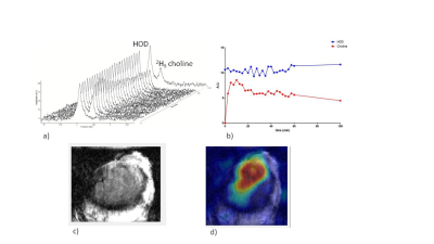 |
Simultaneous recording of the uptake and conversion of glucose and choline in tumors by deuterium MR
Andor Veltien1, Sjaak van Asten1, Nia Ravichandran1, Robin de Graaf2, Henk de Feyter2, Jeannette Oosterwijk3, Egbert Oosterwijk3, and Arend Heerschap1
1Medical Imaging, Radboud UMC, Nijmegen, Netherlands, 2Radiology and biomedical imaging, Yale University, New Haven, CT, United States, 3Urology, Radboud UMC, Nijmegen, Netherlands
Increased glucose and choline uptake are hallmarks of cancer. In this study we demonstrated that it is possible to follow the uptake and to image the presence of [2H9]choline in tumors after a bolus administration of this compound. In addition we demonstrated the successful performance of DMI simultaneously of [2H9]choline and of [6,6 2H2]glucose. As DMI of glucose uptake and its metabolic conversions has been shown to be feasible in patients, the simultaneous performance of choline DMI next to glucose DMI is an important complementary extension with clinical potential.
|
|||
2350.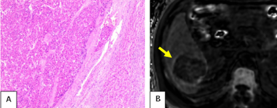 |
Evaluation of Microvascular Invasion of Hepatocellular Carcinoma by Using R2* mapping of Chemical shift encoded MRI
Ting Jiang1, Diego Hernando2, Scott B. Reeder2, and Jin Wang1
1Radiology, The Third Affiliated Hospital of Sun Yat-Sen University, Guangzhou, China, 2University of Wisconsin, Madison, WI, United States
Hepatocellular carcinoma (HCC) is the third most common cause of cancer-related mortality. Microvascular invasion (MVI) of HCC is a major prognostic factor that influences treatment strategy and long-term survival, but it is difficult to identify MVI until the tumor is surgically removed and analyzed histologically. Both DCE-MRI and Chemical shift encoded (CSE) MRI can be used to evaluate of HCC in clinical practice. Using CSE-MRI, we found a high percentage of elevated peritumoral R2* on quantitative R2* maps with MVI-positive patients compared to MVI-negative patients. CSE-MRI has the potential to assess MVI of HCC preoperatively.
|
|||
2351.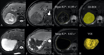 |
R2* value derived from multi-echo Dixon technique can aid discrimination between benign and malignant focal liver lesions
Guangzi Shi1, Hong Chen1, Weike Zeng1, Mengzhu Wang2, and Jun Shen1
1Radiology, Sun Yat-Sen Memorial Hospital, Sun Yat-Sen University, Guangzhou, China, 2Department of MR Scientific Marketing, Siemens Healthineers, Guangzhou, China
R2* derived from multi-echo Dixon imaging is a potential biomarker to differentiate malignant from benign FFLs. R2* value of malignant FLLs was significantly higher than that of the benign FLLs. The multi-echo Dixon sequence is easy to perform and required only a single breath-hold of 16 s to image the entire liver, which holds a good potential for clinical application.
|
|||
2352.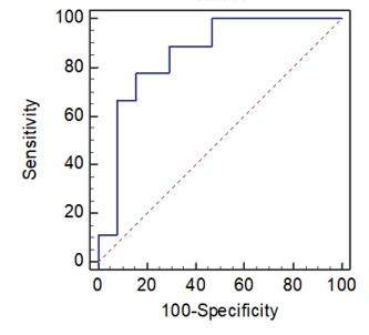 |
Preliminary Exploration and Analysis of MRI-based radiomics for Assessment of MSI-high metastatic colorectal cancer
Fu Shen1, Xiaolu Ma1, and Fangying Chen1
1Changhai Hospital, Shanghai, China
In the current study, we evaluated Imaging and genomic data for patients with Microsatellite instability high (MSI-H) metastatic colorectal carcinoma (mCRC) to determine whether Radiomics Based on MRI could be used to select the patients with MSI-H status. Using the pretreatment MRI data, we developed a radiomics model with excellent performance for individualized, noninvasive prediction of MSI-H in patients with mCRC and potentially guide treatment.
|
|||
2353.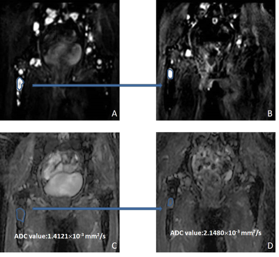 |
Clinical value of whole-body diffusion-weighted imaging in evaluation of multiple myeloma
Lei Zhang1, Bei Zhang1, and Xiuzheng Yue2
1First Hospital of Jilin University, Changchun, China, 2Philips Healthcare, Beijing, China
Whole-body diffusion-weighted imaging (WB-DWI) in the evaluation of bone marrow infiltration in multiple myeloma, and it will be a helpful supplement to the evaluation of multiple myeloma.
|
|||
2354.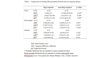 |
Prediction of early treatment response in patients with newly diagnosis multiple myeloma using WB-MRI: comparative study with and without anemia
Huazheng Dong1, Wenyang Huang2, Xiaodong Ji3, Shuang Xia3, Zhiwei Shen4, Dehui Zou2, Wen Shen3, and Zhiyi Song1
1Tianjin Academy of Traditional Chinese Medicine Affiliated Hospital, Tianjin, China, Tianjin, China, 2State Key Laboratory of Experimental Hematology, Institute of Hematology and Blood Diseases Hospital, CAMS and PUMC, Tianjin 300020, China, Tianjin, China, 3Tianjin First Central Hospital Affiliated To Nankai University, Tianjin, China, Tianjin, China, 4Philips healthcare,Beijing, China, Beijing, China
For patients with newly diagnosed multiple myeloma, red marrow reconversion caused by anemia is common,its hyperintense signal on DWI image could affect the evaluation , because it is very similar with the diffusion signal in focal lesions. We aim to evaluate the efficacy prediction of early treatment response in MM patients with and without anemia based on the tumor burden score (TBS), ADC value and signal fat fraction (sFF). We found that early treatment response of MM could be predicted in patients without anemia using TBS, ADC and sFF, anemia is a factor which must be taken into consideration in evaluation.
|
|||
2355.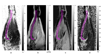 |
MR Imaging of Murine Tibia for Co-Clinical Studies of Myelofibrosis
Ghoncheh Amouzandeh1, Kevin A Heist1, Dariya I Malyarenko1, Youngsoon Jang1, Tanner Robison1, Christopher Bonham1, Cyrus Amirfazli1, Scott D Swanson1, Gary D Luker1, Brian D Ross1, and Thomas L Chenevert1
1Radiology, University of Michigan, Ann arbor, MI, United States
This study assesses the use of non-invasive MRI to monitor evolution and treatment of Myelofibrosis (MF) in both clinical and pre-clinical settings to quantify disease progression and treatment efficacy. Based on a limited number of studies, MRI can detect changes in the bone marrow fat distribution and fibrosis caused by the progression and treatment of MF. This work seeks quantitative measures such as proton density fat fraction, magnetization transfer and diffusion weighted imaging to investigate proportions of fat, cellularity and fibrosis in bone marrow caused by MF.
|
|||
2356.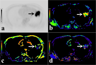 |
Using 18F-FDG PET-MR to compare and analyze the value of SUVmax and IVIM parameters in evaluating lung squamous and adenocarcinoma
Pengyang Feng1, Nan Meng2, Zhun Huang1, Ting Fang2, Fangfang Fu3, Yaping Wu3, Wei Wei3, Yan Bai3, Jianmin Yuan4, Yang Yang 5, Hui Liu 6, and Meiyun Wang*1,2
1Department of Radiology, Henan University People’s Hospital & Henan Provincial People’s Hospital, Zhengzhou, China, 2Department of Radiology, Zhengzhou University People’s Hospital & Henan Provincial People’s Hospital, Zhengzhou, China, 3Department of Radiology, Henan Provincial People’s Hospital, Zhengzhou, China, 4Central Research Institute, UIH Group, Shanghai, China, 5Central Research Institute, UIH Group, Beijing, China, 6UIH America, Inc, Houston, TX, United States
PET-MR is a new multi-modal imaging system that organically integrates PET and MRI. SUVmax is the most commonly used semi-quantitative index to measure how much 18F-FDG is taken up by the lesion. Intravoxel incoherent motion (IVIM), including D, D*, f values, provide parameters for the movement of tissue water molecules and parameters for the degree of tissue perfusion. Our results show that SUVmax and IVIM have similar diagnostic performance in the diagnosis of lung squamous and adenocarcinoma.
|
|||
2357.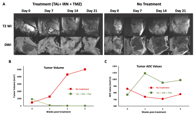 |
Multimodal MRI/MRS evaluation of treatment response in a patient-derived xenograft model of Ewing sarcoma
Puneet Bagga1, Jeffrey Steinberg2, Walter Akers2, Matthew Scoggins1, Zoltan Patay1, Beth McCarville1, Burkhard Hoeckendorf3, Khaled Khairy3, Michael Dyer3, and Beth Stewart3
1Department of Diagnostic Imaging, St Jude Children's Research Hospital, Memphis, TN, United States, 2Center for In Vivo Imaging and Therapeutics (CIVIT), St Jude Children's Research Hospital, Memphis, TN, United States, 3Department of Developmental Neurobiology, St Jude Children's Research Hospital, Memphis, TN, United States
A recent development in multi-modality treatment with poly ADP-ribose polymerase inhibitors (PARPi’s) combined with DNA damaging agents (temozolomide), is increasingly recognized as an important approach for curing or prolonging survival in patients with recurrent Ewing sarcoma (EWS). In this study, we employed multimodal quantitative imaging methods to predict treatment response in EWS. We used T2-weighted MRI, diffusion-weighted MRI, and 1H MRS in a patient derived xenograft model of EWS. We envisage that multi-parametric MR indices will be able to evaluate the treatment response and predict treatment response in the tumors.
|
The International Society for Magnetic Resonance in Medicine is accredited by the Accreditation Council for Continuing Medical Education to provide continuing medical education for physicians.