Digital Posters
Cardiovascular Parameter Mapping
ISMRM & SMRT Annual Meeting • 15-20 May 2021

Digital Posters
Cardiovascular Parameter Mapping
| Concurrent 3 | 13:00 - 14:00 |
3597.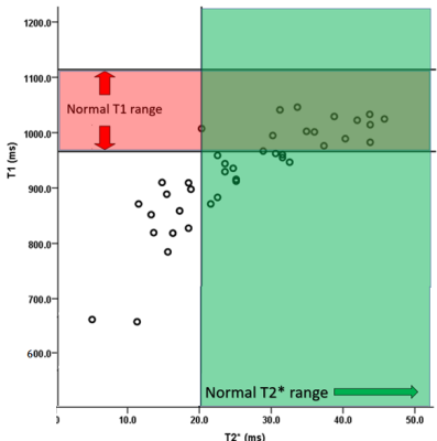 |
T2* VERSUS NATIVE T1, T2 MAPPING IN PATIENTS WITH SUSPECTED MYOCARDIAL IRON OVERLOAD
Rosh Varghese Georgy1, Elizabeth Joseph1, Aparna Irodi1, Binita Riya Chacko1, Leena Vimala Robinson1, and Roshan Samuel Livingstone1
1Department of Radiology, Christian Medical College, Vellore, India
Parametric techniques like native T1 and T2 mapping showed a strong positive correlation with T2* in the non-invasive assessment of cardiac iron overload. T1 mapping was shown to be superior to T2 mapping in the diagnosis of cardiac iron overload. 30% of the study population had normal T2* values, but low T1 values. T1 mapping may be more sensitive in the detection of patients with early/mild cardiac iron overload, who are being missed by T2*.
|
|||
3598. |
Free-breathing, motion-resolved myocardial T1 mapping using inversion-recovery radial FLASH and model-based reconstruction
Xiaoqing Wang1,2, Sebastian Rosenzweig1,2, Moritz Blumenthal1, Zhengguo Tan1,2, Nick Scholand1,2, and Martin Uecker1,2,3,4
1University Medical Center Göttingen, Göttingen, Germany, 2Partner Site Göttingen, German Centre for Cardiovascular Research (DZHK), Göttingen, Germany, 3Cluster of Excellence "Multiscale Bioimaging: from Molecular Machines to Networks of Excitable Cells" (MBExC), University of Göttingen, Göttingen, Germany, 4Campus Institute Data Science (CIDAS), University of Göttingen, Göttingen, Germany
In this work, we develop a motion-resolved model-based reconstruction for free-breathing multi-phase myocardial T1 mapping using a free-running inversion-recovery radial FLASH sequence. Initial results on an experimental phantom and two healthy subjects have demonstrated that the proposed method could achieve motion-resolved T1 mapping at a spatial resolution of 1.33 × 1.33 × 6 mm3 with good accuracy, precision and reproducibility.
|
|||
3599.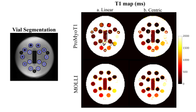 |
ProMyoT1: Open-source Inversion recovery myocardial T1 mapping sequence for fast prototyping
Andreia S Gaspar1, Nuno A da Silva2, and Rita G Nunes1
1Institute for Systems and Robotics - Lisboa and Department of Bioengineering, Instituto Superior Técnico, Universidade de Lisboa, Lisboa, Portugal, 2Hospital da Luz Learning Health, Lisboa, Portugal
T1 mapping is a crucial technique for myocardial tissue characterization. T1-mapping MOLLI sequences are widely used in the clinic but fast prototyping for concept testing and improvements is challenging and time consuming as it requires knowledge of the vendor specific programming environment. The use of an open-source framework would enable faster sequence prototyping, and facilitate reproducibility studies of new T1 mapping techniques. The aim of this work is to develop an open-source Prototype of Myocardial T1 mapping (ProMyoT1) using Pulseq to implement an inversion recovery T1 mapping sequence based on the MOLLI inversion/triggering scheme.
|
|||
3600. |
A novel MOLLI T1 analysis method with data matching for reduced T1 underestimation
Yuta Endo1, Haruna Shibo1, Makoto Amanuma1, Kuninori Kobayashi1, and Shigehide Kuhara1
1Faculty of Health Sciences, Kyorin University, Mitaka-shi, Tokyo, Japan
Myocardial T1 mapping using the modified Look-Locker inversion recovery (MOLLI) method shows good precision of T1 measurements, but their underestimation has long remained a problem in accurate T1 estimation. We developed a novel T1 analysis method by applying a data-matching strategy to the MOLLI method. The analysis method we propose estimates the T1 map by matching the MOLLI signals of a subject to dictionary data pixel-wise, which are the Look-Locker signals calculated in advance for various tissue parameters. The proposed method sufficiently reduces T1 underestimation of the MOLLI method and obtains more accurate T1 values.
|
|||
3601.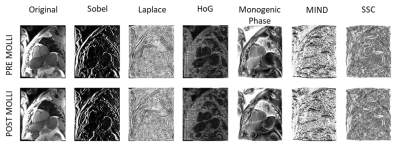 |
Evaluation of various image descriptor for motion correction between pre- and post-injected cardiac T1 maps, based on a demon algorithm
Habib Rebbah1, Anaîs Bernard1, Julien Rouyer1, and Timothé Boutelier1
1Research & Innovation, Olea Medical, La Ciotat, France
The registration of pre- and post-injected T1 maps is a multimodal image registration problem. To employ algorithms based on intensity preservation assumption, one need to use image descriptor. By comparing six of them, we aim to choose the most adequate one to our issue. We conducted our analyzes using a STEMI-patients database. The Sobel filter emerge as the most accurate and precise descriptor. The study of the results shows that the position of the acquired slice has no effect on the performances of the algorithm, while the size of the lesion and the cross-slice motion are inversely correlated to them
|
|||
3602. |
Multi-Echo GRASP for Cardiac T2*-Relaxometry
Thomas Lottner1, Joannes Fischer1, Simon Reiss1, Lars Bielak1, Timo Heidt2, Ali Caglar Özen1,3, Julien Thielmann2, Constantin von zur Mühlen2, and Michael Bock1
1Department of Radiology, Medical Physics, Faculty of Medicine, University Medical Center Freiburg, University of Freiburg, Freiburg, Germany, 2University Heart Center Freiburg, Faculty of Medicine, University of Freiburg, Freiburg, Germany, 3German Consortium for Translational Cancer Research Partner Site Freiburg, German Cancer Research Center (DKFZ), Heidelberg, Germany
Iron accumulation is a side effect from regular blood transfusions, which are a common treatment for diseases like hemochromatosis and sickle cell disease. T2* mapping can be a useful tool in therapy planning as it allows for monitoring of the iron accumulation in the heart. Multi-echo-GRASP offers a method of T2* mapping for all phases of the cardiac cycle. The acquisition can be performed in free breathing which is beneficial for patients with an impaired cardiac and/or respiratory capacity.
|
|||
3603.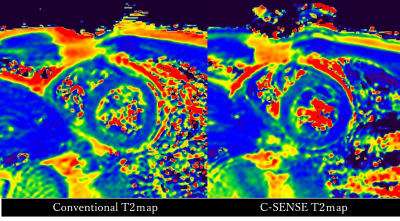 |
Improvement of multi-echo gradient-spin-echo (mGraSE) myocardial T2 mapping utilizing Compressed SENSE reconstruction framework
Isao Shiina1, Michinobu Nagao2, Masami Yoneyama3, Yasuhiro Goto4, Kazuo Kodaira4, takumi ogawa4, Mamoru Takeyama4, Isao Tanaka4, and Shuji Sakai2
1Radiological Services, Tokyo Women's Medical University Hospital, tokyo, Japan, 2Department of Diagnostic Imaging and Nuclear Medicine, Tokyo Women's Medical University Hospital, Tokyo, Japan, 3Philips Electronics Japan, Tokyo, Japan, 4Department of Radiological Services, Tokyo Women's Medical University Hospital, Tokyo, Japan
Myocardial T2 mapping using a multi-echo gradient-spin-echo (mGraSE) is widely used in clinical practice for quantitatively evaluating myocardial tissue properties with single breath-hold scan, but the limited scan time during the breath-hold period often results in poor signalto-noise ratio. Compressed SENSE has recently been developed to accelerate the acquisition time. Although C-SENSE is basically applied for non-EPI scans, mGraSE can also be applied the C-SENSE reconstruction framework. mGraSE myocardial T2 mapping with C-SENSE demonstrated improved image quality with higher image uniformity entire the shot-axis myocardium compared with conventional SENSE.
|
|||
3604.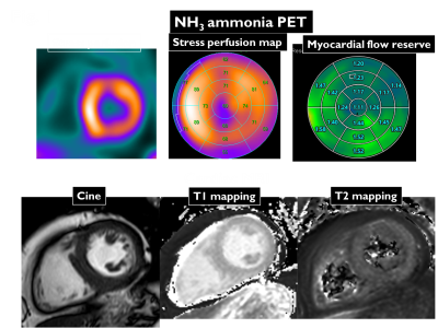 |
Native T1 and T2 mapping cardiovascular magnetic resonance for detection of cardiac allograft vasculopathy after heart transplantation
Yurie Shirai1, Michinobu Nagao1, Noriko Kikuchi1, Atsushi Yamamoto1, Yuka Matsuo1, Risako Nakao1, Kiyoe Ando1, Eri Watanabe1, Shinichi Nunoda1, Masami Yoneyama2, and Syuji Sakai1
1Tokyo Women’s Medical University, Tokyo, Japan, 2Philips Japan, Tokyo, Japan
We investigate the diagnostic performance of native T1 and T2 mapping cardiovascular magnetic resonance imaging (CMR) for cardiac allograft vasculopathy (CAV) after heart transplantation, using myocardial flow reserve (MFR) estimated by 13N-ammonia (NH3) positron emission tomography (PET) as reference. Non-contrast native T1 mapping is a minimally invasive and effective method for monitoring CAV. In contrast, the effectiveness of T2 mapping could not be demonstrated.
|
|||
3605.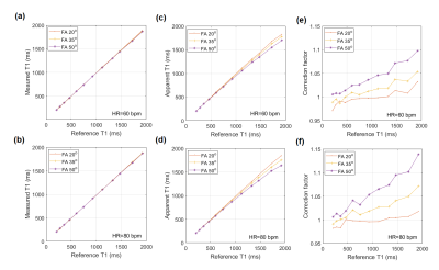 |
The suggested possibility of the T2rho contrast in the modified Look-Locker MRI technique used for the T1 quantification
Seonghwan Yee1, Lorna Browne1, and Justin Honce1
1Radiology, University of Colorado Anschutz Medical Campus, Aurora, CO, United States
The modified Look-Locker imaging (MOLLI) technique is widely used in cardiac imaging for T1 mapping. When the MOLLI technique is performed, the B1 field, perpendicular to the magnetization, is continually applied during the steady state imaging. Hence, the relaxation of the magnetization during this time may be linked to the T2rho (the T2 relaxation time constant in the rotating frame) contrast, sensitive to the iron content. Here, the theoretical background and a preliminary phantom imaging results are presented to explore the possibility of extracting T2rho contrast from the MOLLI sequence.
|
|||
3606.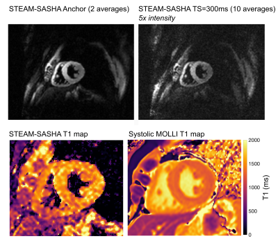 |
Right ventricular T1 mapping using a novel STEAM-based approach: STEAM-SASHA
Malte Roehl1,2, Peter D Gatehouse1,2, Pedro F Ferreira1,2, Sonya V Babu-Narayan1,2, David N Firmin1,2, Dudley J Pennell1,2, Sonia Nielles-Vallespin1,2, and Andrew D Scott1,2
1National Heart and Lung Institute, Imperial College London, London, United Kingdom, 2Cardiovascular Magnetic Resonance Unit, Royal Brompton Hospital, London, United Kingdom
Here we propose a new method to reduce the influence of blood and epicardial fat on right ventricular T1 mapping. This is achieved by leveraging the excellent blood and fat suppression provided by the stimulated echo acquisition mode (STEAM) EPI sequence in a novel saturation recovery based (SASHA) single-shot T1 mapping sequence. The novel (STEAM-SASHA) approach is evaluated and compared to a standard modified Look-Locker imaging sequence in both phantom and in vivo measurements in the septum and right ventricle.
|
|||
3607.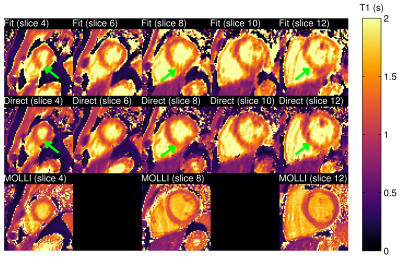 |
Three-dimensional cardiac T1 mapping using subspace and sparsity constrained direct estimation
Thibault Marin1, Paul K. Han1, Yue Zhuo1, Yanis Djebra1,2, Fang Liu1, Georges El Fakhri1, and Chao Ma1
1Massachusetts General Hospital, Harvard Medical School, Boston, MA, United States, 2LTCI, Telecom Paris, Institut Polytechnique de Paris, Paris, France
Cardiac T1 mapping is a powerful MR imaging technique for quantitative assessment of microstructural changes in myocardial tissues. Existing methods are limited in terms of spatial coverage and through-plane resolution due to limitations in acquisition speed and the presence of cardiac and respiratory motion. This work presents a direct reconstruction framework, which allows estimation of 3D T1 maps from sparsely sampled k-space data using physical modeling through the Bloch equation, low-rank constraints on the dynamic images and sparsity constraints on the estimated T1 maps.
|
|||
3608.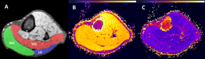 |
Altered T1 and T2 relaxation times in leg muscles are linked to hemodynamic and ambulatory parameters in patients with Peripheral Artery Disease.
Constance J Mietus1, Yue Gao1, Mariano G Uberti2, Nicholas G Lambert1, Panagiotis Koutakis3, Evlampia Papoutsi3, Jonathan R Thompson1, Holly K DeSpiegelaere4, Michael D Boska2, Sara A Myers5, George P Casale1, Iraklis I Pipinos1,4, and Balasrinivasa R Sajja2
1Department of Surgery, University of Nebraska Medical Center, Omaha, NE, United States, 2Department of Radiology, University of Nebraska Medical Center, Omaha, NE, United States, 3Department of Biology, Baylor University, Waco, TX, United States, 4Department of Surgery and VA Research Service, VA Nebraska-Western Iowa Health Care System, Omaha, NE, United States, 5Department of Biomechanics, University of Nebraska Omaha, Omaha, NE, United States
Understanding the quantitative MRI features of the myopathy of Peripheral Artery Disease (PAD) may aid in grading disease severity, treatment response, and potentially predicting favorable response to exercise or revascularization surgery. In this study we explored the relationship between T1 and T2 relaxation times and measurements of hemodynamic and ambulatory performance in patients with PAD. T1 relaxation time positively correlated with ankle brachial index and peak plantar flexion and inversely correlated with claudication onset time. T2 relaxation time correlated positively with peak plantar flexion and inversely correlated with ischemic window, claudication onset time, and peak walking time.
|
|||
3609.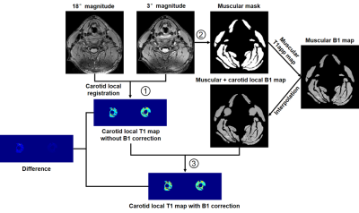 |
Carotid T1 mapping with Muscle Referenced B1 Mapping Correction Using Variable Flip Angle Imaging without Extra Scan
Huiyu Qiao1, Shuo Chen1, Zihan Ning1, Hualu Han1, Rui Shen1, and Xihai Zhao1
1Center for Biomedical Imaging Research, Department of Biomedical Engineering, School of Medicine Tsinghua University, Beijing, China
Carotid T1 mapping calculated by variable flip angle imaging usually needs an additional B1 mapping to correct nominal flip angle. However, B1 mapping techniques are difficult to implement in clinical settings. This study proposed a muscle referenced B1 mapping for a more accurate carotid T1 mapping using variable flip angle imaging but without an additional B1 mapping scan. The proposed muscle referenced B1 mapping could effectively adjust the B1 inhomogeneity, particularly for the B1 inhomogeneity between left and right carotid vessel walls. Furthermore, the T1 mapping with muscle referenced B1 mapping correction showed the capability of identifying the intraplaque hemorrhage.
|
|||
3610.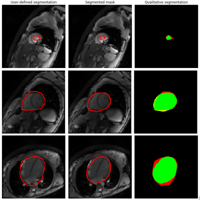 |
Improving T1 mapping robustness by automatic segmentation of myocardial tissue in MOLLI series
María A Iglesias1, Daniel Lorenzatti2, José T Ortiz2, Susanna Prat2, Adelina Doltra2, Rosario J Perea2, Teresa M Caralt2, Oscar Camara1, Gaspar Delso3, and Marta Sitges2
1Universitat Pompeu Fabra, Barcelona, Spain, 2Hospital Clínic de Barcelona, Barcelona, Spain, 3GE Healthcare, Barcelona, Spain
In this study, an image processing pipeline based on Deep Learning is presented to identify myocardial tissue in MOLLI series. The main goal is to provide an automated tool to evaluate the impact of motion correction on cardiac T1 mapping.
|
|||
3611.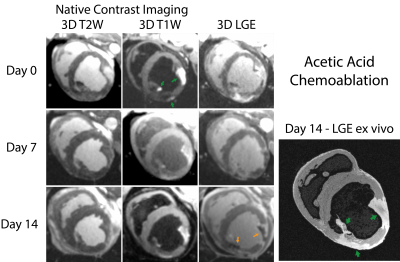 |
Visualization of Coronary Myocardial Chemoablation: Comparison of Ethanol and Acetic Acid
Daniel A Herzka1, Rajiv A Ramasawny1, Chris G. Bruce1, Delaney R. McGuirt1, William H. Schenke1, Jaffar M. Khan1, Adrienne E. Campbell-Washburn1,2, Aravindan A Kolandaivelu1,3, Toby A Rogers1,4, and Robert J. Lederman1
1NHLBI, Division of Intramural Research, National Institutes of Health, Bethesda, MD, United States, 2Biophysics and Biochemistry Branch, Division of Intramural Research, NHLBI, NIH, Bethesda, MD, United States, 3Division of Cardiology, Department of Medicine, Johns Hopkins University School of Medicine, Baltimore, MD, United States, 4Department of Cardiology, Medstar Washington Hospital Center, Washington, DC, United States
In this work, we visualize chemoablation lesions created by intracoronary injection as used in alcohol septal ablation for the treatment of hypertrophic obstructive cardiomyopathy. 3D native contrast and gadolinium-enhanced MRI were examined. Two chemoablation agents were used: standard ethanol and a potential alternative agent, glacial acetic acid. In swine, both 3D native contrast and gadolinium-enhanced imaging clearly delineated lesion extent acutely and up to two weeks post ablation. Acutely, chemoablation with ethanol induced a 22% increase in T1 within lesion cores and acetic acid yielded a 36% decrease.
|
|||
 |
3612.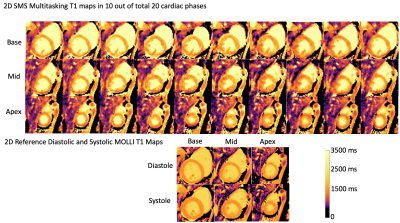 |
Simultaneous Multi-slice Cardiac MR Multitasking for Motion-Resolved, Non-ECG, Free-Breathing Joint T1-T2 Mapping
Xianglun Mao1, Hsu-Lei Lee1, Sen Ma1, Zhehao Hu1,2, Fei Han3, Yibin Xie1, Debiao Li1,2, and Anthony G Christodoulou1,2
1Biomedical Imaging Research Institute, Cedars-Sinai Medical Center, Los Angeles, CA, United States, 2Department of Bioengineering, University of California in Los Angeles, Los Angeles, CA, United States, 3Siemens Medical Solutions Inc., Los Angeles, CA, United States
Cardiac MR Multitasking has been shown to acquire simultaneous T1 and T2 maps without ECG or breath-holds. Simultaneous multi-slice (SMS) acquisition has the potential to increase the clinical value of CMR by decreasing the exam time. In this work, we developed an SMS, non-ECG and free-breathing MR Multitasking technique and demonstrated that it had consistent T1, T2 measurements when compared with the overall-slower series of reference protocols (MOLLI, T2-prep FLASH).
|
||
3613.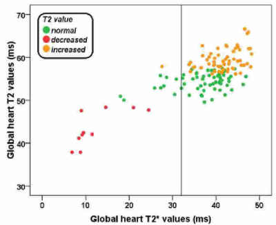 |
Myocardial tissue characterization by T2 mapping in thalassemia major
Antonella Meloni1, Nicola Martini1, Rita Laura Borrello2, Vincenzo Positano1, Laura Pistoia1, Calogera Gerardi3, Mauro Murgia4, Valentina Carrai5, Monica Benni6, Sara Gentili7, Roberto Pedrinelli2, and Alessia Pepe1
1MRI Unit, Fondazione G. Monasterio CNR-Regione Toscana, Pisa, Italy, 2Università degli Studi di Pisa, Pisa, Italy, 3Presidio Ospedaliero "Giovanni Paolo II" - Distretto AG2 di Sciacca, Sciacca (AG), Italy, 4Ospedale San Martino di Oristano, Oristano, Italy, 5Azienda Ospedaliero - Universitaria Careggi, Firenze, Italy, 6Policlinico S. Orsola "L. e A. Seragnoli", Bologna, Italy, 7Ospedale "San Donato", Arezzo, Italy
The T2 mapping does not offer any advantage over the T2* technique in terms of sensitivity for myocardial iron overload assessment. However, more than half of patients with thalassemia major had an increased T2 value, that may be caused by the presence of myocardial inflammation and/or edema.
|
|||
3614.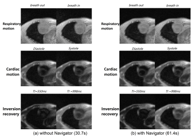 |
Self-Navigated Spiral T1 CMR Multitasking
Jingyuan Lyu1, Qi Liu1, Zhongqi Zhang2, Jian Xu1, and Weiguo Zhang1
1UIH America, Inc., Houston, TX, United States, 2United Imaging Healthcare, Shanghai, China
This abstract presents a new approach to accelerated T1 mapping of the heart under free-breathing and without ECG. Compared with traditional radial sampling trajectory, spiral sampling offers the possibility to get ride of navigator data in the framework of “multitasking”, though at an extra cost of increased susceptibility to system imperfections such as gradient delay.
|
|||
3615.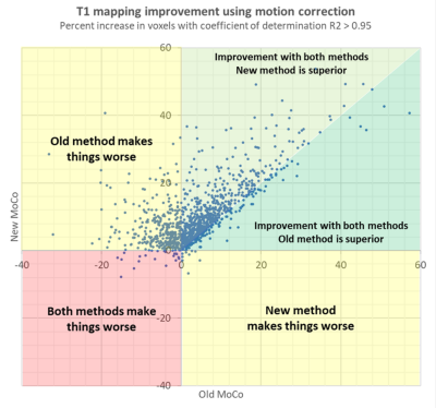 |
Clinical validation of a dedicated motion correction algorithm for cardiac MOLLI series using a quantitative metric
Gaspar Delso1, Laura Farre2, Daniel Lorenzatti3, Santi Sotes3, Adelina Doltra3, Susanna Prat3, Rosario J Perea3, Teresa M Caralt3, José T Ortiz3, and Marta Sitges3
1GE Healthcare, Barcelona, Spain, 2Universitat de Barcelona, Barcelona, Spain, 3Hospital Clínic de Barcelona, Barcelona, Spain
Cardiac T1-mapping methods often require motion correction. The contrast changes intrinsic to the inversion recovery series often used for this purpose can occasionally cause registration errors, resulting in inaccurate T1 values. It has been shown, using a large database of clinical cases, that accounting for those contrast changes markedly increases the robustness of the correction. A new metric of cardiac anatomy alignment had to be defined, in order to automate the quantitative analysis of the database. This metric was shown to correlate with the visual scoring of misalignment in MOLLI series.
|
|||
3616.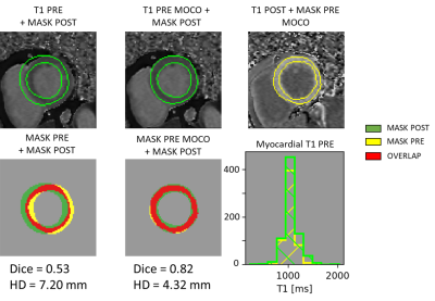 |
Evaluation of cardiac pre- and post-T1 maps registration for extracellular volume computation
Habib Rebbah1, Anaïs Bernard1, Julien Rouyer1, and Timothé Boutelier1
1Department of Research & Innovation, Olea Medical, La Ciotat, France
The myocardial ECV involves manual segmentation on pre- and post-injection T1 maps, which is a time-consuming process. An initial registration of the two maps represents a suitable solution to reduce the analyze duration. Here, we propose to compare the ECV obtained after the registration against the manual computed one using a STEMI-patients database. If the difference between the two approaches was significant, the bias remained thin (less than 1% for the remote part and 3% for the infarct part). These results and the speed of the registration (1s per slice) tip the scales in favor of its use
|
The International Society for Magnetic Resonance in Medicine is accredited by the Accreditation Council for Continuing Medical Education to provide continuing medical education for physicians.