Digital Posters
Cardiovascular Tissue Characterization: Beyond Relaxometry
ISMRM & SMRT Annual Meeting • 15-20 May 2021

Digital Posters
Cardiovascular Tissue Characterization: Beyond Relaxometry
| Concurrent 3 | 13:00 - 14:00 |
3617.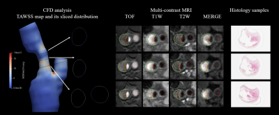 |
Time-averaged wall shear stress: a potential indicator for carotid intra-plaque hemorrhage
Rui Shen1, Huiyu Qiao1, Zihan Ning1, Dongye Li2, Dandan Yang1, and Xihai Zhao1
1Center for Biomedical Imaging Research, Tsinghua University, Beijing, China, 2Department of Radiology, Sun Yat-Sen Memorial Hospital, Sun Yat-Sen University, Guangzhou, China
We investigated the relationship between the TAWSS and presence of plaque components in histology combined with plaque burden metrics interpreted from multi-contrast MR images among patients scheduled for CEA within one week. Based on 18 patients with 60-slice histological and MR images, correlation analysis revealed that there is an association between the presence of IPH and TAWSS. In addition, our findings indicated that TAWSS can improve the performance of plaque burden metrics, MWT in predicting presence of IPH.
|
|||
3618.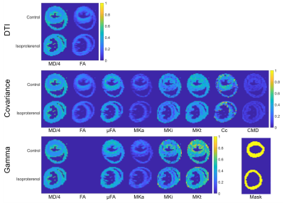 |
Multidimensional Diffusion MRI in the Ex Vivo Mouse Heart
Irvin Teh1, Samo Lasič2,3, Henrik Lundell3, Beata Wereszczyńska1, Matthew Budde4, Erica Dall'Armellina1, Nadira Yuldasheva1, Filip Szczepankiewicz5,6,7, and Jürgen E. Schneider1
1Leeds Institute of Cardiovascular and Metabolic Medicine, University of Leeds, Leeds, United Kingdom, 2Random Walk Imaging, Lund, Sweden, 3Danish Research Centre for Magnetic Resonance, Centre for Functional and Diagnostic Imaging and Research, Copenhagen University Hospital Hvidovre, Copenhagen, Denmark, 4Department of Neurosurgery, Neurobiology, and Anatomy, Medical College of Wisconsin, Milwaukee, WI, United States, 5Clinical Sciences, Lund University, Lund, Sweden, 6Harvard Medical School, Boston, MA, United States, 7Brigham and Women's Hospital, Boston, MA, United States
Multidimensional diffusion MRI, specifically, tensor-valued encoding is a promising technique for improving specificity in microstructural measurements in the myocardium beyond that achievable with DTI. Tensor-valued encoding data combining linear and spherical tensor encoding were acquired in ex vivo mouse hearts at 7T, including an isoproterenol-induced model of hypertrophy. Covariance and gamma fitting methods were employed to reconstruct parameter maps reflecting the isotropic and anisotropic components of the diffusion signal kurtosis. The results were consistent across both methods, and highlight the potential of multidimensional diffusion MRI for improving specificity in cardiac diffusion MRI.
|
|||
3619.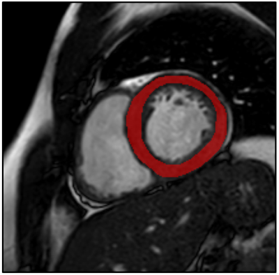 |
Conventional balanced SSFP magnetic resonance images reveal patterns of clinically suspected myocarditis using texture analysis
Evin Ina Papalini1, Christian Polte2, and Kerstin Magdalena Lagerstrand1
1Institute of Clinical Sciences, Sahlgrenska Academy, University of Gothenburg, Gothenburg, Sweden, Gothenburg, Sweden, 2Institute of Medicine, Sahlgrenska Academy, University of Gothenburg, Gothenburg, Sweden, Gothenburg, Sweden
Myocarditis is a common inflammatory disease in the myocardium, associated with acute heart failure, chronic dilated cardiomyopathy and sudden cardiac death. Current clinical diagnosis is based on magnetic resonance imaging, including administration of gadolinium-based contrast agents. We propose that conventional balanced steady-state-free-precession magnetic resonance imaging images reveals quantitative diagnostic features based on texture analysis. Our results showed that the texture features, in specific Variance, Gradient Mean and Sum Average, were able to significantly separate patients with and without myocarditis using conventional balanced steady-state-free-precession magnetic resonance imaging images.
|
|||
3620.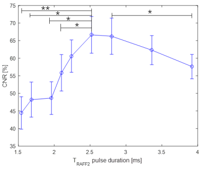 |
Imaging of cardiac skeleton without contrast agents
Yi Li1, Jiri Mares1, and Timo Liimatainen1
1Research Unit of Medical Imaging, Physics and Technology, University of Oulu, Oulu, Finland
We applied Relaxation along Fictitious Field with rank n (RAFFn) to clinical 3T scanner to study RAFFn contrast between cardiac skeleton and myocardium with multiple RAFFn refocusing times i.e. pulse durations in ex vivo porcine hearts. We found the relationship between relaxation times and RAFFn pulse duration in both cardiac skeleton and myocardium. Furthermore, the optimal pulse duration to gain maximum contrast was close to 2.5 ms. RAFF2 and T2 maps demonstrated higher contrast between cardiac skeleton and myocardium tissues when compared to T1 and T1ρ.
|
|||
3621.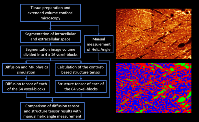 |
Microstructure-Based Simulation of Myocardial Diffusion Using Extended Volume Confocal Microscopy
Alexander James Wilson1, Kevin M Moulin2, Gregory B Sands3, and Daniel B Ennis2
1Radiology, Stanford University, Palo Alto, CA, United States, 2Radiology, Stanford University, Stanford, CA, United States, 3Auckland Bioengineering Institute, University of Auckland, Auckland, New Zealand
Aiming to improve noninvasive assessment of tissue microstructure, a physics-based simulation of diffusion tensor imaging (DTI) was used to compare helix angles between DTI and structure tensor (ST) analyses of a confocal myocardial image stack. Rodent myocardium was imaged using extended volume confocal microscopy producing a high-resolution image volume, which was segmented into intracellular and extracellular compartments. The image volume was divided into voxel-blocks, which served as the DTI simulation voxels. The DTI acquisition showed good agreement with both the ST and manually measured helix angles. This proof-of-concept work demonstrates the feasibility of direct comparison of confocal images with DTI.
|
|||
3622.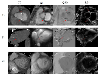 |
Quantitative Susceptibility Mapping for Mitral Annulus Calcification Detection via Validation of Computed Tomography/Echocardiography
Jiahao Li1,2, Hannah Mitlak3, Lakshmi Nambiar3, Romina Tafreshi3, Jiwon Kim3, Yi Wang1,2, Jonathan W. Weinsaft3, and Pascal Spincemaille2
1Biomedical Engineering, Cornell University, Ithaca, NY, United States, 2Radiology, Weill Cornell Medicine, New York, NY, United States, 3Medicine, Weill Cornell Medicine, New York, NY, United States
Mitral annulus calcification is common in patients with mitral regurgitation and impacts prognosis and response to mitral valve interventions. While cardiac MRI is widely used to assess MR, identification of MAC is a key gap in current cardiac MRI. Quantitative susceptibility mapping is an emerging MRI tissue characterization approach that is sensitive to calcium because it is strongly diamagnetic. Here, we demonstrate the feasibility of using cardiac QSM for detecting MAC in patients using Computed Tomography and Echocardiography as reference.
|
|||
3623.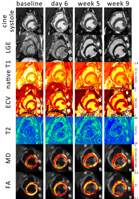 |
Microstructural CMR imaging in a longitudinal pig model of acute to chronic myocardial infarction
Christian T Stoeck1, Constantin von Deuster1, Maximilian Fuetterer1, Malgorzata Polacin1,2, Conny F Waschkies1, Robbert JH van Gorkum1, Mareike Kron3, Thea Fleischmann3, Nikola Cesarovic3,4, Miriam Weisskopf3, and Sebastian Kozerke1
1Institute for Biomedical Engineering, University and ETH Zurich, Zurich, Switzerland, 2Institute of Diagnostic and Interventional Radiology, University Hospital Zurich, Zurich, Switzerland, 3Division of Surgical Research, University Hospital Zurich, Zurich, Switzerland, 4Institute of Translational Cardiovascular Technologies, ETH Zurich, Zurich, Switzerland
In this work we compare non-contrast enhanced imaging methods such as native T1, T2 mapping and cardiac diffusion tensor imaging, to ECV measurements in a longitudinal porcine study of myocardial infarction. T2 is elevated only during the acute stage. During the transition from acute to chronic stage T1 remains unchanged and ECV as well as MD progressively increase while FA decreases. During the acute stage, T2 mapping provides highest contrast whereas diffusional metrics showed larger changes during the chronic stage compared to native relaxometry showing the potential of DTI for probing dynamic myocardial changes upon an ischemic event.
|
|||
3624.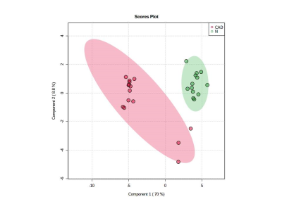 |
Metabolic changes in coronary artery disease assessed using 1H NMR Metabolomics
Pawan Kumar1, Uma Sharma1, Rajeev Narang2, and Sujeet Mewar1
1Nuclear Magnetic Resonance and MRI Facility, All India Institute of Medical Sciences, New Delhi, India, 2Cardiology, All India Institute of Medical Sciences, New Delhi, India
Coronary artery disease (CAD) is caused by building up of plaque in the arteries of the heart, which narrows the lumen of arteries. Present study investigated the metabolic profile of blood plasma using 1H-NMR spectroscopy for distinguishing coronary artery disease (CAD) patients from healthy control (N). The results showed significant differences in the concentration of several metabolites like lactate, pyruvate, choline, acetate and alanine in the blood plasma of CAD patients in comparison to healthy controls suggesting alterations of several metabolic pathways including glycolysis, gluconeogenesis which may be related to the development of CAD.
|
|||
3625.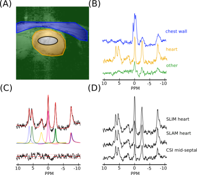 |
Increased SNR and improved reproducibility for cardiac 31P MRS at 7T using compartmentalized spectroscopy
Andrew Tyler1,2, Justin Y C Lau1, Jane Ellis1, Jack J Miller1,2,3, Paul A. Bottomley4, Christopher T Rodgers1,5, Damian J Tyler1,2, and Ladislav Valkovic1,6
1Oxford Centre for Clinical Cardiac Magnetic Resonance Research, University of Oxford, Oxford, United Kingdom, 2Department of Physiology, Anatomy & Genetics, University of Oxford, Oxford, United Kingdom, 3Department of Physics, University of Oxford, Oxford, United Kingdom, 4The Division of MR Research, Johns Hopkins Medicine, Baltimore, MD, United States, 5Wolfson Brain Imaging Centre, University of Cambidge, Cambridge, United Kingdom, 6Department of Imaging Methods, Institute of Measurement Science, Slovak Academy of Sciences, Bratislava, Slovakia
The intrinsically low SNR and long acquisition times of 31P spectroscopy make translation to clinical practice very challenging, even at ultra-high fields. In this study, 12 healthy volunteers were scanned twice at 7T with a short 31P CSI and reduced k-space acquisitions for reconstruction with the SLAM and SLIM algorithms. PCr/ATP ratio and PCr SNR were computed for each scan and coefficients of reproducibility and variability were calculated. Compared to a Fourier based reconstruction of the short 31P acquisition, SNR was significantly improved and PCr/ATP was maintained when SLAM and SLIM reconstructions were used.
|
|||
3626.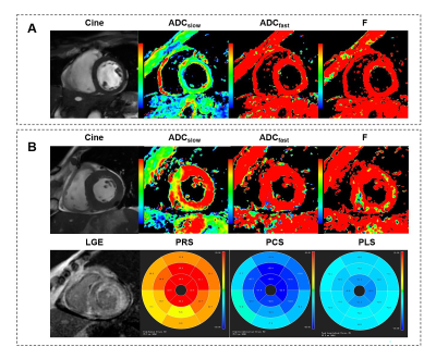 |
Evaluating the Myocardial Diffusion Status in Cardiac Amyloidosis: A Novel Intravoxel Incoherent Motion Diffusion-weighted MR Imaging Study
Mengdi Jiang1, Xianghua Huang2, Guifen Yang3, Weiqiang Dou4, and Yong Shen5
1Department of Medical Imaging, Jinling Hospital, Medical School of Nanjing University, NanJing, China, 2National Clinical Research Center of Kidney Disease, Jinling Hospital, Medical School of Nanjing University, NanJing, China, 3Department of Nuclear Medical, Jinling Hospital, Medical School of Nanjing University, NanJing, China, 4GE Healthcare,MR Research China, BeiJing, China, 5GE Healthcare,MR Enhanced Application China, BeiJing, China
The purpose of this study was to evaluate the diagnostic value of myocardial diffusion and mechanical properties by using IVIM-DWI imaging, feature tracking and native T1 in patients with cardiac amyloidosis (CA). The relationships of strain, native T1, IVIM-derived parameters (ADCslow, ADCfast and F) and late gadolinium enhancement (LGE) were analyzed based on six mid-ventricle subregions. Significantly different IVIM related parameters were found in patients than healthy controls. Furthermore, IVIM parameters also showed significant correlation with peak strain and nativeT1. With these findings, IVIM parameters has proven as effective biomarkers in the diagnosis of cardiac microcirculation in CA patients.
|
|||
3627.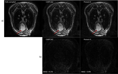 |
3D wave Cardiac Magnetic Resonance for myocardial scar tissue characterization
Quentin Lebret1,2, Pierre Bour1,2, Valéry Ozenne1,2, Nestór Pallares-Lupon1,2, Richard Walton1,2, and Bruno Quesson1,2
1IHU Liryc, Electrophysiology and Heart Modeling Institute, fondation Bordeaux Université, Pessac, France, 2Univ. Bordeaux, INSERM, Centre de recherche CardioThoracique de Bordeaux, U1045, Bordeaux, France
3D MRI of the myocardium after an ischemic attack could provide an accurate scar segmentation, much needed by the clinicians. However, this type of acquisition, usually obtained by late gadolinium enhancement (LGE), is very time consuming. We combined a wave acquisition with an inversion pulse and a variable density Poisson undersampling strategy to accelerate 3D cardiac imaging. Retrospectively subsampled images of a sheep heart were successfully reconstructed with an acceleration factor of 4, opening the path to a fast high-resolution 3D LGE acquisition.
|
|||
3628.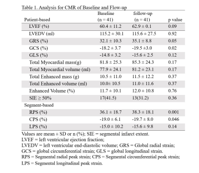 |
Quantification of strain analysis in coronary chronic total occlusion: A cardiovascular magnetic resonance imaging follow-up study
Lijun Zhang1, Jinfan Tian2, Xueyao Yang2, Jing An 3, Yi He4, and Xiantao Song2
1Department of Radiology, Beijing AnZhen Hospital, Capital Medical University, Beijing, China, 2Department of Cardiology, Beijing AnZhen Hospital, Capital Medical University, Beijing, China, 33Siemens Shenzhen Magnetic Resonance Ltd, Beijing, China, 4Department of Radiology, Beijing Friendship Hospital, Capital Medical University, Beijing, China
This study investigated the benefits of percutaneous coronary intervention (PCI) in patients with chronic total occlusions (CTOs) by using cardiac magnetic resonance imaging (CMR)-feature tracking (CMR-FT).The results showed the infarct size and LVEF did not increase significantly from baseline to 1 year of follow-up.However, global peak strains improved over time, and GCS showed significant treatment effect of CTO-PCI in the entire CMR population,which indicated improvement in left ventricular function.
|
|||
3629.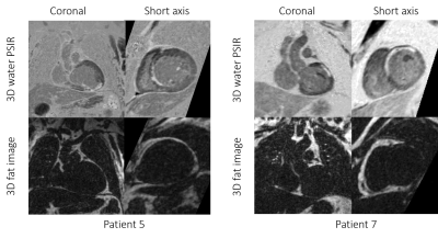 |
3D Whole Heart Grey-blood PSIR Slow Infusion Imaging for High-resolution Isotropic LGE Imaging
Alina Psenicny1, Reza Hajhosseiny1, Giorgia Milotta2, Karl P Kunze3, Radhouene Neji1,3, Amedeo Chiribiri1, Pier Giorgio Masci1, Claudia Prieto1, and René M Botnar1
1School of Biomedical Engineering and Imaging Sciences, King's College London, London, United Kingdom, 2Wellcome Centre for Human Neuroimaging, UCL Queen Square Institute of Neurology, University College London, London, United Kingdom, 3MR Research Collaborations, Siemens Healthcare Limited, Frimley, United Kingdom
A free-breathing water/fat motion corrected 3D grey blood phase sensitive inversion recovery (PSIR) late gadolinium enhancement (LGE) technique that achieves whole heart coverage and provided excellent agreement with 2D grey blood LGE MRI has been recently proposed. However, contrast washout due to the relatively long scan time of ~10 minutes may impact scar detection and limit spatial resolution. We therefore sought to investigate the feasibility of high-resolution slow infusion motion-corrected 3D grey blood PSIR-LGE in comparison with a conventional clinically used breath-held 2D grey blood PSIR-LGE MRI technique. Here we report first qualitative and quantitative results.
|
|||
3630.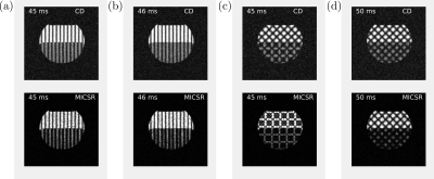 |
An Off-Resonance Insensitive Orthogonal CSPAMM Sequence (ORI-O-CSPAMM)
Hernán Mella1,2,3, Hui Wang4,5, and Sergio Uribe2,3,6
1Department of Electrical Engineering, Pontificia Universidad Católica de Chile, Santiago, Chile, 2Biomedical Imaging Center, Pontificia Universidad Católica de Chile, Santiago, Chile, 3Millennium Nucleus for Cardiovascular Magnetic Resonance, ANID - Millennium Science Initiative Program, Santiago, Chile, 4Philips, Cincinnati, OH, United States, 5Department of Radiology, Cincinnati Children’s Hospital Medical Center, Cincinnati, OH, United States, 6Department of Radiology, Pontificia Universidad Católica de Chile, Santiago, Chile
We propose the Off-Resonance Insensitive Orthogonal CSPAMM sequence (ORI-O-CSPAMM). ORI-O-CSPAMM allows the acquisition of a CSPAMM grid in half of the acquisition time, removing off-resonance effects during the tagging preparation. Additionally, ORI-O-CSPAMM allows Magnitude Image CSPAMM Reconstruction (MICSR), which improves the tagging contrast through the cardiac cycle and remove spurious phases gained during the readout.
|
|||
3631.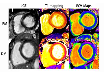 |
Assessment of myocardial involvement characteristics by cardiac MR imaging in patients with polymyositis and dermatomyositis
Changjing Feng1, Wangyan Liu1, Xiaoxuan Sun1, Qiang Wang1, Xiaomei Zhu1, Xiaoyue Zhou2, Yi Xu1, and Yinsu Zhu1
1The First Affiliated Hospital of Nanjing Medical University, Nanjing, China, 2Siemens Healthineers Ltd., Shanghai, China
Cardiac involvement is frequently observed in polymyositis (PM) and dermatomyositis (DM) but typically remains subclinical. Cardiac MR tissue characterization parameters could serve as early detection markers for myocardial involvement in PM/DM patients without overt LV dysfunction. PM and DM patients showed a different positive segment distribution of myocardial involvement, where PM patients was more serious than that in DM patients. Above all, CMR tissue characterization parameters can detect myocardial involvement characteristics between PM and DM and compare the differences between them.
|
|||
3632.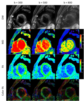 |
Assessment of different b values in motion-controlled myocardium spin echo diffusion tensor imaging in vivo
Yuli Huang1, Xinyang Wu2, Lifei Ma2, Haipeng Dong3, and Qian Jiang2
1Philips Healthcare (Suzhou) Co., Ltd, Suzhou, China, 2Philips (China) Investment Co., Ltd., Shanghai, China, 3Ruijin Hospital, Shanghai Jiaotong University School of Medicine, Shanghai, China
Diffusion MR is challenging in myocardium because of respiratory motion, cardiac pulsation, and field inhomogeneity in thorax. Diffusion tensor imaging using SE-EPI with respiratory navigation and cardiac triggering of 3 b values (300, 500, 800 s/mm2) was performed in 9 subjects. Sufficient image quality was obtained with b values of 300 and 500 s/mm2. Significant difference was found in SNR, CNR, MD, and FA between various b values while not between myocardium segments. Intermediate b values are recommended to achieve a balance of image quality and diffusion sensitivity . Sufficient myocardium quiescent duration is extremely important for optimal diffusion image quality.
|
|||
3633.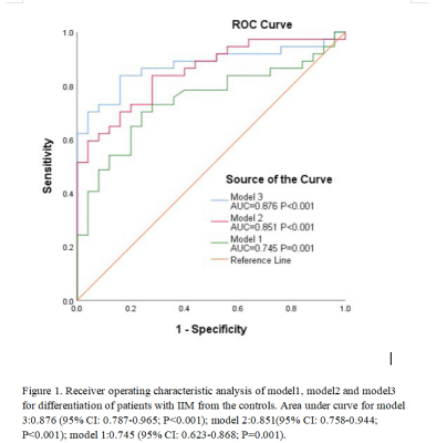 |
Early Cardiac Involvement Detected by CMR Feature Tracking in Idiopathic Inflammatory Myopathy with Preserved Ejection Fraction
Wangyan Liu1, Yinsu Zhu1, and Yi Xu1
1the first affiliated hospital of Nanjing medical university, Nanjing, China
We applied CMR-FT technique to evaluate LV and LA performance in IIM patients. The main findings were as follow: (1) compared with controls, the damaged LV strain in IIM patients mainly involved global and regional LV PS in longitudinal direction; (2) LA reservoir function and conduit function were impaired in IIM patients; (3) LA volumetric parameters and function parameters showed significant difference between IIM patients and the controls. (4) Combination of global LV longitudinal PS, LAVpre-ac index and Apical LV circumferential PS can improve diagnostic accuracy. Therefore, CMR-FT can help early diagnosis of cardiac involvement of IIM patients.
|
|||
3634.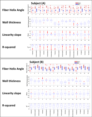 |
A Framework to extract and visualize the myofiber helix angle locally and globally from the cardiac diffusion tensor images
Mehrzad Tartibi1, Randall Lee2, Christopher Nguyen3, Jaume Coll-Font3,4, Youngho Seo2, and Qizhi Fang2
1DelBeat Inc., Berkeley, CA, United States, 2University of California San Francisco, San Francisco, CA, United States, 3Massachusetts General Hospital, Boston, MA, United States, 4Harvard Medical School, Boston, MA, United States
The DTI measurement contains measurement noise dominantly exaggerated at the myocardium boarders. Accurate fiber orientation will improve the accuracy of mathematical modeling. It may shed light on the short-term and long-term effects of scar tissue on the myofiber's morphology locally and globally. We have developed a filter based on the three measured DTI quantities of helix angle, fractional anisotropy, and mean diffusivity. This filter's result enabled us to automatically segment the myocardium's primary structural part and remove the trabeculae and papillary muscles. Hence, found a linear correlation between the fiber helix angle and the ventricle wall thickness.
|
|||
3635.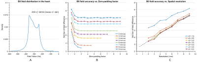 |
Optimization of B0 Simulation Strategy in the Human Heart based on CT Images at limited Field of View
Yun Shang1, Sebastian Theilenberg1, Laura M. Schreiber2,3, and Christoph Juchem1,4
1Department of Biomedical Engineering, Columbia University, New York, NY, United States, 2Chair of Cellular and Molecular Imaging, Comprehensive Heart Failure Center, University Hospital Wuerzburg, Wuerzburg, Germany, 3Department of Cardiovascular Imaging, Comprehensive Heart Failure Center, University Hospital Wuerzburg, Wuerzburg, Germany, 4Department of Radiology, Columbia University Medical Center, New York, NY, United States
B0 simulation in the heart based on thoracic CT images is a powerful tool to investigate cardiac B0 conditions in the general population for the development of an optimized cardiac B0 shimming strategy. Thoracic CT scans typically have a limited field of view posing a challenge to the accuracy of field simulations. Here we present a systematic analysis of errors introduced by B0 computations in the human heart from CT-based susceptibility distributions of limited field of view and present strategies to resemble B0 conditions in the human heart to achieve elevated accuracy with model-based addition of selected anatomical features.
|
|||
3636.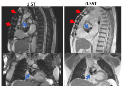 |
A Comparison of Metal Artifacts in Cardiovascular MRI at 0.55T and 1.5T
W. Patricia Bandettini1, Christine Mancini2, Sujata M. Shanbhag2, Jennifer Lynn Henry2, Margaret M. Lowery2, Marcus Y. Chen2, and Adrienne E. Campbell-Washburn2
1NIH/NHLBI, Bethesda, MD, United States, 2NATIONAL INSTITUTES OF HEALTH/NHLBI, BETHESDA, MD, United States
Low-field MRI may offer reduced artifacts in patients with implanted metallic devices. In this early comparison, we sought to evaluate the appearance of artifacts seen on cardiac MR at a 0.55T versus 1.5T for common cardiovascular devices, including sternal wires, valves, surgical clips, and cardiac implantable electronic devices (CIEDs). For smaller discrete implants, the artifact profile was reduced at 0.55T, as expected. Whereas, CIED generators contributed significant artifacts at both field strengths, illustrating the complex relationship with material properties, sequence details and field strength. Overall, lower field may offer some advantage for cardiac imaging of patients with common implanted devices.
|
The International Society for Magnetic Resonance in Medicine is accredited by the Accreditation Council for Continuing Medical Education to provide continuing medical education for physicians.