Digital Posters
Velocity & Flow: Applications
ISMRM & SMRT Annual Meeting • 15-20 May 2021

| Concurrent 4 | 15:00 - 16:00 |
| 2063. | TKE measurement based on 4D Flow MRI can predict distal aortic expansion after artificial blood vessel replacement for type A aortic dissection
SAYAKA SHIRAI1, Tetsuro Sekine1, Kenichiro Takahashi1, Jiro Kurita1, Yosuke Ishii1, and Shinichiro Kumita1
1Nippon Medical School Hospital, Tokyoto Bunkyoku, Japan
To investigate whether turbulent kinetic energy measurement around anastomosis can be used as a predictor for residual aortic expansion after aortic replacement for type A aortic dissection. The TKEpeak of AoR group was significantly higher than those of volunteers (15.12 ± 7.06 [3.51–28.38] vs. 4.50 ± 1.64 [2.57–8.13] mJ, p < 0.001). There was a strong correlation between the annual expansion rate and distal TKEpeak (R²=0.818).
|
|||
2064.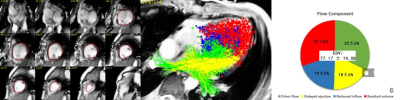 |
Attenuation of left ventricular blood flow kinetic energy and direct flow in repaired Fontan patients: a 4D flow MRI study
Liwei Hu1, Xiaodan Zhao2, Rongzhen Ouyang1, Shuang Leng2, Yong Zhang3, Liang Zhong2,4, and Yumin Zhong1
1Shanghai Children's Medical Center, Shanghai, China, 2National Heart Centre Singapore, Singapore, Singapore, 3GE Healthcare, Shanghai, China, 4Duke-NUS Medical School, National University of Singapore, singapore, Singapore
Four-dimensional flow (4D flow) magnetic resonance imaging (MRI) enables the quantification of blood flow such as kinetic energy (KE) and flow components within the ventricular. Both KE and flow components is associated with ventricular diastolic and systolic function. This study aimed to quantify left ventricular (LV) KE and flow components in repaired Fontan patients in comparison to normal volunteers. The data showed that LV diastolic and systolic KE and direct flow were significant reduced, and residual flow increased in repaired Fontan patients.
|
|||
2065.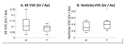 |
Assessment of sex differences in ventricular-vascular coupling of left ventricular and aortic flow derived from 4D Flow MRI in healthy adults
Cody Johnson1, Ryan Pewowaruk2, David Rutkowski1, Amanda Wolfinger1, and Alejandro Roldán-Alzate1,2,3
1Radiology, University of Wisconsin-Madison, MADISON, WI, United States, 2Biomedical Engineering, University of Wisconsin-Madison, MADISON, WI, United States, 3Mechanical Engineering, University of Wisconsin-Madison, Madison, WI, United States
To gain insight into male-female differences in cardiovascular conditions, understanding healthy, sex differences is critical. We analyzed 20 healthy subjects with cardiac 4D flow MRI data (10 male and 10 female) to quantify LV and aortic flow, and ventricular vascular coupling (VVC) of KE and vorticity. The sex difference found in LV flow were not found in aortic flow. The VVC of LV-to-aortic flow was similar for men and women. The analysis methods and results of this study may be of further use in understanding ventricular vascular coupling of flow variables in various cardiovascular conditions.
|
|||
2066.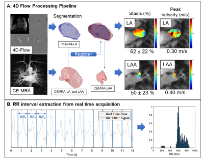 |
Associations of Cardiac Rhythm with left atrial and left atrial appendage hemodynamics measured with 4D flow in Atrial Fibrillation
Amanda L DiCarlo1, Justin Baraboo1,2, Patrick McCarthy3, Rishi Arora4, Rod Passman4, Philip Greenland4, Daniel C Lee1,4, Daniel Kim1,2, and Michael Markl1,2
1Radiology, Northwestern University, Chicago, IL, United States, 2Biomedical Engineering, Northwestern University, Chicago, IL, United States, 3Cardiac Surgery, Northwestern University, Chicago, IL, United States, 4Cardiology, Northwestern University, Chicago, IL, United States
Atrial fibrillation (AF) is the most common cardiac arrhythmia, characterized by irregular heart rhythm which can lead to stroke due to thrombus formation in the left atrium (LA) or left atrial appendage (LAA). Reduced velocities and increased stasis measured with 4D flow MRI are risk metrics correlated with LA thrombus formation and potential predictors of stroke. This study analyzed the relationship between heart rate variability derived from a real time MRI acquisition and 4D flow hemodynamic risk metrics. LA peak velocity and stasis were significantly correlated with heart rate variability, suggesting a link between rhythm status and thrombus risk.
|
|||
2067.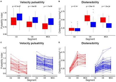 |
Pulsatility attenuation along the carotid siphon in pseudoxanthoma elasticum
Rick J. van Tuijl1, Jonas W. Bartstra1, Pim A. de Jong1, Willem P. T. M. Mali1, Irene C. van der Schaaf1, Ynte M. Ruigrok2, Gabriël J. E. Rinkel2, Birgitta K. Velthuis1, Wilko Jason Spiering3, and Jaco J. M. Zwanenburg1
1Radiology, UMC Utrecht, Utrecht, Netherlands, 2Neurology, UMC Utrecht, Utrecht, Netherlands, 3Vascular Medicine, UMC Utrecht, Utrecht, Netherlands
Arterial internal elastic lamina calcifications in pseudoxanthoma elasticum (PXE) may reduce the normal attenuation by the carotid siphon. This could contribute to increased intracranial pulsatility seen in PXE patients. We compared velocity pulsatility and distensibility along the internal carotid artery and in the middle cerebral artery between 50 PXE patients and 40 age and sex-matched controls, using 2D phase-contrast velocity mapping on 3T MRI. Distensibility was lower and pulsatility higher in patients. However, pulsatility attenuation over the siphon was similar between patients and controls. Siphon dysfunction does, therefore, not explain the increased intracranial arterial pulsatility in PXE.
|
|||
2068.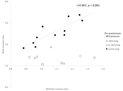 |
4D Flow MRI evaluation and intraoperative MCA pressure measurements before and after STA-MCA bypass surgery
Tetsuro Sekine1, Erika Orita1, Takahiro Ando1, Yasuo Murai1, and Shinichiro Kumita1
1Nippon Medical School, Tokyo, Japan The purpose of this study was to clarify the intracranial hemodynamics before and after superficial temporal artery to middle cerebral artery (STA-MCA) bypass surgery by comparing flow parameters obtained by time-resolved 3-dimensional phase-contrast (4D flow) MRI and intraoperative MCA pressure measurement. The visual and quantitative assessment of 4D flow MRI revealed that intracranial blood flow changes complementarily after STA-MCA bypass surgery. The sum of intracranial BFV can be used for the evaluation of treatment outcome after the surgery. |
|||
2069. |
Abdominal Aortic Aneurysm Prevents Efficient Blood Flow Delivery to Common Iliac Arteries: A study of Hemodynamic Effect of EVAR by 4D Flow.
Masataka Sugiyama1, Yasuo Takehara1, Shinji Naganawa2, Satoshi Goshima3, Atsushi Nozaki4, Tetsuya Wakayama4, and Marcus Alley5
1Department of Fundamental Development for Advanced Low Invasive Diagnostic Imaging, Nagoya University, Graduate School of Medicine, Nagoya, Japan, 2Department of Radiology, Nagoya University, Graduate School of Medicine, Nagoya, Japan, 3Department of Radiology, Hamamatsu University School of Medicine, Hamamatsu, Japan, 4Applied Science Laboratory Asia Pacific, GE Healthcare Japan, Hino, Japan, 5Department of Radiology, Stanford University School of Medicine, Palo Alto, CA, United States
4D Flow MRI were performed for 12 abdominal aortic aneurysm patients who underwent EVAR before and after the treatment. Peak systolic blood flow in the common iliac arteries has significantly increased after EVAR (right: p=0.016, left: p=0.016). The aneurysm might have stored blood like a reservoir in systole and have inhibited the delivery of blood flow to the iliac arteries. The stent placement of EVAR may have repaired the deformed blood flow path and improved the efficiency of blood flow delivery to the periphery. 4D Flow have shown its power for analyzing the hemodynamic effects of EVAR.
|
|||
2070.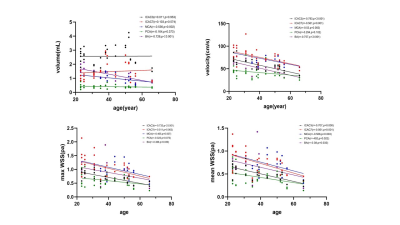 |
Age and anatomical location related hemodynamic changes assessed by 4D flow MRI in the Cerebral Arterial of healthy adults
Laiyang Ma1, Chuang Wu1, Na Han1, Jing Zhang1, and Kai Ai2
1LanZhou University Second Hospital, LanZhou, China, 2Philips Healthcare, Xi'an, China
The purpose of this study was to evaluate the hemodynamic changes in the cerebral arterial of healthy adults among different ages and anatomical locations by using 4D flow MRI. We measured blood flow volume, velocity, wall shear stress (WSS) of cerebral arterial. The hemodynamic changes among different groups were significantly distinct. The young group showed significantly higher in velocity and WSS. Velocity and WSS were decreased with age. The proximal posterior cerebral artery (pro-PCA) had lower volume, velocity and WSS than other parts. 4D Flow MRI could quantify the change of hemodynamics parameters in the cerebral arterial of healthy adults.
|
|||
2071.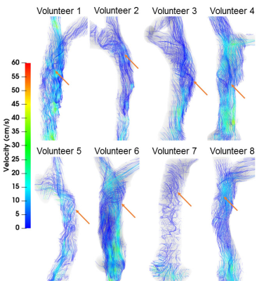 |
Helical Flow of Superior Vena Cava Helical Flow in Healthy Young Males: a 4D Flow MRI Study
Huaxia Pu1, Haoyao Cao2, Yubo Fan3, Jinge Zhang1, Zhenlin Li1, Zhan Liu2, Liqing Peng1, Tinghui Zheng2, Xiaoyue Zhou4, and Ning Jin5
1Department of Radiology, West China Hospital, Sichuan University, Chengdu, China, 2Department of Applied Mechanics, Sichuan University, Chengdu, China, 3Key Laboratory for Biomechanics and Mechanobiology of the Ministry of Education, School of Biological Science and Medical Engineering, Beihang University, Beijing, China, 4MR Collaboration, Siemens Healthineers Ltd, Shanghai, China, 5Siemens Medical Solutions USA, Chicago, IL, United States
The existence of helical flow in the superior vena cava (SVC) is not well understood in vivo. 4D flow MRI data of the SVC and brachiocephalic veins (BVs) were acquired in 8 healthy young males. SVC hemodynamic parameters, including velocity, pathlines, streamlines, and flow waveforms, were measured using a specialized post-processing software. Helical flow was present in the normal SVC. The flow pathlines from the right and left BVs formed helical flows, respectively, with two development types: twining and untwining. These findings may elucidate the occurrence and development of potential SVC disease.
|
|||
2072.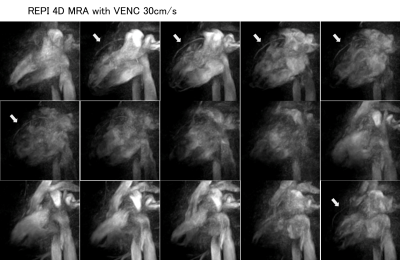 |
Non-contrast 4D Dynamic Coronary MRA using Retrospective EPI (REPI) 4D-Flow Sequence
Yasuhiro Goto1, Michinobu Nagao1, Masami Yoneyama2, Yasutomo Katsumata2, Isao Shiina3, Kazuo Kodaira1, Takumi Ogawa1, Yutaka Hamatani3, Mamoru Takeyama3, Isao Tanaka1, and Shuji Sakai1
1Women's Medical University Hospital, tokyo, Japan, 2Philips Japan, tokyo, Japan, 3Tokyo Women's Medical University Hospital, tokyo, Japan
Since the REPI 4D MRA images the entire cardiac cycle, it may be possible to visualize the flow of the pulmonary artery, pulmonary vein, left ventricle, left ventricle, and aorta in addition to morphological evaluation of the coronary artery.The purpose of this study was to examine mainly LAD about the possibility of the practical use of REPI 4D MRA. REPI 4D MRA with choice of the VENC=30 cm/s could well visualize the coronary arteries from proximal to distal of the LAD.
|
|||
2073.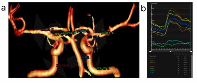 |
Anatomical location related hemodynamic changes assessed by 4D flow MRI in the intracranial arteries of young secondary hypertensive patients
Na Han1, Chuang Wu1, Laiyang Ma1, Yurong Ma1, Jing Zhang1, and Kai Ai2
1Department of Magnetic Resonance, Lanzhou University Second Hospital, Lanzhou, China, 2Philips Healthcare, Xi'an, China
The multi-parameter analysis of 4D flow MRI can identify the changes of hemodynamic parameters in different anatomical location of intracranial arteries. This study evaluating the hemodynamic changes (volume, velocity, wall shear stress) in the intracranial arteries of young secondary hypertensive patients among different anatomical locations (ICA-C3, ICA-C7, proximal MCA, proximal PCA, middle BA) using 4D flow MRI. The proximal PCA had significantly lower volume, velocity and wall shear stress than the values determined for other locations. The wall shear stress at the anatomical location of ICA-C3 was decreased in young secondary hypertensive patients.
|
|||
2074.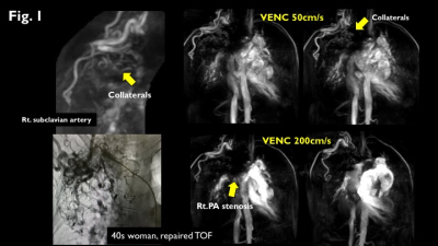 |
Multi-VENC 4D flow MRI demonstrates pulmonary stenosis and arterial-pulmonary collateral in congenital conotruncal anomaly
Michinobu Nagao1, Yumi Shiina1, Yasuhiro Goto1, Isao Shiina1, Kazuo Kodaira1, Masami Yoneyama2, Takashi Namiki2, Yuka Matsuo1, Atsushi Yamamoto1, Kei Inai1, and Shuji Sakai1
1Tokyo Women's Medical University, Tokyo, Japan, 2Philips Japan, Tokyo, Japan
We propose a free-breathing 4D flow sequence with echo-planar imaging (EPI) and rapid reconstruction to facilitate clinical use. Pulmonary stenosis or atresia is a common anatomic component of congenital conotruncal anomaly. Multi-VENC 4D flow MRI with EPI can simultaneously visualize the pulmonary artery stenosis and associated collaterals of slow flow formed by peripheral arteries. Consequently, all sources of pulmonary blood supply and of the size and morphology of the pulmonary arteries can be completely delineated.
|
|||
2075.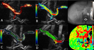 |
Evaluation of Portal System Flow with liver 4D-Flow and APTw MRI in Response to a Meal Challenge
Lihua Chen1, Ailian Liu1, Jiazheng Wang2, Yishi Wang2, and Qingwei Song1
1The First Affiliated Hospital of Dalian Medical University, Dalian, China, 2Philips Healthcare, Beijing, China
Liver MRI was conducted with both 4D-Flow and APTw imaging before and after a meal challenge. After the meal, significantly increased blood flow velocity and volume were observed in portal vein (PV) and superior mesenteric vein (SMV) and significantly decreased blood flow was observed in splenic vein (PV), while no change was observed in APT values (MTRasym) in liver parenchyma after the meal.
|
|||
2076.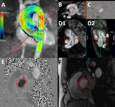 |
First Experiences Utilizing Whole-Chest 4D-Flow for Everyday Clinical Use: A Step from Bench to Bedside
Maurice Pradella1,2, Michael B Scott1, Brad D Allen1, Ryan Avery1, and Michael Markl1,3
1Department of Radiology, Northwestern University, Chicago, IL, United States, 2University Hospital Basel, University of Basel, Basel, Switzerland, 3Department Biomedical Engineering, Northwestern University, Chicago, IL, United States
We present initial results of our novel full chest, free-breathing 4D-flow protocol which was designed for clinical use. In 25 patient who required imaging of aortic and pulmonic flow, it proved to be feasible for two reasons: mean acquisition times of 4D-flow was 12.5min versus 9.7min for 2D phase contrast (2D-PC) imaging, resulting in only a minor mean measurement time increase of about three minutes. Second, flow measurements for aortic and pulmonic flow for were consistent for both 4D-flow and 2D-PC. We will continue with our evaluation in a larger cohort to confirm these initial results.
|
|||
2077.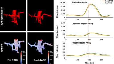 |
4D flow MRI for assessment of blood flow in hepatic arteries of patients with HCC treated with transarterial chemoembolization: a pilot study
Rihab Mansour1, Narges Salehi2, William Tanguay1,3, Guillaume Gilbert4, Catherine Huet1, Gilles Soulez1, Pierre Perreault1, Damien Olivé 1, An Tang1,3, and Samuel Kadoury1,2
1Centre de recherche du Centre hospitalier de l'Université de Montréal (CRCHUM), Montréal, QC, Canada, 2École Polytechnique, Montréal, QC, Canada, 3Department of Radiology, Radio-Oncology and Nuclear Medicine, Université de Montréal, Montréal, QC, Canada, 4MR Clinical Science, Philips Healthcare Canada, Markham, ON, Canada
4D flow MRI measurements in the abdominal aorta, common hepatic artery, and proper hepatic arteries are feasible and provide repeatable values before and after transarterial chemoembolization in patients with hepatocellular carcinoma. Blood flow showed no statistically significant change before and after transarterial chemoembolization in the abdominal aorta, common hepatic artery, and proper hepatic artery based on a Wilcoxon test (P = 0.87, 0.72, and 0.86 respectively).
|
|||
2078.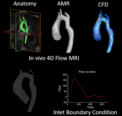 |
Computationally Enhanced 4D Flow MRI for the Assessment of Pre- and Post-Coarctation Repair Aorta Flow Dynamics
Labib Shahid1, James Rice1, Haben Berhane2, Cynthia Rigsby3, Joshua Robinson3, Lindsay Griffin3, Michael Markl2, and Alejandro Roldán-Alzate1
1University of Wisconsin-Madison, Madison, WI, United States, 2Northwestern University, Chicago, IL, United States, 3Ann & Robert H. Lurie Children's Hospital of Chicago, Chicago, IL, United States
CMR is used for diagnosis of COA and post-intervention assessment. Current limitations of 4D flow MRI include insufficient spatio-temporal resolution, inability to image low velocity regions, and distortion of image due to stent. The presented MRI-based computational enhancement method addresses stated limitations. Patient specific flow velocities defined the boundary conditions for numerical simulations. CFD simulations with AMR were run on segmented aorta geometries. Comparison between in-vivo and CFD results show good agreement, and residual complex flows post-repair compared to healthy control. Computational enhancement of 4D flow MRI can be used as a predictive tool in future clinical settings.
|
|||
2079.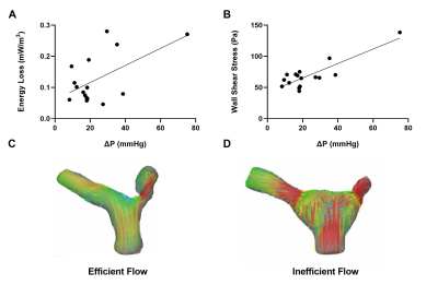 |
4-D Flow CMR Reveals Inefficient PA Flow Correlates with Afterload in Repaired Transposition of the Great Arteries.
Marc Delaney1, Vincent Cleveland2, Paige Mass2, Francesco Capuano3, Yue-Hin Loke4, and Laura Olivieri4
1Pediatrics, Children's National Hospital, Washington, DC, United States, 2Sheikh Zayed Institute for Pediatric Surgical Innovation, Children's National Hospital, Washington, DC, United States, 3Industrial Engineering, Universita di Napoli Federico II, Naples, Italy, 4Pediatric Cardiology, Children's National Hospital, Washington, DC, United States
Repair of D- transposition of great arteries (DTGA) involves pulmonary artery (PA) manipulation that alters shape and flow patterns. Many patients experience increased right ventricular afterload and the etiology remains unclear. We examined the contribution of PA flow separation to afterload in these patients using 4D flow CMR imaging of a mock circulatory system incorporating 3D-printed PA replicas. We found that 2 distinct markers of flow inefficiency correlated with afterload. These data emphasize the utility of 4D flow CMR in quantifying abnormal blood flow and identifying important early clinical predictors of complications in DTGA.
|
|||
2080.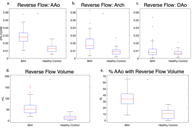 |
Reverse Flow and Reverse Flow Volume is Associated with Aortic Dilatation in Bicuspid Aortic Valve Disease
Elizabeth Weiss1, Kelly Jarvis1, Chris Malaisrie2, Patrick McCarthy2, Robert Bonow3, James Carr1, Cynthia Rigsby4, and Michael Markl1
1Radiology, Northwestern University, Chicago, IL, United States, 2Cardiothoracic Surgery, Northwestern University, Chicago, IL, United States, 3Cardiology, Northwestern University, Chicago, IL, United States, 4Radiology, Lurie Children's Hospital, Chicago, IL, United States
Bicuspid Aortic Valve (BAV) patients often develop aortic dilation which can lead to life-threatening complications. Voxel-wise reverse flow was calculated to assess deranged flow in BAV patients with dilation and in healthy volunteers. In BAV patients, the mean reverse flow was significantly increased in the ascending aorta and aortic arch. The reverse flow volume (volume with reverse flow>0.03mL/cycle) was also increased. Linear regression demonstrated that these reverse flow metrics were significant contributors to mid-ascending aorta diameter even when age was accounted for. We demonstrated that reverse flow is a significant component of deranged flow in BAV disease.
|
|||
2081.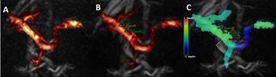 |
Assessing Portal Flow using Four-dimensional Flow MRI in Healthy Volunteers – A Reproducibility Study
Lihua Chen1, Ailian Liu1, Jiazheng Wang2, Yishi Wang2, and Qingwei Song1
1The First Affiliated Hospital of Dalian Medical University, Dalian, China, 2Philips Healthcare, China, Beijing, China
Because of the development of compressed sensing (CS) technology in both spatial and temporal related data acquisition, four-dimensional flow MRI scan can be accomplished in a clinically acceptable time. We evaluated the highly accelerated four-dimensional flow magnetic resonance imaging in portal system hemodynamics studies with Compressed SENSE (CS-SENSE). There was no statistically significant difference in flow velocity, volume rate, axial-WSS of portal system when compared among different CS, measurements from the two observers matched well with each other.
|
|||
2082.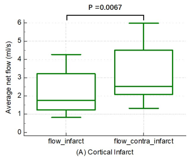 |
Lesion Patterns and Blood Flow Lateralization in Atherosclerotic Middle Cerebral Artery Disease
Wenwen Chen1, Xiaowei Song2, Shuo Chen1, Mingzhu Fu1, Hanyu Wei1, Duoduo Hou2, Le Chen2, Miaoqi Zhang1, Jian Wu2, and Rui Li1
1Center for Biomedical Imaging Research, Department of Biomedical Engineering, School of Medicine, Tsinghua University, Beijing, China, 2Beijing Tsinghua Changgung Hospital, Beijing, China
Intracranial atherosclerosis disease (ICAD) can induce blood flow lateralization (BFL). However, the relationship of infarct patterns (IPs) with BFL was rarely explored. This study investigated the relationship between BFL and IPs in middle cerebral artery (MCA) using 4D flow MRI. Twenty-eight patients with unilateral MCA infarction were included. Average net flow (Flowavg) of infarction side and the contralateral side of MCA-M1 were compared in various IPs. Flowavg is significantly different between two sides of MCA in cortical infarct, but not significant in subcortical lesions. This suggests that cortical infarct may be the specific pattern for BFL in MCA.
|
The International Society for Magnetic Resonance in Medicine is accredited by the Accreditation Council for Continuing Medical Education to provide continuing medical education for physicians.