Digital Posters
Neurovascular MRA & Vessel Wall
ISMRM & SMRT Annual Meeting • 15-20 May 2021

Digital Posters
Neurovascular MRA & Vessel Wall
| Concurrent 3 | 19:00 - 20:00 |
1631.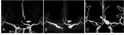 |
Usefulness of 3D Radial Stack-of-stars Ultra-short TE MRA after Treatment of Cerebral Aneurysms with Metallic Devices
Daichi Murayama1, Takayuki sakai1, Masami Yoneyama2, and Shigehiro Ochi3
1Radiology, Eastern Chiba Medical Center, Chiba, Japan, 2Philips Japan, Tokyo, Japan, 3Eastern Chiba Medical Center, Chiba, Japan
We combined 3D radial sequences using stack-of-stars acquisition with uTE sampling to enable short-time imaging of local areas. This study was to apply uTE sequences using 3D radial stack-of-stars collection to MRA to determine its usefulness as a follow-up MRA after treatment of cerebral aneurysms metallic devices. The range of signal reduction around the treated area was significantly smaller in the uTE-MRA than in the TOF-MRA. uTE-MRA improved the visualization of peri-aneurysm vessels and neck remnant in the metallic devices. uTE-MRA using 3D radial stack-of-stars collection is useful for follow-up MRA after treatment of cerebral aneurysms metallic devices.
|
|||
1632.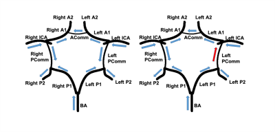 |
Identification of Blood Flow Directions in the Circle of Willis using iSNAP dynamic MRA
Josh Liu1, Zhensen Chen1, Niranjan Balu1, and Chun Yuan1
1Electrical and Computer Engineering, University of Washington, Seattle, WA, United States
Blood flow directions in vessels of the Circle of Willis (CoW) may have important implications for stroke risk. In this study, an algorithm is developed to identify the blood flow direction within vessel segments of the Circle of Willis (CoW) using the dynamic MRA component of the recently developed sequence iSNAP. The CoW of twenty-two patients were analyzed using the algorithm, and an ~85% accuracy was achieved with 3D Phase Contrast as the reference.
|
|||
1633. |
Developmental trajectories of cerebral blood flow and neurodevelopmental score in preterm children from 28 days to 13 years
Peiyao Chen1, Chao Jin1, Xianjun Li1, Miaomiao Wang1, Congcong Liu1, Xiaoyu Wang1, Fan Wu1, Yuli Zhang1, Cong Tian1, Mengxuan Li1, Xiaocheng Wei2, and Jian Yang1
1First Affiliated Hospital of Xi 'an Jiaotong University, Xi'an, Shaanxi, China, 2MR Research China, GE Healthcare, Beijing, China
Preterm children often accompany undesirable neurodevelopmental outcomes. Adequate cerebral blood flow (CBF) is essential to brain development and function. However, little is known about relationship among preterm birth, CBF and neurodevelopmental outcome. This study aims to investigate the effect of preterm birth on CBF and explore the relationship between CBF and neurodevelopmental score. Our results suggest that the effects of preterm birth on intellectual development are long lasting and extend even into late childhood. After controlling for age, CBF shows positive correlations with mental and psychomotor development indexes in left frontal and occipital lobes in preterm children.
|
|||
1634.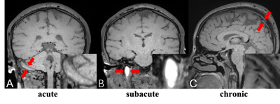 |
The Current Status of Black-Blood Thrombus Imaging in Assessment of Cerebral Venous Thrombosis in a Real-word Clinical Practice
Xiaoxu Yang1, Qi Yang2, Jiangang Duan2, Zhaoyang Fan3, Fang Wu4, and Xunming Ji4
1Chaoyang Hospital, Beijing, China, 2Chaoyang Hospitao, Beijing, China, 3Cedars Sinai Medical Center, Los Angeles, CA, United States, 4Xuanwu Hospital, Beijing, China
We evaluated diagnostic and staging performance of BTI in patients with suspected CVST compared with conventional imaging methods in a real-world study. Our results verified high diagnostic value of BTI in a large patient cohort and BTI provided another advantage of accurate staging (acute, subacute and chronic), achieving more thrombosed segments of definite stage, which is important for clinical strategy.
|
|||
1635.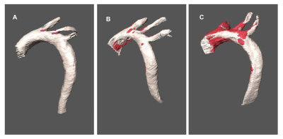 |
Using High Resolution MR microscopy to assess atherosclerosis in ApoE-KO mice fed diets of varying macronutrient content
Courtney Whalen1, Sean Gullette2, A. Catharine Ross1, Rita Castro1,3, and Thomas Neuberger4,5
1Nutritional Sciences, The Pennsylvania State University, University Park, PA, United States, 2Huck Institute of the Life Science, The Pennsylvania State University, University Park, PA, United States, 3University of Lisbon, Lisbon, Portugal, 4Huck Institute of the Life Science, University Park, PA, United States, 5Biomedical Engineering, The Pennsylvania State University, University Park, PA, United States
A ketogenic, low carbohydrate and extremely high fat, diet has been shown to positively impact CVD risk factors. Nevertheless, its effect on atherosclerosis has not been measured. Here, we used 14T MR microscopy to assess atherosclerotic plaque volume, localization, and regional plaque burden in apoE-KO mice fed a ketogenic diet compared to those fed a high fat diet or a control diet. Results show that a ketogenic diet had a protective effect against atherosclerosis compared to a high fat diet as measured by total plaque volume and plaque burden, this effect was not specific to individual regions of the aorta.
|
|||
1636.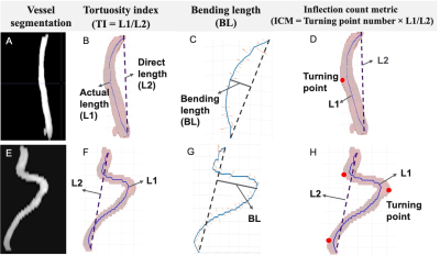 |
Morphological and hemodynamic alterations of major brain feeding arteries across the lifespan
Zhe Sun1,2, Dengrong Jiang3, Marco Muccio1, Chenyang Li1,2, Zixuan Lin3, Peiying Liu3, Hanzhang Lu3, and Yulin Ge1
1Radiology, NYU Langone Health, New York, NY, United States, 2Vilcek Institute of Graduate Biomedical Science, New York University School of Medicine, New York, NY, United States, 3Radiology, Johns Hopkins University School of Medicine, Baltimore, MD, United States
Vascular dysfunction is frequently found in neurodegenerative and cerebrovascular diseases and closely related to age. Bilateral internal carotid arteries (ICAs) and vertebral arteries (VAs) are the key feeding vessels of the brain, which may provide important indices of intracranial health status. We aimed to evaluate age-related morphological and hemodynamic changes of the vessels using TOF MRA and phase contrast MRI. More tortuous appearance as well as reduced hemodynamics were observed with aging; Blood velocity was found to be associated with the morphology after adjusting age and gender, indicating that high blood velocity and wall shear stress could induce arterial tortuosity.
|
|||
1637.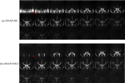 |
Improving iSNAP by using FOCI inversion RF pulse for simultaneous measurement of multiple intracranial vascular imaging contrasts
Kaiyu Zhang1, Zhensen Chen1,2, Rui Li2, Huijun Chen2, Niranjan Balu1, and Chun Yuan1,2
1Vascular Imaging Lab and BioMolecular Imaging Center, Department of Radiology, University of Washington, Seattle, WA, United States, 2Center for Biomedical Imaging Research, Tsinghua University, Beijing, China
Multi-contrast images including hemodynamic information in one sequence have significant impact in evaluating intracranial vessels. In this work, we proposed to use FOCI inversion pulse combined with the multi-contrast sequence iSNAP to further improve image quality of dynamic and static MRA. Promising results were obtained from both phantom and in vivo data.
|
|||
1638.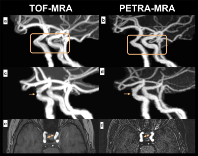 |
MRA based on pointwise encoding time reduction with radial acquisition improves uniformity of cavernous segment of internal carotid artery
Tongtong Sun1, Yu Zhang1, Tongtong Li1, Yuan Luo2, Jinxia Zhu3, Cheng Cheng4, and Hongyan Ni5
1Radiology, First Central Clinical College, Tianjin Medical University, Tianjin, China, 2Radiology, China-Japan Friendship Hospital, Beijing, China, 3MR Collaboration, Siemens Healthcare, Beijing, China, 4MR Application, Siemens Healthcare, Beijing, China, 5Radiology, Tianjin First Central Hospital, Tianjin, China
This study compared the differences between PETRA-MRA and TOF-MRA on the imaging of the cavernous segment of the ICA with qualitative analysis (image quality) and quantitative analysis (SNR, CNR, CR, and uniformity). The results showed that PETRA-MRA significantly improved the uniformity and CR of the cavernous segment of ICA, although it is inferior in both SNR and CNR. This suggests that PETRA-MRA could provide a reasonable alternative to reduce the loss of signal from curved vessels.
|
|||
1639.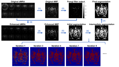 |
Vessel segmentation on intracranial noncontract enhanced dynamic MR angiography using spatial-temporal correlation between voxels
Cheng Zhong1, Niranjan Balu2, Chun Yuan2, and Zhensen Chen2,3
1Fudan University, Shanghai, China, 2Department of Radiology, University of Washington, Seattle, WA, United States, 3Center for Biomedical Imaging Research, School of Medicine, Tsinghua University, Beijing, China
A novel method that exploits spatial-temporal correlation between pixels was developed to segment the dynamic MRA (dMRA) of intracranial arteries. Preliminary results on 3 healthy volunteers demonstrate that the proposed method can effectively identify distal vessels with low signal intensity.
|
|||
1640.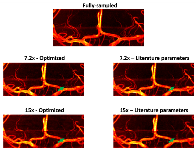 |
An investigation into the optimal undersampling parameters for 3D TOF MRA at 7T using compressed sensing reconstruction
Matthijs de Buck 1, Peter Jezzard1, and Aaron Hess2
1Wellcome Centre for Integrative Neuroimaging, FMRIB Division, Nuffield Department of Clinical Neurosciences, University of Oxford, Oxford, United Kingdom, 2Oxford Centre for Clinical Magnetic Resonance Research, Department of Cardiovascular Medicine, University of Oxford, Oxford, United Kingdom
3D TOF-MRA at 7T can be greatly accelerated using compressed sensing reconstruction of undersampled acquisitions, but the achieved reconstruction quality depends on the undersampling pattern. In this work, optimal parameters for variable-density Poisson disk undersampling were established. For all acceleration factors, optimal reconstructions were achieved by using small fully-sampled calibration areas of 12x12 lines and a polynomial order of around 2. The optimal undersampling parameters were the same for all acceleration factors, but the importance of using optimized undersampling parameters was found to increase for higher acceleration factors.
|
|||
1641.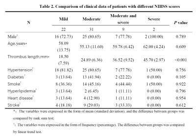 |
Quantitative evaluation of thrombus length and its relationship with the severity and prognosis of acute cerebral infarction using CUBE imaging
Chao Zhang1, Xinyi Wang1, and Weiqiang Dou2
1The First Affiliated Hospital of Shandong First Medical University, Jinan, China, 2GE Healthcare, MR Research China, Beijing, China
This study aimed to quantitatively evaluate thrombus length and its relationship with the severity and prognosis of acute cerebral infarction using high resolution CUBE imaging. National institute of health stroke scale (NIHSS) score and modified rankin scale (mRS) score were used to quantify the degree of neurological impairment and the prognosis of patients. The length of cerebral artery thrombosis was measured with CUBE imaging. Strong relationship was revealed between the thrombus length and NIHSS or mRS score. We therefore demonstrated that thrombus length measurement based on CUBE MRI can effectively evaluate the severity and prognosis of acute cerebral infarction.
|
|||
1642.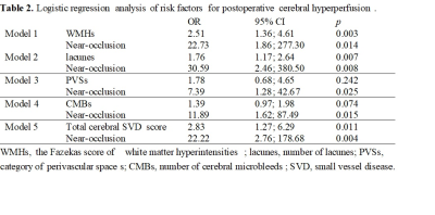 |
Preoperative cerebral small vessel disease is independently associated with cerebral hyperperfusion after carotid endarterectomy
Xiaoyuan Fan1, Zhichao Lai1, Tianye Lin1, Juan Wei2, and Feng Feng1
1Peking Union Medical College Hospital, Beijing, China, 2GE Healthcare, Beijing, China
Whether preoperative cerebral small vessel disease and carotid near-occlusion are associated with cerebral hyperperfusion (CH) after carotid endarterectomy has not been reported. This study demonstrated that preoperative Fazekas score ≥ 3, number of lacunes ≥ 2 combined with the presence of carotid near-occlusion can accurately predict CH after carotid endarterectomy. This encourages preoperative MRI scans and suggests we should pay particular attention to patients with CSVD and carotid near-occlusion.
|
|||
1643.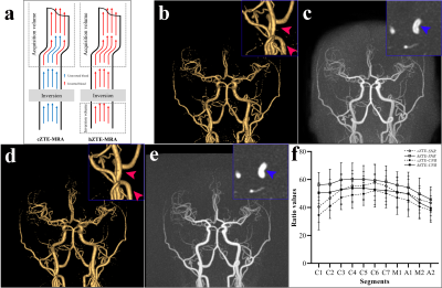 |
Can hybrid ASL-tagged ZTE MRA be an effective candidate in the evaluation of intracranial artery diseases? A clinical feasibility study
Song'an Shang1, Weiqiang Dou2, and Jingtao Wu3
1Nanjing First Hospital, Nanjing Medical University, Nanjing, China, 2GE Healthcare, MR Research China, Beijing, China, 3Northern Jiangsu People’s Hospital, Yangzhou, China
Flow related artifacts in continuous arterial spin labeling (cASL) zero-echo-time (ZTE) magnetic resonance angiography (MRA) could influence the visualization of vasculature. This study aimed to investigate the clinical feasibility of ZTE-MRA with hybrid arterial-spin-labeling (hASL) tagged for assessing the intracranial artery diseases assessment. Our clinical results indicated that relative to conventional ZTE-MRA, hASL-ZTE-MRA was more robust and superior in the depiction of intracranial artery diseases and follow-up assessment in a clinical population while maintaining a ZTE-derived nature.
|
|||
1644. |
Vessel-selective 4D MRA Based on ASL for Treatment Evaluation in Patients with Bypass Surgery: Comparison with 3D TOF MRA and DSA
Maoxue Wang1, Yi Wang1, Yongbo Yang1, Yongbo Yang1, Ming Li1, Jilei Zhang1, and Bing Zhang1,2
1The Affiliated Drum Tower Hospital of Nanjing University Medical School, Nanjing, China, 2Institute of Brain Science, Nanjing University Nanjing, Nanjing, China
A comparison of 4D MRA and 3D TOF MRA in patients with moyamoya vasculopathy after revascularization.
|
|||
1645. |
Evaluation of optimized 4D ultrashort TE MR Angiography using Variable Inversion Time
Haruyuki Fukuchi1,2,3, Toshiya Akatsu4, Kei Fukuzawa3, Nao Takano4, Yutaka Ikenouchi2, Michimasa Suzuki2, Kohji Kamagata2, Akihiko Wada2, Osamu Abe1, and Shigeki Aoki2
1Radiology, Department of Radiology, Graduate School of Medicine, University of Tokyo, Tokyo, Japan, 2Department of Radiology, Juntendo University Graduate School of Medicine, Tokyo, Japan, 3Department of Radiology, Toranomon Hospital, Tokyo, Japan, 4Department of Radiology, Juntendo University Hospital, Tokyo, Japan
We developed and refined a novel method, Variable TI, to improve the visibility of ASL based UTE 4D-MRA. From numerical simulations and the flow phantom studies we found the signal tendency and blood signal skipping phenomena. With refined parameters we got 50% of signal enhancement comparing to conventional method. We conduct volunteer study with the refined Variable TI UTE 4D-MRA and this novel method offered a higher signal intensity and improved visualization of arteries in late phase compared to the conventional method. The Variable TI technique can improve clinical usability of 4D-MRA.
|
|||
1646.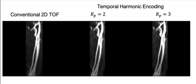 |
Removing pulsatile flow artifacts in 2D TOF MRA with Temporal Harmonic Encoding
Tzu-Cheng Chao1, Guruprasad Krishnamoorthy2, and James G. Pipe1
1Department of Radiology, Mayo Clinic, Rochester, MN, United States, 2Philips Healthcare, Gainesville, FL, United States
2D TOF MRA provides important diagnostic information around carotid bifurcation. However, the image is prone to artifacts caused by pulsatile flow. The present work utilized Temporal Harmonic Encoding scheme to reduce these artifacts by extracting only the static signals for MRA. Meanwhile, the harmonic components containing dynamic information could also be used as a reference.
|
|||
1647.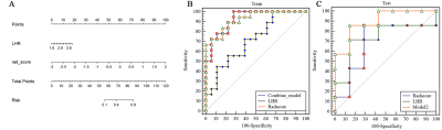 |
High resolution magnetic resonance imaging-based texture analysis for the assessment of carotid atherosclerotic plaques vulnerability
Sihan Chen1, Changsheng Liu1, Xixiang Chen1, Ling Ma2, and Yunfei Zha1
1Radiology, Renmin Hospital of Wuhan University and Hubei General Hospital, Wuhan, China, 2Advanced Application Team, GE Healthcare, Wuhan, China
This study aimed to assess the vulnerability of carotid atherosclerotic plaques using the combination of radiomics nomogram and clinical high-risk factors. Texture features were extracted from high-resolution multi-contrast magnetic resonance imaging. Radscore was applied to build a diagnostic model with the consideration of hrMRI texture features and patient demography to assess the power in differentiating vulnerable and stable lesions. The radscore showed a better diagnostic performance. The combination model of texture and clinical information had the best performance in assessing lesion vulnerability. This study demonstrated that hrMRI texture features provided incremental value for the carotid atherosclerotic vulnerability assessment.
|
|||
1648.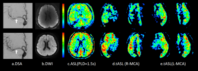 |
Can 3D pseudo-continuous territorial arterial spin labeling effectively diagnose patients with recanalization of unilateral MCA stenosis?
Xinyu Wang1, Weiqiang Dou2, Xinyi Wang1, Kunjian Chen1, Huimin Mao1, and Yu Guo1
1Radiology department, The First Affiliated Hospital of Shandong First Medical University, Jinan, China, 2MR Research China, GE Healthcare, Beijing, China
In this study, the main purpose was to explore the feasibility of 3D pseudo-continuous territory arterial spin labeling (tASL) in evaluating unilateral middle cerebral artery (MCA) recanalization. We included 47 patients with unilateral MCA stenosis diagnosed by DSA and performed tASL examination. Increased relative cerebral blood flow (rCBF) values were found in the blood supply area of MCA postoperatively than pre-operatively. Good prognosis was also revealed to closely associate with Alberta Stroke Program Early CT Score (ASPECTS) scores based on tASL perfusion images. Therefore, tASL can be used as one valuable method to diagnose the effect of vascular recanalization.
|
|||
1649.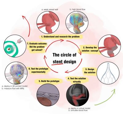 |
The circle of stent design for intracranial aneurysm treatment: role of MRI.
Mariya Stanislavovna Pravdivtseva1, Prasanth Velvaluri2, Fritz Wodarg1, Eckhard Quandt2, Rodrigo Lima de Miranda3, Jan-Bernd Hövener1, Philipp Berg4, and Olav Jansen1
1Department of Radiology and Neuroradiology, Section Biomedical Imaging, Molecular Imaging North Competence Center (MOIN CC), University Medical Center Schleswig-Holstein (UKSH), Kiel University, Kiel, Germany, Kiel, Germany, 2Chair for Inorganic functional materials, University of Kiel, Kiel, Germany, Kiel, Germany, 3Acquandas GmbH, Kasierstrasse 2, Kiel, Germany, Kiel, Germany, 4Research Campus STIMULATE, University of Magdeburg, Magdeburg, Germany, Magderburg, Germany
Intracranial aneurysms are a life-threatening disease, which can be treated with flow diverter stents. However, complications during or after this endovascular treatment might occur. Therefore, there is a need to improve the current minimally-invasive technologies. In the present study, we proposed and utilized the concept of the iterative stent design focusing on 1) fast prototyping using microsystem technologies and 2) in vitro testing the flow reduction in real-world conditions using 4D flow MRI. As a result, the flow volume entering the aneurysm in the presence of a stent was reduced by 10 %, and then by 85 %.
|
|||
1650. |
Clinical feasibility study of extracranial carotid arteries magnetic resonance angiography using PETRA-MRA
Yue Qin1, Xin Li1, Yinhu Zhu1, Dayong Jin1, Yifan Qian1, Juan Tian1, Liyao Liu1, Yanqiang Qiao1, and Shaoyu Wang2
1XIAN DAXING HOSPITAL, Xi'an, China, 2MR Scientific Marketing, Siemens Healthineers, Xi'an, China
Magnetic resonance angiography (MRA) is increasingly used as a non-invasive method to assess carotid arteries. TOF-MRA, because it is dependent on the flow of magnetized blood into the volume being imaged, is highly susceptible to conditions that disrupt laminar flow. Depiction of the carotid region using Silent imaging was often superior to that obtained with conventional techniques, where the ultrashort TE minimized flow-related signal dropout. This study aimed to evaluate the feasibility of PETRA-MRA in imaging the vascular structures of the extracranial carotid arteries. We found that PETRA-MRA showed significantly higher image quality and better visualized extracranial carotid arteries.
|
The International Society for Magnetic Resonance in Medicine is accredited by the Accreditation Council for Continuing Medical Education to provide continuing medical education for physicians.