Digital Posters
Towards Real-Time Cardiac MRI
ISMRM & SMRT Annual Meeting • 15-20 May 2021

| Concurrent 2 | 15:00 - 16:00 |
2870. |
Real-time cardiac MRI using spiral read-outs and a Variational Network for data-driven reconstruction
Jonas Kleineisel1, Philipp Eirich1,2, Julius F. Heidenreich1, Herbert Köstler1, Thorsten A. Bley1, and Tobias Wech1
1Department of Diagnostic and Interventional Radiology, University of Würzburg, Würzburg, Germany, 2Comprehensive Heart Failure Center Würzburg, Würzburg, Germany
Spiral sampling patterns are well suited for fast MR applications like cardiac imaging, yet additional undersampling is necessary to achieve real-time temporal resolution. To still transform these data into images of high quality, a Variational Network is presented. The proposed model uses U-Nets for regularizing a gradient descent scheme, to reconstruct dynamic image series comparable to a segmented fully sampled cine investigation. The acquisition time for an entire stack from base to apex was below one minute; the overall reconstruction time was about 6 minutes.
|
|||
2871.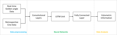 |
Deep-learning cardiac MRI for quantitative assessment of ventricular volumes
Yifan Qi1, Fusheng Wang1, J. Jane Cao2, and Yulee Li2
1Computer Science, Stony Brook University, Stony Brook, NY, United States, 2St. Francis Hospital, DeMatteis Center for Cardiac Research and Education, Greenville, NY, United States
The presented work introduces a deep-learning cardiac MRI approach to quantitative assessment of ventricular volumes from raw MRI data without image reconstruction. As the information required for volumetric measurements is less than that for image reconstruction, ventricular function may be assessed with less MRI data than conventional image-based methods. This offers the potential to improve temporal resolution for quantitatively imaging cardiac function.
|
|||
2872. |
Deep generative manifold model: a novel approach for free breathing dynamic MRI
Qing Zou1, Abdul Haseeb Ahmed1, Prashant Nagpal1, Rolf Schulte2, and Mathews Jacob1
1University of Iowa, Iowa City, IA, United States, 2GE Global Research, Munich, Germany
Cardiac disease is a frequent comorbidity and cause of death in subjects with compromised pulmonary function (e.g. COPD). High-resolution and free breathing cine imaging sequences are necessary for the evaluation of cardiac function in such subjects. Several self-gated and manifold algorithms were introduced for free-breathing MRI with good success. The main focus of this work is to further reduce the scan time of current manifold methods. We introduce a novel unsupervised deep generative manifold framework to recover free-breathing cine data from around 7seconds/slice, which will facilitate the acquisition of the whole volume in under two minutes.
|
|||
2873.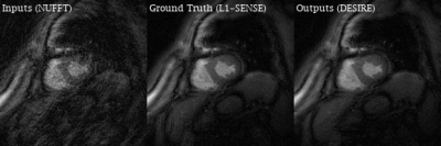 |
Free-breathing High-resolution Spiral Real-time Cardiac Cine Imaging using DEep learning-based rapid Spiral Image REconstruction (DESIRE)
Junyu Wang1, Ruixi Zhou1, and Michael Salerno1,2,3
1Biomedical Engineering, University of Virginia, Charlottesville, VA, United States, 2Medicine, University of Virginia, Charlottesville, VA, United States, 3Radiology, University of Virginia, Charlottesville, VA, United States
Cardiac real-time cine imaging is valuable for patients who cannot hold their breath or have irregular heart rhythms. Spiral acquisitions, which provide high acquisition efficiency, make high-resolution cardiac cine real-time imaging feasible. However, the reconstruction for under-sampled non-Cartesian real-time imaging is time-consuming, and hence cannot provide rapid feedback. We sought to develop a DEep learning-based rapid Spiral Image REconstruction technique (DESIRE) for spiral real-time cardiac cine imaging with free-breathing, to provide fast and high-quality image reconstruction and make rapid online reconstruction feasible. High image quality was demonstrated using the proposed technique for healthy volunteers and patients.
|
|||
2874. |
Rapid De-aliasing of Undersampled Real-Time Phase-Contrast MRI Images using Generative Adversarial Network with Optimal Loss Terms
Huili Yang1,2, Amanda Lynn DiCarlo1,2, Daming Shen1,2, Hassan Haji-Valizadeh1,2, Michael Markl1,2, and Daniel Kim1,2
1Department of Biomedical Engineering, Northwestern University, Evanston, IL, United States, 2Department of Radiology, Northwestern University, Chicago, IL, United States
While compressed sensing is a proven method for highly accelerating cardiovascular MRI, its lengthy reconstruction time hinders clinical translation. Deep learning is a promising method to accelerate reconstruction processing. We propose a generative adversarial network (GAN) with optimal loss terms for rapid reconstruction of 28.8-fold accelerated real-time phase-contrast MRI. Our results show that GAN reconstructs images 613 times faster than compressed sensing without significant loss in peak and mean velocity measurements and image sharpness.
|
|||
2875.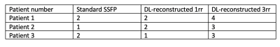 |
Deep Learning Algorithms May Aid In The Evaluation Of Cine Images In Patients With Atrial Arrhythmia – A Case Series
Ria Garg1,2, Elizabeth Hillier2,3,4, Andrew Coristine2,3,5, and Matthias G. Friedrich2,3
1Cardiovascular imaging, RI-McGill University Health Center, Montreal, QC, Canada, 2Faculty of Medicine, McGill University Health Center, Montreal, QC, Canada, 3RI-McGill University Health Center, Montreal, QC, Canada, 4Faculty of Medicine, University of Alberta, Edmonton, Alberta, AB, Canada, 5MR applications and workflow, GE Healthcare, Montreal, QC, Canada
Cardiovascular magnetic resonance cine imaging is utilised to give comprehensive information on ventricular function. Arrhythmia obstructs acquisition of these images, where the use of faster acquisition protocols with deep learning reconstruction methods may aid in solving the problem. We evaluated cine images of three patients in atrial arrhythmia, acquired using standard method; fast, variable density spatiotemporal sampling acquisition (VD kt) of one(1rr) or three(3rr) heart beats; and deep learning reconstruction of the same (DL-1rr,DL-3rr). Our results showed that undersampled techniques combined with deep-learning algorithms result in image quality improvements with no significant difference in quantitative values between all acquisition techniques.
|
|||
2876.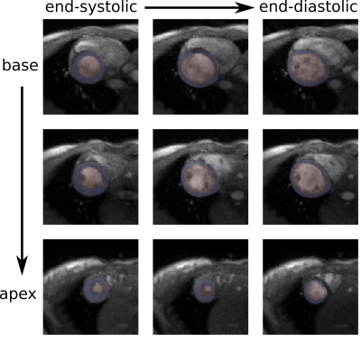 |
Generating Cardiac Segmentation Masks of Real-Time Images from Self-Gated MRI to Train Neural Networks
Martin Schilling1, Sebastian Rosenzweig1,2, Moritz Blumenthal1, and Martin Uecker1,2,3
1Institute for Diagnostic and Interventional Radiology, University Medical Center Göttingen, Göttingen, Germany, 2DZHK (German Centre for Cardiovascular Research), Göttingen, Germany, 3Campus Institute Data Science (CIDAS), University of Göttingen, Göttingen, Germany
Cardiac segmentation is essential for analyzing cardiac function. Manual labeling is relatively slow, so machine learning methods have been proposed to increase segmentation speed and precision. These methods typically rely on cine MR images and supervised learning. However, for real-time cardiac MRI, ground truth segmentations are difficult to obtain due to lower image quality compared to cine MRI. Here, we present a method to obtain ground truth segmentation for real-time images on the basis of self-gated MRI (SSA-FARY).
|
|||
2877.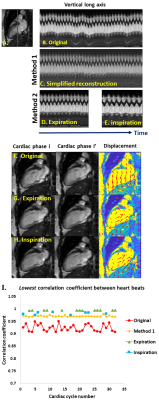 |
Small and Large Respiratory Motions from Free-Running Cardiac Magnetic Resonance Corrected by Two Post-Processing Strategies
Ummul Afia Shammi1, Zhijian Luan2, Jia Xu3, Aws Hamid4, Joanne Cassani4, Talissa A. Altes4, Robert P. Thomen4, and Steven R Van Doren5
1Biomedical, Biological & Chemical Engineering, University of Missouri, Columbia, MO, United States, 2Institute for Data Science and Informatics, University of Missouri, Columbia, MO, United States, 3Biochemistry, University of Missouri, Columbia, MO, United States, 4Radiology, University of Missouri, Columbia, MO, United States, 5Biochemistry, Institute for Data Science, University of Missouri, Columbia, MO, United States
Two new methods of automatic, retrospective suppression of breathing motion appear to be effective for real-time cardiac MR (CMR) scans of free-breathing healthy volunteers. This may obviate the need for multiple breath-holds during CMR exams, allowing for a quicker, more comfortable experience for cardiac patients. The first method demonstrated corrects smaller breathing motions and has the advantage of correcting all cardiac cycles. The second method demonstrated successfully compensates the respiratory excursions of largest amplitude, common in short axis scans, by extracting cardiac cycles at end-inspiration or end-expiration.
|
|||
2878.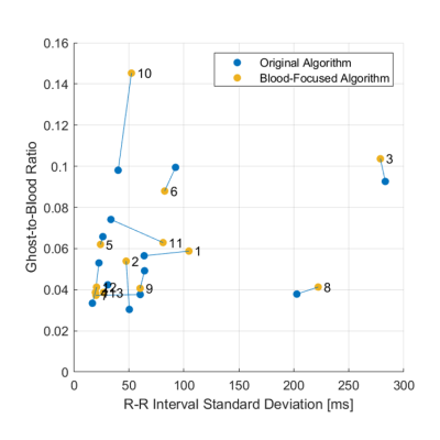 |
Blood-Focused Dynamic Inversion Times for 3D LGE Imaging: Initial Patient Demonstration
Jack Allen1,2, George Mathew1, Miriam Conway1, Sophie Jenkins1, David Firmin1,2, Jennifer Keegan1, Sonya V. Babu-Narayan1, and Peter Gatehouse1
1Cardiovascular Biomedical Research Unit, Royal Brompton and Harefield NHS Trust, London, United Kingdom, 2National Heart and Lung Institute, Imperial College London, London, United Kingdom
3D Late Gadolinium-Enhanced (LGE) imaging is used to assess scarring in patients with Atrial Fibrillation (AF). Acquiring image data during every cardiac cycle allows a reasonable total scan duration but exacerbates ghosting artefacts caused by variable heart rates, such as those of patients with AF. Dynamic Inversion Time (TI) methods improve image quality by modifying the TI for each cardiac cycle. We present the initial stage of a patient study to validate a recently-proposed blood-focused dynamic TI algorithm. No improvement was found in comparison to the original algorithm. Future comparisons will include more patients with high R-wave interval variability.
|
|||
2879.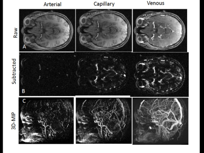 |
Rapid Time-Resolved 4D MRA of the Brain Using Golden-Angle Radial Sparse Parallel (GRASP) MRI
Adam E. Goldman-Yassen1, Maria J. Borja2, Anna Derman2, Eytan Raz2, Duan Chen2, Siddhant Dogra2, Kai Tobias Block2, and Seena Dehkharghani2,3
1Department of Radiology, Children's Healthcare of Atlanta, Atlanta, GA, United States, 2Department of Radiology, New York University Langone Medical Center, New York, NY, United States, 3Department of Neurology, New York University Langone Medical Center, New York, NY, United States
GRASP is a fast and flexible MRI technique combining compressed sensing, parallel imaging, and golden-angle radial sampling. GRASP MRI obtained as part of routine clinical care without specific angiographic parameters can be displayed as 4D MRA images with high spatial and temporal resolution. It exhibits excellent performance and high reader confidence in dynamic angiography, using a highly flexible plug-and-play reconstruction and visualization pipeline. Further exploration of diagnostic accuracy in disease-specific applications is warranted.
|
|||
2880.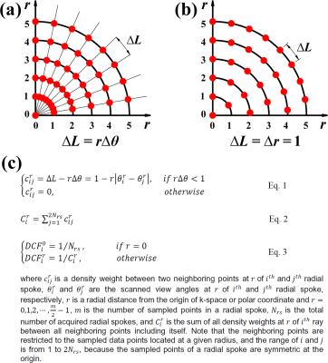 |
Optimized Density Compensation Function for Filtered Backprojection and Compressed Sensing Reconstruction in Radial k-space MRI
KyungPyo Hong1, Amanda L DiCarlo1, Aggelos K Katsaggelos1,2,3, Florian A Schiffers3, Cynthia K Rigsby1,4, Hassan Haji-Valizadeh5, and Daniel Kim1,6
1Radiology, Northwestern University Feinberg School of Medicine, Chicago, IL, United States, 2Electrical and Computer Engineering, Northwestern University, Evanston, IL, United States, 3Computer Science, Northwestern University, Evanston, IL, United States, 4Medical Imaging, Ann & Robert H. Lurie Children's Hospital of Chicago, Chicago, IL, United States, 5Internal Medicine, Cardiovascular Division, Beth Israel Deaconess Medical Center and Harvard Medical School, Boston, MA, United States, 6Biomedical Engineering, McCormick School of Engineering, Northwestern University, Evanston, IL, United States While conventional density compensation function(DCF) performs sufficiently well for filtered backprojection(FBP) and radial k-space MRI when the Nyquist sampling condition is met and/or evenly-spaced view angles are used, it may perform poorly when sub-sampling and/or irrational-view angles are used. We propose an optimized DCF for the aforementioned conditions by calculating the density weights based on geometric properties of radial k-space sampling in a discrete environment, regardless of scan conditions such as data sizes and view angles. Compared with standard DCF, the optimized DCF produces higher signal-to-noise ratio(SNR) in FBP (phantom) and more accurate flow metrics in 48-fold accelerated, phase-contrast MRI. |
|||
2881.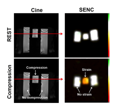 |
Optimized fast strain-encoding MRI with echo-planar readout
V Emre Arpinar1, L Tugan Muftuler1, Andrew Nencka1, Kevin Koch1, and El-Sayed H Ibrahim1
1Medical College of Wisconsin, Milwaukee, WI, United States
Myocardial strain imaging by MRI showed to be an early marker of changes in regional cardiac function before global cardiac function is affected. However, conventional MRI tagging techniques has the limitations of limited spatial resolution and complicated post-processing. Strain-encoding (SENC) showed to be a valuable technique for quantifying myocardial strain with high spatial resolution and simple post-processing. In this study, we provide preliminary results of echo-planar imaging (EPI)-based fast SENC imaging along with imaging artifacts and approaches to minimize their effects. The results showed the feasibility of EPI-based SENC with improved image quality and ultrafast acquisition.
|
|||
2882.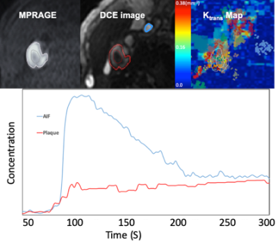 |
3D DCE MRI of carotid plaque by dynamic T1 mapping using Single Reference Variable Flip Angle 3D Pseudo Golden Angle Stack of Stars Acquisition
Seong-Eun Kim1, J Scott McNally1, John A Roberts1, Matthew Alexander 1, Hediyeh Baradaran 1, Gerald S Treiman2, and Dennis L Parker1
1UCAIR, Department of Radiology and Imaging Sciences, University of Utah, Salt Lake City, UT, United States, 2Department of Veterans Affairs, VASLCHCS, Salt Lake City, UT, United States Dynamic contrast‐enhanced (DCE) MRI has been used for quantitative assessment of the neovascular architecture and perfusion properties in the carotid artery wall5. While DCE-MRI has great potential in plaque component characterization, it is presently limited by SNR, contrast, available resolution, and motion artifacts for carotid plaque application. In this work, we propose a new dynamic MRI method capable of high spatial and temporal resolution, and dynamic T1 mapping-based quantification of contrast agent concentration for the assessment of carotid intraplaque hemorrhage to overcome the barriers that prevent DCE-MRI from being the clinical standard for carotid IPH evaluation. |
|||
2883.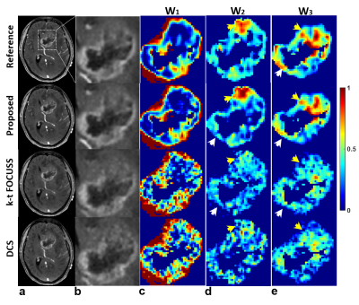 |
Functional Segmentation and Reconstruction for High-Definition DCE MRI Exploiting Vascular Heterogeneity priors
Joon Sik Park1, Seung-Hong Choi2, Chul-Ho Sohn2, and Jaeseok Park1,3
1Biomedical Engineering, Sungkyunkwan University, Gyunggido, Korea, Republic of, 2Seoul National University Hospital, Seoul, Korea, Republic of, 3Intelligent Precision Healthcare Convergence, Sungkyunkwan University, Gyunggido, Korea, Republic of
In this work, we propose a novel, joint functional segmentation and reconstruction directly from largely incomplete DCE measurements exploiting multi-scale vascular heterogeneity priors to achieve high-definition whole brain DCE MRI. Experimental studies were performed in patients with brain tumor to investigate the feasibility of the proposed method in jointly visualizing vascular structures and functions from a single measurement after administering contrast agents. We demonstrated that the proposed method outperforms conventional methods in delineating angiographic structures, functional segment maps, and corresponding perfusion-permeability maps
|
|||
 |
2884.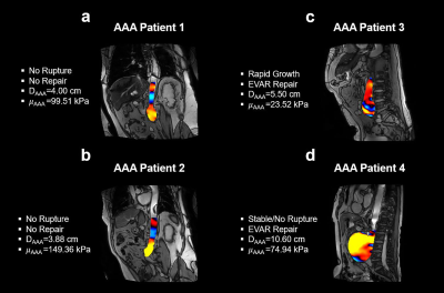 |
In Vivo Aortic MR Elastography in Abdominal Aortic Aneurysm Patients: A Potential Biomarker for Predicting Aneurysmal Events
Huiming Dong1,2, Brian Raterman1, Mariah Eisner3, Guy Brock3, Richard D White1, Jean Starr4, Mounir Haurani4, Michael Go4, Patrick Vaccaro4, and Arunark Kolipaka1,2
1Radiology, The Ohio State University Wexner Medical Center, Columbus, OH, United States, 2Biomedical Engineering, The Ohio State University, Columbus, OH, United States, 3Biomedical Informatics, The Ohio State University Wexner Medical Center, Columbus, OH, United States, 4Surgery, The Ohio State University Wexner Medical Center, Columbus, OH, United States
Abdominal aortic aneurysm (AAA) can result in life-threatening rupture. AAA diameter remains the only clinically useful parameter to predict growth and rupture risk. However, some high-risk AAAs can be overlooked due to their small diameters. AAA stiffness is associated with its extracellular matrix remodeling and thus potentially provides more relevant rupture prediction. Therefore, this study aims to estimate AAA stiffness using aortic MRE in patients with and without aneurysmal events. This is the first study to demonstrate that AAA stiffness and AAA/remote normal (AAA/RN) stiffness ratio are significantly lower in patients with aneurysmal events.
|
||
2885.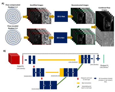 |
Deep Artifact Suppression For Real Time Phase Contrast Cardiac Magnetic Resonance imaging.
Olivier Jaubert1,2, Grzegorz Kowalik2, Javier Montalt-Tordera2, Simon Arridge1, Jennifer Steeden2, and Vivek Muthurangu2
1Department of Computer Science, UCL, London, United Kingdom, 2Centre for Cardiovascular Imaging, UCL, London, United Kingdom
Real-time spiral phase contrast MR is practical for free-breathing assessment of flow. However, for sufficient spatial and temporal resolutions it requires high acceleration rates leading to long reconstruction times. Here we propose to train a 3D U-Net with complex convolutions to accurately reconstruct phase data and flow curves from highly undersampled data. Prospectively acquired in-vivo data were reconstructed with similar image quality but ~4.6x faster than compressed sensing reconstructions which could improve workflow.
|
|||
2886.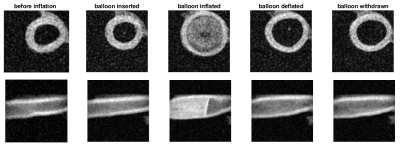 |
Feasibility of high resolution low-field MRI of balloon angioplasty in ex vivo porcine arteries with realistic blood flow
Eline Huizing1, Richte C.L. Schuurmann2, Frank F.J. Simonis3, Çağdaş Ünlü1, Henri G.D. Leuvenink4, and Jean-Paul P.M. de Vries2
1Department of Surgery, Northwest Clinics, Alkmaar, Netherlands, 2Department of Surgery, University Medical Center Groningen, Groningen, Netherlands, 3University of Twente, Enschede, Netherlands, 4Surgical Research Laboratory, University Medical Center Groningen, Groningen, Netherlands
Optimal treatment parameters of balloon angioplasty for revascularization such as inflation time and pressure are subject to debate. Limited data exists because accurate assessment of vascular function is challenging and therefore the influence procedure parameters have on arterial wall damage is largely unknown. This study shows that portable MRI at 0.125x0.125x2 mm³ resolution can be used for assessment of diameter and wall thickness during balloon angioplasty in ex vivo porcine arteries. Even after deflation and removal of the balloon the arterial diameter remained enlarged; this was coupled to a decrease in wall thickness.
|
|||
2887.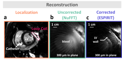 |
Feasibility of motion-resolved high resolution cardiac MRI using a local receiver and MR-tracking micro-coils.
Marylène Delcey1,2,3,4, Pierre Bour1,3,5, Isabelle Saniour6, Dounia El Hamrani1,3,5, Valery Ozenne1,3,5, Marie Poirier-Quinot6, and Bruno Quesson1,3,5
1IHU Liryc, Electrophysiology and Heart Modeling Institute, Fondation Bordeaux Université, Pessac, France, 2Univ. Bordeaux, Centre de recherche Cardio-Thoracique de Bordeaux, U1045, Pessac, France, 3INSERM, Centre de recherche Cardio-Thoracique de Bordeaux, U1045, Bordeaux, France, 4Siemens Healthcare SAS, Saint-Denis, France, 5Univ. Bordeaux, Centre de recherche Cardio-Thoracique de Bordeaux, U1045, Bordeaux, France, 6IR4M, UMR8081, Université Paris-Sud/CNRS, Université Paris-Saclay, Orsay, France In patients presenting cardiac electrical dysfunction, sub-millimetric resolution of CMR would help improving characterisation of the arrhytmogenic substrate. Here, a local coil was placed directly near the anatomic region to image to overpass the lack of sensitivity and selectivity of current conventionnal coils and reduce the voxel size. We implemented a motion compensation method that exploits the signal of micro-coils embodied on a catheter to retrospectively sort radial k-space data prior to ESPIRIT image reconstruction. Using this approach, we present high resolution (300µm), motion-resolved, image of the heart obtained in vivo in pig. |
|||
2888.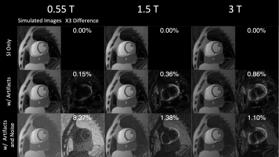 |
Realistic Simulation of Real-Time Cardiac Cine and First Pass Perfusion on High Performance 0.55T System
Ye Tian1, Nam G. Lee2, and Krishna S. Nayak1
1Ming Hsieh Department of Electrical and Computer Engineering, University of Southern California, Los Angeles, CA, United States, 2Biomedical Engineering, University of Southern California, Los Angeles, CA, United States
We present a realistic cardiac MRI simulation framework, including concomitant effects, off-resonance, realistic coil-array and noise level. The general framework is flexible to different field strengths, sequences, and different motion patterns and ischemia models can be introduced. We demonstrate three usages of the framework, in comparing artifact/noise level at different field strengths, in optimization of real-time 3D trajectories and reconstruction, and capturing myocardial perfusion deficits. Real-time 3D cine and first-pass perfusion with high-resolution whole-heart coverage were tested feasible on a high-performance 0.55T system by simulation.
|
|||
2889.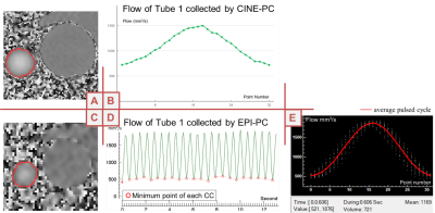 |
Accuracy of “real-time” Echo-Planar Imaging Phase Contrast MRI
Pan Liu1, Sidy Fall2, and Olivier Baledent2,3
1University of Picardie Jules Verne/CHIMERE EA 7516, Amiens, France, 2Facing Faces Institute/CHIMERE EA 7516, Amiens, France, 3Medical image processing department, University hospital Amiens, Amiens, France
Compared with CINE phase contrast MRI (CINE-PC), echo-planar imaging phase contrast (EPI-PC) can achieve real-time quantification of blood flow, with lower SNR. In this study, the pulsating model of the simulated cerebral vasculature was used to verify the accuracy of EPI-PC. The imaging time of EPI-PC was 62ms/image at 1.2*1.2mm2 spatial resolution and FOV=100*60mm2. The reconstructed EPI-PC flow curve was extracted by homemade post-processing software. After comparison with CINE-PC flow curve, it was concluded that EPI-PC can provide an average flow with less than 3% error, and its flow curve will be similar to the CINE-PC flow curve in shape.
|
The International Society for Magnetic Resonance in Medicine is accredited by the Accreditation Council for Continuing Medical Education to provide continuing medical education for physicians.