Digital Posters
Cardiac Structure & Function: From Macro- to Micro-
ISMRM & SMRT Annual Meeting • 15-20 May 2021

Digital Posters
Cardiac Structure & Function: From Macro- to Micro-
| Concurrent 2 | 15:00 - 16:00 |
 |
2890. |
A fast navigator (fastNAV) for prospective respiratory motion correction in first-pass myocardial perfusion imaging
Ronald Mooiweer1, Radhouene Neji2, Sarah McElroy1, Muhummad Sohaib Nazir1, Reza Razavi1, Amedeo Chiribiri1, and Sébastien Roujol1
1School of Biomedical Engineering and Imaging Sciences, King's College London, London, United Kingdom, 2MR Research Collaborations, Siemens Healthcare, Frimley, United Kingdom
A fast respiratory navigator (fastNAV) was developed for dynamic contrast enhanced CMR perfusion imaging by combining spatially non-selective saturation with slice-selective tip-up and slice-selective excitation pulses. A calibration scan was developed to enable the estimation of subject-specific tracking factors. Prospective motion correction using fastNAV was applied to perfusion imaging in 10 patients under free-breathing conditions. Compared to conventional perfusion imaging, fastNAV reduced the effect of respiratory motion while no difference in image quality was observed.
|
||
2891.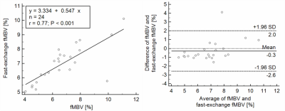 |
Optimization of a multi-dose ferumoxytol-enhanced T1 MRI protocol for estimation of fractional myocardial blood volume
Caroline Colbert1,2,3, Michael A. Thomas3,4, Ran Yan2,5, Hengjie Liu1,2, Peng Hu1,2,5, and Kim-Lien Nguyen1,2,3
1Physics and Biology in Medicine Graduate Program, UCLA David Geffen School of Medicine, Los Angeles, CA, United States, 2Department of Radiology, UCLA David Geffen School of Medicine, Los Angeles, CA, United States, 3Division of Cardiology, UCLA David Geffen School of Medicine, Los Angeles, CA, United States, 4Department of Radiology, Northwestern School of Medicine, Chicago, IL, United States, 5Department of Bioengineering, UCLA Samueli School of Engineering, Los Angeles, CA, United States
We aimed to evaluate the feasibility of an abbreviated MRI protocol for estimation of fractional myocardial blood volume (fMBV). Four normal swine were imaged with the MOLLI sequence at baseline and following seven ferumoxytol doses. We estimated fMBV using our full dataset, after application of retrospective dose under-sampling, and with the fast-exchange approximation. A four-acquisition protocol with compartmental modelling may have potential to accurately estimate fMBV in studies of myocardial perfusion.
|
|||
2892.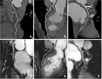 |
High-resolution 3T post-contrast whole-heart coronary MRA with image-based navigation: stenosis identification compared to CTA.
Lu Lin1, Jian Wang1, René M Botnar 2, Claudia Prieto2, Jing An3, Michaela Schmidt4, Christoph Forman4, Karl philipp kunze5, Zhengyu Jin1, and Yining Wang1
1Peking union medical college hospital, Beijing, China, 2School of Biomedical Engineering and Imaging Sciences, King's College London, London, United Kingdom, 3Siemens Shenzhen Magnetic Resonance Ltd., Shenzhen, China, 4Siemens Healthcare GmbH, Erlangen, Germany, 5Siemens Healthcare Limited, London, United Kingdom
This study sought to evaluate the clinical feasibility, image quality and diagnostic accuracy of a prototype 3T post-contrast high-resolution whole-heart coronary MR angiography (CMRA) sequence with image-navigator (iNAV) based motion correction and 100% respiratory scan efficiency integrated into clinical cardiac MR scan before LGE imaging. The results showed CMRA imaging completed on average in 5.2minutes with promising image quality. CMRA showed good inter-modality agreement compared to computed tomography angiography (CTA) and high negative predictive value (NPV), which allows effective exclusion of coronary artery disease.
|
|||
2893.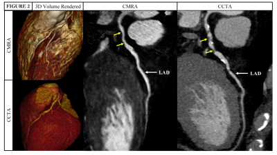 |
Non-contrast, high spatial resolution coronary magnetic resonance angiography versus coronary computed tomography angiography
Reza Hajhosseiny1, Imran Rashid1, Aurélien Bustin1, Camila Munoz1, Gastao Cruz1, Muhummad Sohaib Nazir1, Karine Grigoryan1, Tevfik F. Ismail1, Rebecca Preston2, Radhouene Neji1,3, Karl Kunze1,3, Reza Razavi1, Amedeo Chiribiri1,
Pier Giorgio Masci1, Ronak Rajani1, Claudia Prieto1, and René M. Botnar1
1School of Biomedical Engineering and Imaging Sciences, King's College London, London, United Kingdom, 2Department of Radiology, Guy’s and St Thomas’ NHS Foundation Trust, London, United Kingdom, 3MR Research Collaborations, Siemens Healthcare Limited, Frimley, United Kingdom
Conventional coronary magnetic resonance angiography (CMRA) is limited by long and unpredictable acquisition-times, low spatial resolution and motion related image quality degradation. To overcome these challenges, we have proposed a highly undersampled acquisition with image-based navigators and non-rigid motion correction to enable high resolution (0.9mm3) CMRA with ≈11min acquisition time. In 30 patients with suspected coronary artery disease (CAD) who also underwent a coronary computed tomography angiography (CCTA), our CMRA framework achieved excellent image quality across all coronary artery segments, with a per patient sensitivity of 100%, specificity of 68% and negative predictive value of 100% for identifying/excluding significant CAD.
|
|||
2894.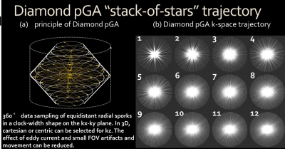 |
Improved free-breathing zoomed whole heart coronary MRA using 3D stack-of-stars radial sequence with diamond pseudo-golden angle sampling
Takashige Yoshida1, Takashige Yoshida1, Masami Yoneyama2, Jihun Kwon2, Kohei Yuda3, Yuki Furukawa3, and Nobuo Kawauchi3
1radiology, Tokyo metropolitan police hospital, Tokyo, Japan, 2Philips Japan, Tokyo, Japan, 3Tokyo metropolitan police hospital, Tokyo, Japan
One of the problems of whole heart coronary MRA is the prolongation of acquisition time, and the radial sampling technique is able to obtain the image of inconspicuous artifacts such as aliasing and motion. Furthermore, the improved sequence of 3D d-Vane is possible to adopt a self-navigator without extend scan time. Hence the small FOV whole heart coronary MRA with diamond pseudo golden angle radial sampling showed improved efficiency with maintaining the image quality.
|
|||
2895.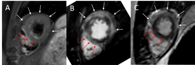 |
Title: 3T contrast-enhanced coronary magnetic resonance angiography in the assessment of myocardial hyperemia in acute myocarditis.
Zhiyong Chen1, Bin Sun1, Yunjing Xue1, Qing Duan1, ZhongShuai Zhang2, and Jing An3
1Radiology, Union Hospital, School of Medical Technology and Engineering, Fujian Medical University, Fuzhou, China, 2Diagnostic imaging, Siemens Healthcare, Shanghai, China., Shanghai, China, 3Siemens Shenzhen Magnetic Resonance Ltd., Shenzhen, China., Shenzhen, China
this study demonstrated the feasibility of CE-CMRA for detection of myocardial hyperemia in patients with suspected acute myocarditis. Furthermore, this approach could serve as a helpful supplementary tool to assess coronary artery significant stenosis. The results of the present study suggested that CE-CMRA may potentially improve accuracy of evaluating the original Lake Louise Criteria to identify acute myocarditis in combination with T2-STIR and LGE imaging.
|
|||
2896.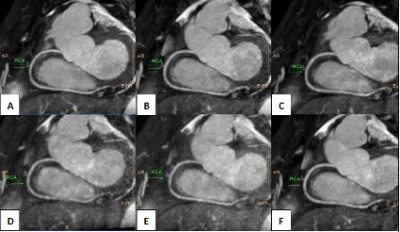 |
Clinical impact of compressed sensing on whole-heart coronary MRI
Yang Wu1, Xiaojing Ma1, Weiwei Wang1, Feng Xiong1, Ke Yu1, Jiazheng Wang2, Peng Sun2, and Qingping gu2
1MRI department, Wuhan Asia Heart General Hospital, WuHan, China, 2philips, healthcare, Beijing, China
Diagnosis of Coronary artery disease using MRI remains a challenge due to the long scan times, limited spatial resolution, and low signal-to-noise of MRI technique. Compressed Sensing (CS) is a novel technique to accelerate the imaging and improve detection of coronary stenosis by coronary MRI. Results of this study indicate that there was no statistical difference in coronary artery image quality from the scanning with CS acceleration factors (AF) of 2, 4, 6. Since CS AF of 6 provides the shortest scan time and favorable image quality, this might be an optimal setting for clinical whole-heart coronary MRI.
|
|||
2897.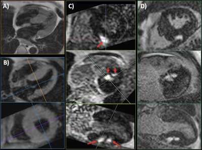 |
Breath-hold Multi-contrast 3D Dark Blood MRI of the Heart and Great Vessels with Gated Unbalanced T1 Relaxation-Enhanced Steady-State (uT1RESS)
Robert R Edelman1,2, Nondas Leloudas3, Jianing Pang4, and Ioannis Koktzoglou3,5
1Radiology, NorthShore University HealthSystem, EVANSTON, IL, United States, 2Feinberg School of Medicine, Northwestern University, Chicago, IL, United States, 3Radiology, NorthShore University HealthSystem, Evanston, IL, United States, 4Siemens Medical Solutions USA, Chicago, IL, United States, 5Pritzker School of Medicine, University of Chicago, Chicago, IL, United States
We recently described a novel class of steady-state pulse sequence called T1 Relaxation-Enhanced Steady-State (T1RESS). The unbalanced variant, called uT1RESS, generates dark blood images without the need to apply additional preparation modules. We sought to determine whether an electrocardiogram (ECG)-gated implementation of uT1RESS could be used to generate dark blood images for evaluation of the heart and great vessels. A pilot study of healthy volunteers and patients undergoing cardiac MRI was approved by the hospital institutional review board. Initial results suggest that ECG-gated uT1RESS shows promise for volumetric multi-contrast evaluation of the heart and great vessels.
|
|||
2898.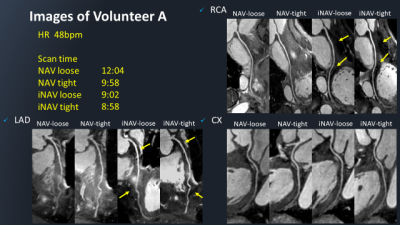 |
Whole heart coronary MRA using image-based 2D navigator (iNav) and conventional Nav system: comparison of image quality and scan time
Kazuo Kodaira1, Michinobu Nagao2, Masami Yoneyama3, Yasutomo Katsumata3, Takumi Ogawa1, Yutaka Hamatani1, Isao Shiina1, Yasuhiro Goto1, Mamoru Takeyama1, Isao Tanaka1, and Shuji Sakai2
1Department of Radiological Services, Tokyo Women's Medical University Hospital, tokyo, Japan, 2Department of Diagnostic imaging & Nuclear Medicine, Tokyo Women's Medical University Hospital, tokyo, Japan, 3Philips Japan, tokyo, Japan
Diaphragmatic one-dimensional navigation (NAV) is the conventional approach for CMRA respiratory motion compensation. This method has limitations such as the cumbersomeness of setting NAV and giving stresses to patients by wrapping the compression band around the chest/abdomen for minimizing respiratory artifacts. Image-based 2D navigator (iNAV) can solve this limitation because it directly corrects the translational movement of the heart in two directions (R-L and F-H) by using 2D real-time imaging. We investigate the feasibility of iNAV with loosely wrapped abdominal compression band compared to the conventional NAV approach.
|
|||
2899.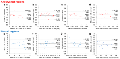 |
Reproducibility between 3D turbo spin-echo and 2D dual inversion recovery turbo spin-echo for coronary vessel wall imaging in Kawasaki disease
Koji Matsumoto1, Hajime Yokota2, Takafumi Yoda 1, Ryota Ebata3, Hiroki Mukai1, Yoshitada Masuda1, and Takashi Uno2
1Department of Radiology, Chiba University Hospital, Chiba, Japan, 2Diagnostic Radiology and Radiation Oncology, Graduate School of Medicine, Chiba University, Chiba, Japan, 3Department of Pediatrics, Chiba University Hospital, Chiba, Japan
We performed qualitative and quantitative evaluations of cross-sectional coronary vessel wall images collected by 3D-TSE and 2D-DIR-TSE in Kawasaki disease (KD) and assessed the reproducibility of both. Coronary vessel walls were evaluated separately in aneurysmal and normal regions. 3D-TSE was comparable to 2D-DIR-TSE in visual assessment and has reproducibility of 3D of lumen area (LA), wall area (WA), normalized wall index (NWI = WA / (LA + WA) ), and lumen-cardiac wall contrast measurements. 3D-TSE coronary vessel wall imaging can collect wide FOV and accurate cross-sectional images, and is feasible for following up the coronary arteries of KD.
|
|||
2900.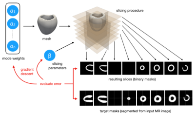 |
Rapid Personalisation of Left Ventricular Meshes using Differentiable Rendering
Thomas Joyce1, Stefano Buoso1, Christian T Stoeck1, and Sebastian Kozerke1
1University and ETH Zurich, Zurich, Switzerland
We present a novel method for volumetric left ventricle mesh personalisation from cardiac MR images. The proposed method does not require any ground-truth mesh training data. Additionally, it corrects for slice misalignment and can propagate these correction back to the original image data. The method is expressive enough to capture diverse morphology, and is also differentiable, allowing for direct inclusion in deep-learning pipelines. We demonstrate that our mesh personalisation approach works robustly on both healthy and pathological anatomy.
|
|||
2901.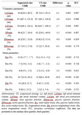 |
Single-shot compressed sensing versus segmented cine cardiac magnetic resonance in assessing left atrial volume and strain
yang chen1, Pan pan Xu1, Xiao yue Zhou2, Yi Xu1, and Xiao mei Zhu1
1the First Affiliated Hospital of Nanjing Medical University, Nanjing, China, 2Siemens Healthineers Ltd, Shanghai, China
Left atrial (LA) size is a useful measurement in predicting adverse cardiovascular outcomes. The current study compared LA volume accuracy and strain analysis between conventional segmented cine cardiac magnetic resonance (CMR) and single-shot compressed sensing (CS) cine CMR in 31 and 30 patients with and without left ventricular (LV) diastolic dysfunction, respectively. In both techniques, LA passive ejection fraction and passive radial and longitudinal strain showed diagnostic efficacy for LV diastolic dysfunction, and volume accuracies were not significantly different between techniques. CS cine CMR can reliably assess LA volume and strain in patients who are at risk for cardiovascular diseases.
|
|||
| 2902. | Global circumferential strain based on cardiac magnetic resonance is associated with ventricular arrhythmias in hypertrophic cardiomyopathy
Cailing Pu1, Jingle Fei1, Yan Wu1, Chengbin He1, and Hongjie Hu1
1Radiology, Sir Run Run Shaw Hospital, Zhejiang University School of Medicine, Hangzhou, China
Myocardial strain parameters detected by tissue tracking on cardiac magnetic resonance (CMR-TT) were helpful for early prediction of myocardial damage in patients with hypertrophic cardiomyopathy (HCM). Global circumferential strain (GCS) and late gadolinium enhancement (LGE) percentage were reliable and independent predictors for ventricular arrhythmias (VAs) in HCM. For patients who can’t undergo the LGE scan, reduced GCS may have potential value to identify HCM patients with the risk of VAs.
|
|||
2903.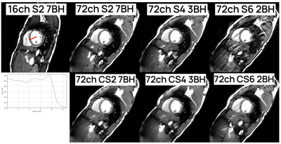 |
Double breath-hold whole heart cine cardiac MR imaging using a new 72-channel local receive array
Hugo Klarenberg1, Mark Gosselink2, Martijn Froeling2, Tim Leiner2, Aart J. Nederveen3, Adrianus J. Bakermans3, Hildo J. Lamb4, S. Matthijs Boekholdt5, and Gustav J. Strijkers1
1Biomedical Engineering & Physics, Amsterdam UMC, Amsterdam, Netherlands, 2Radiology, University Medical Center Utrecht, Utrecht, Netherlands, 3Radiology and Nuclear Medicine, Amsterdam UMC, Amsterdam, Netherlands, 4Radiology, Leiden University Medical Center, Leiden, Netherlands, 5Cardiology, Amsterdam UMC, Amsterdam, Netherlands
Currently, a standard short-axis cardiac CINE protocol consists of ~7 breath-holds which is time-consuming, costly and patient-unfriendly. We investigated whether it is feasible to accurately quantify biventricular morphology and function in 3 to 2 breath-holds using a new 72‑channel receive array and (compressed-)SENSE acceleration. To that end, we performed a comparison between a 16 channel local array with a SENSE-factor 2 and the new coil with (compressed‑) SENSE factors of 2, 4 and 6. In all cases, left ventricular ejection fractions were similar with acceptable image quality decline for higher acceleration, facilitating cardiac function assessment in only 2 breath-holds.
|
|||
2904.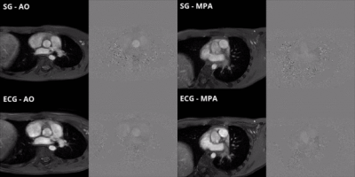 |
Cardiac Self-Gating for Free-Breathing Phase-Contrast MRI
Marcus Hott1,2,3, Henrik von Kleist4, Jihye Jang2,5, Andrew Powell1,2, and Mehdi Hedjazi-Moghari1,2
1Department of Pediatrics, Harvard Medical School, Boston, MA, United States, 2Department of Cardiology, Boston Children's Hospital, Boston, MA, United States, 3Department of Informatics, Technical University of Munich, Munich, Germany, 4Munich School for Data Science, Munich, Germany, 5Philips Healthcare, Gainesville, FL, United States
We implemented an end-to-end cardiac self-gating algorithm for a novel free-breathing 2D cine phase-contrast sequence on a clinical MRI scanner. We validated our technique against clinically performed ECG-gated cine phase-contrast scan. Strong agreement with no statistically significant difference was shown between the systemic and pulmonary blood flow measurements calculated between the standard ECG-gating and self-gating scans. Our approach eliminates the need for an ECG signal and allows for blood flow measurements where an ECG is not accessible such as fetal cardiac MRI.
|
|||
2905.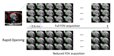 |
Visualizing Human Aortic Valve Opening and Closing with Sub-Millisecond Temporal Resolution Over a Reduced Field-of-View
Zheng Zhong1,2, Qingfei Luo1, Kaibao Sun1, Guangyu Dan1,2, Muge Karaman1,2, and Xiaohong Joe Zhou1,2,3,4
1CMRR, University of Illinois at Chicago, Chicago, IL, United States, 2Bioengineering, University of Illinois at Chicago, Chicago, IL, United States, 3Neurosurgery, University of Illinois at Chicago, Chicago, IL, United States, 4Radiology, University of Illinois at Chicago, Chicago, IL, United States
An imaging technique, coined epi-based Sub-millisecond Periodic Event Encoded Dynamic Imaging, or epi-SPEEDI, has been shown capable of visualizing the rapid opening and closing of human aortic valve with sub-millisecond temporal resolution. However, the acquisition time of epi-SPEEDI was relatively long (8-10 breath holds). Herein, we report an alternative SPEEDI technique with a reduced FOV, which we call rFOV-SPEEDI, to shorten the scan times. This technique has been successfully applied to visualization of human aortic valve opening and closing with a temporal resolution of 0.6 ms, together with a scan time reduction of 32.5%.
|
|||
2906.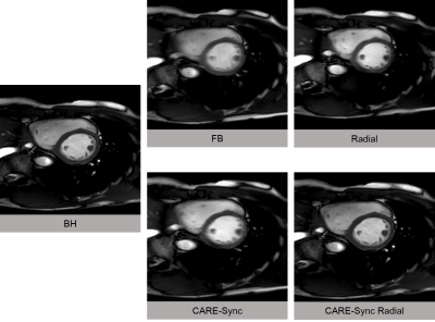 |
Free breathing respiratory triggered retrospectively cardiac gated (CARE-Sync) bSSFP cine imaging with radial acquisition
Isao Shiina1, Michinobu Nagao2, Masami Yoneyama3, Yasutomo Katsumata3, Yasuhiro Goto4, Kazuo Kodaira4, Takumi Ogawa4, Yutaka Hamatani1, Mamoru Takeyama4, Isao Tanaka4, and Shuji Sakai2
1Radiological Services, Tokyo Women's Medical University Hospital, tokyo, Japan, 2Department of Diagnostic Imaging and Nuclear Medicine, Tokyo Women's Medical University Hospital, Tokyo, Japan, 3Philips Electronics Japan, Tokyo, Japan, 4Department of Radiological Services, Tokyo Women's Medical University Hospital, Tokyo, Japan
we investigate the clinical usefulness of respiratory-triggered cardiac cine radial MRI. Compared with the breath-hold scan, the left ventricular function values obtained from the breath-triggered scan showed a better correlation than the conventional free-breathing scan.
|
|||
2907.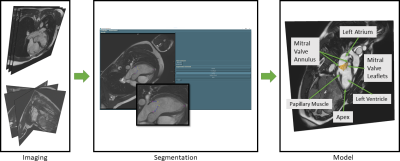 |
Precision of Mitral Valve Segmentations from Standard and Rotational Long Axis Cardiac Cine MRI
Chiara Manini1, Lennart Tautz1,2, Alireza Khasheei3, Titus Kühne1,4, Christoph Kolbitsch5, Jeanette Schulz-Menger1,4, and Anja Hennemuth1,2,4
1ICM, Charité - Universitätsmedizin Berlin, Berlin, Germany, 2Fraunhofer MEVIS, Bremen, Germany, 3Deutsches Herzzentrum Berlin, Berlin, Germany, 4German Center for Cardiovascular Research (DZHK), Partner Site Berin, Germany, 5Physikalisch-technische Bundesanstalt, Berlin, Germany
Diagnostic assessment of the mitral valve considers pathological alterations in anatomy as well as their effect on cardiac function. The goal of our work was to evaluate, whether the mitral valve anatomy could be assessed with diagnostic precision based on MRI imaging using standard (SLA) or rotational long axis (RLA) cine MRI. In a preliminary study, 6 healthy volunteers underwent imaging in a 1.5T scanner with both approaches. We generated 3D models for the quantitative assessment of the shape of the valves and achieve a higher precision based on the RLA image data.
|
|||
2908.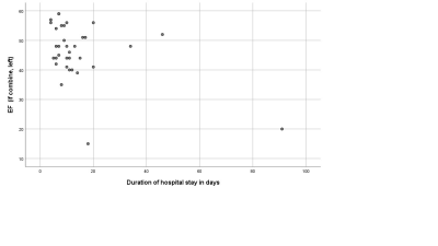 |
Cardiac Magnetic Resonance and hemodynamic predictors of postoperative Fontan Outcomes
Jyothsna Akam Venkata1,2, Mohamed Abdelghafar Hussein1,3, Joshua Greer1, Robert Jaquiss4, Gerald Greil1, Jeanne Dillenbeck1, Surendrenath R Veeram Reddy 1, Jenifer Hernandez1, and Tarique Hussain1
1Dept of Pediatrics, Division of Pediatric Cardiology, UT Southwestern Medical Center, Dallas, TX, United States, 2University of Mississippi Medical Center, Jackson, MS, United States, 3Kafrelsheikh University, Kafr Elsheikh, Egypt, 4Department of Pediatric Cardiothoracic Surgery, UT Southwestern Medical Center, Dallas, TX, United States
Pre-Fontan cardiac assessment with CMR in conjunction with cardiac catheterization has increasingly become the standard of care. Cardiac catheterization and CMR performed for pre-Fontan assessment at a single institution between April 2017 and June 2020 were analyzed. Our cohort consisted of 43 patients with median age of 3.7 years(range 1.8-14) at the time of CMR. Median age at Fontan operation was 4 years (2.4-14). We found significant correlation between CMR derived ventricular end diastolic, end systolic volume, ejection fraction and cardiac catheterization derived ventricular end-diastolic and mean superior vena cava pressure, and duration of post-operative hospital stay after Fontan operation.
|
|||
2909.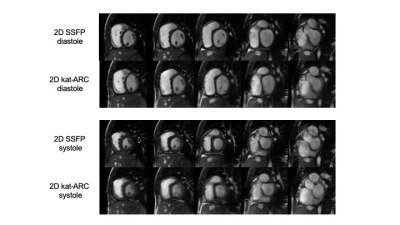 |
Reliability of respiratory-triggered 2D cine kat-ARC for the assessment of biventricular function in patients with repaired tetralogy of Fallot
Makoto Orii1, Tsuyoshi Sugawara1, Hidenobu Takagi1, Martin A Janich2, Atsushi Nozaki3, and Kunihiro Yoshioka1
1Iwate Medical University, Yahaba, Japan, 2GE Healthcare, Munich, Germany, 3GE Healthcare, Tokyo, Japan
To assess the image quality, reproducibility, and accuracy of two-dimensional (2D)-cine k-adaptive-t Autocalibrating Reconstruction for Cartesian sampling (2D kat-ARC) for the quantification of biventricular volumes and function compared to 2D balanced steady state free precession (2D SSFP) in patients with repaired tetralogy of Fallot (TOF). In this study, we were able to find good reproducibility of biventricular volumes and ejection fraction (EF) between 2D SSFP and 2D kat-ARC sequences despite some drawbacks including an overestimation of right ventricular (RV) end-systolic volume and an underestimation of RV stroke volume and RVEF compared to 2D SSFP.
|
The International Society for Magnetic Resonance in Medicine is accredited by the Accreditation Council for Continuing Medical Education to provide continuing medical education for physicians.