Digital Posters
Imaging of Heart Failure: Tissue Characterization
ISMRM & SMRT Annual Meeting • 15-20 May 2021

Digital Posters
Imaging of Heart Failure: Tissue Characterization
| Concurrent 3 | 13:00 - 14:00 |
0979.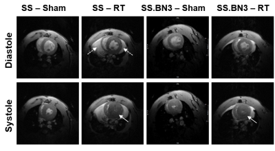 |
Myocardial contractility distinguishes the effect of radiation therapy on different rat models
El-Sayed H Ibrahim1, Luba Frank1, Sherry-Ann Brown1, Jason Rubenstein1, Rachel Schlaak1, Elizabeth Gore1, and Carmen Bergom1
1Medical College of Wisconsin, Milwaukee, WI, United States
Radiation therapy (RT) is used by more than 50% of all cancer patients, where the risk of RT-induced cardiotoxicity reaches 33%. In this study, we investigate the role of cardiac contractility by MRI to differentiate the response of two different rat models to RT. The results showed the value of myocardial strain for early detection of RT-induced cardiotoxicity and exploring the effect of genetic profile and duration post-RT on cardiotoxicity. In conclusion, MRI strain imaging allows for determining spatial distribution and temporal progression of myocardial contractility post-RT, which would allow for determining the effect of genetic profile on cardiotoxicity development.
|
|||
0980.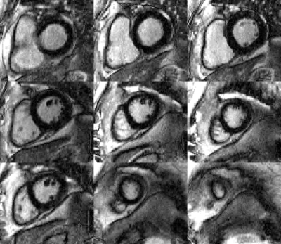 |
Combined simultaneous multi-slice (SMS) bSSFP & compressed sensing (CS) for accelerated late gadolinium enhancement (LGE) imaging in the heart
Ronald Mooiweer1, Sarah McElroy2, Giulio Ferrazzi3, Muhummad Sohaib Nazir2, Carl Evans2, Filippo Bosio2, Nabila Mughal2, Karl P Kunze4, Radhouene Neji4, Peter Speier5, Daniel Stäb6, Pier Giorgio Masci2, Reza Razavi2, Amedeo
Chiribiri2, and Sébastien Roujol2
1King's College London, London, United Kingdom, 2School of Biomedical Engineering and Imaging Sciences, King's College London, London, United Kingdom, 3IRCCS San Camillo Hospital, Venice, Italy, 4MR Research Collaborations, Siemens Healthcare Limited, Frimley, United Kingdom, 5Siemens Healthcare GmbH, Erlangen, Germany, 6MR Research Collaborations, Siemens Healthcare Limited, Melbourne, Australia
SMS-bSSFP combined with compressed sensing was successfully used to acquire LGE images through the use of multiple single-shot dynamic acquisitions in one breath-hold. Instead of 1 slice per breath-hold, 3 slices could be acquired per breathhold.
|
|||
0981.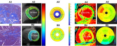 |
Cardiovascular magnetic resonance T1ρ endogenous contrast can detect early myocardial fibrosis in hypertrophic cardiomyopathy
Keyan Wang1, Jingliang Cheng2, Yang Yang3, and Jie Zheng4
1MRI, 1st Affiliated Hospital of Zhengzhou University, Zhengzhou, China, 21st Affiliated Hospital of Zhengzhou University, Zhengzhou, China, 3Icahn School of Medicine at Mount Sinai, New York, NY, United States, 4Mallinckrodt Institute of Radiology, Washington University in St, Washington, WA, United States
The study is to determine clinical utility of T1ρ to detect myocardial fibrosis in patients with hypertrophic cardiomyopathy (HCM) at different subtypes. The results showed that absolute T1ρ (ms) increased progressively and significantly from volunteer to HCM-N and then to HCM-H. ECV has the same trend as T1ρ. There was a strong positive correlation between ECV and T1ρ. If selecting T1ρ relaxation times of 36.5ms as the cutoff value for normal myocardial fibrosis, the sensitivity, specificity and AUC to detect myocardial fibrosis were 83.7%, 91.3%, and 0.937, respectively.T1ρ is able to detect early diffuse fibrosis in HCM and two subtypes.
|
|||
0982.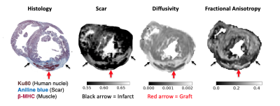 |
Diffusion Tensor Imaging Reveals Myocardial Structure after Stem Cell-derived Cardiomyocyte Therapy in Myocardial Infarction
Moses P. Cook1, Wahiba Dhahri2, Graham A. Wright1,3,4, Michael A. Laflamme2, and Nilesh R. Ghugre1,3,4
1Medical Biophysics, University of Toronto, Toronto, ON, Canada, 2McEwen Centre for Regenerative Medicine, University Health Network, Toronto, ON, Canada, 3Schulich Heart Program, Sunnybrook Health Sciences Centre, Toronto, ON, Canada, 4Physical Sciences Platform, Sunnybrook Research Institute, Toronto, ON, Canada
Cardiac regenerative therapies using pluripotent stem cell-derived cardiomyocytes (PSC-CM, the graft) have shown promise in animal models of myocardial infarction. However, the mechanisms of structural integration of graft with host myocardium and the associated impact on function are less understood. 3D Diffusion Tensor Imaging (DTI) and contrast enhanced MRI were performed at 7T in ex vivo guinea pig hearts to quantify cardiac microstructure post PSC-CM transplantation. DTI parameters within the graft trended towards the healthy non-infarcted state, demonstrating a degree of anisotropic tissue repopulation within the infarct zone. MRI is a valuable marker for treatment efficacy in emerging regenerative therapies.
|
|||
 |
0983. |
Simultaneous dark blood late gadolinium enhancement and MR angiography for atrial scar detection
Dongyue Si1, Yanfang Wu2, Jie Yin2, Rui Guo3, Jingjing Xiao4, Bowei Liu1, Xue Lin2, Peng Gao2, Deyan Yang2, Quan Fang2, Jianwen Luo1, Daniel A. Herzka5, and Haiyan Ding1
1Center for Biomedical Imaging Research (CBIR), Department of Biomedical Engineering, School of Medicine, Tsinghua University, Beijing, China, 2Department of Cardiology, Peking Union Medical College Hospital, Chinese Academy of Medical Sciences & Peking Union Medical College, Beijing, China, 3Department of Medicine, Beth Israel Deaconess Medical Center and Harvard Medical School, Boston, MA, United States, 4Department of Medical Engineering, Xinqiao Hosptial, Army Medical University, Chongqing, China, 5National Heart, Lung, and Blood Institute, National Institutes of Health, Bethesda, MD, United States
Cardiovascular magnetic resonance can provide functional and structural assessment of the left atrium and pulmonary veins, providing highly relevant pre- and post-procedural information in the treatment of atrial fibrillation (AF). In this study, an independent navigator-gated simultaneous dark blood late gadolinium enhancement and MR angiography sequence was developed for atrial scar detection. In 4 post AF ablation subjects, comparison with conventional phase sensitive inversion recovery images indicated that both scar-to-blood and blood-to-myocardium contrast were improved. Excellent correlation was observed between the scar measured by the proposed sequence and the low voltage areas observed with electroanatomical mapping.
|
||
0984.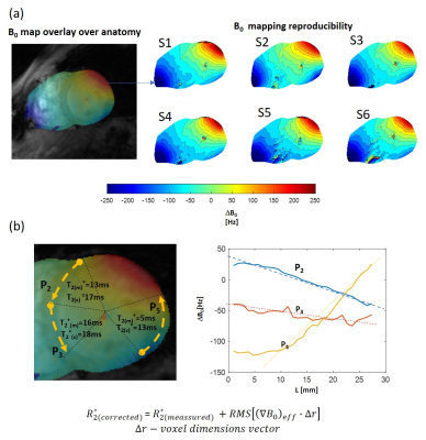 |
Towards Clinical Application of the T2* quantification in the 7T Cardiac MRI: Reproducibility Study in Healthy Volunteers.
Maxim Terekhov1, Theresa Reiter2, Michael Hock1, David Lohr1, Wolfgang Bauer1,2, and Laura M. Schreiber1
1Chair of Molecular and Cellular Imaging, University Hospital Würzburg, Comprehensive Heart Failure Center, Wuerzburg, Germany, 2Department of Internal Medicine I, Cardiology, University Hospital Würzburg, Wuerzburg, Germany
7T cardiac MRI is an emerging technique with the potential to increase the spatial resolution and functional informativity of the cardiac images. T2*-quantification becomes a more sensitive MRI method at 7T compared to the lower B0-fields and may provide valuable information on cardiac tissue remodeling. However, in practice, the value of T2* at 7T as a marker of tissue alterations can be diminished by the suboptimal B0-shimming and problems of acquisition and analysis. We present the results of the reproducibility and quality assurance study with 10 healthy volunteers for the optimization of T2*-measurement protocol for a future clinical application.
|
|||
0985.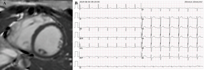 |
Association Between Ventricular Arrhythmias and Late Gadolinium Enhancement in Patients with Apparently Normal Hearts
wen qian1, kai gu2, xiaoyue zhou3, yi xu1, and xiaomei zhu4
1The First Affiliated Hospital of Nanjing Medical University, Nanjing, China, 2Cardiovascular Medicine, The First Affiliated Hospital of Nanjing Medical University, Nanjing, China, 3MR Collaboration, Siemens Healthineers Ltd., Shanghai, China, 4radiology, The First Affiliated Hospital of Nanjing Medical University, Nanjing, China
Cardiac magnetic resonance (CMR) is the gold standard for evaluating myocardial fibrosis. This study investigated the association between the occurrence and morphological features of ventricular arrhythmias (VAs) and left ventricular late gadolinium enhancement (LV-LGE) in patients without structural heart diseases. Seventy-eight patients with apparently normal hearts were enrolled and underwent simultaneous electrocardiogram and CMR examinations. Polymorphic VAs and ST-segment depression indicated a higher probability of having LV-LGE on CMR. The findings can help clinicians screen patients for CMR.
|
|||
0986.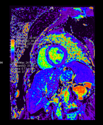 |
Parametric mapping to assess cardiotoxicity in breast cancer patients receiving concurrent anthracyclines and trastuzumab
Divya Susanna Ninan1 and Binita Riya Chacko2
1Radiology, Christian Medical College Vellore, Vellore, India, 2Radiology, Christian Medical College, Vellore, Vellore, India
Novel cardiac MRI mapping techniques such as T1, T2 and ECV parametric mapping have been proven to detect early cardiotoxicity among patients taking chemotherapeutic drugs. We aimed to determine whether these parameters could predict the development of early onset cardiac dysfunction in HER 2 positive breast cancer patients who were on anthracyclines and trastuzumab. We demonstrated statistically and clinically significant drop in left ventricular ejection fraction (LVEF) and right ventricular ejection fraction (RVEF) at four months post chemotherapy. The change in the mapping values were not statistically significant, however there was a trend towards an increase in the ECV.
|
|||
0987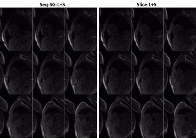 |
High resolution whole-heart coverage multiband first-pass myocardial perfusion MRI using the slice-L+S reconstruction method Video Permission Withheld
Changyu Sun1, Kenneth C. Bilchick2, Daniel Weller3, Michael Salerno1,2,4, and Frederick H. Epstein1,4
1Biomedical Engineering, University of Virginia, Charlottesville, VA, United States, 2Medicine, University of Virginia, Charlottesville, VA, United States, 3Electrical and Computer Engineering, University of Virginia, Charlottesville, VA, United States, 4Radiology, University of Virginia, Charlottesville, VA, United States
k-t undersampled multiband first-pass perfusion imaging provides in-plane and through-plane acceleration and potentially enables high spatial resolution and whole-heart coverage. Our slice-L+S work splits MB data consistency and enforcing L+S into two subproblems wherein MB data, in- and through-plane coil information and L+S properties of slices are systematically utilized. Here we compare slice-L+S to single-band L+S and slice-GRAPPA followed sequentially by L+S (seq-SG-L+S) using 3 retrospectively undersampled datasets. High resolution nine-slice data with multiband factor-3 and rate-3 undersampling were prospectively acquired in 4 patients. Slice-L+S showed better image quality demonstrated by quantitative results and scores from two cardiologists.
|
|||
0988.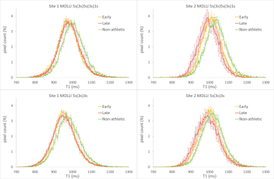 |
Are myocardial MOLLI T1 differences between athletic and non-athletic subjects real or dependent on center differences or MOLLI scheme?
Tom Dresselaers1,2, Ruben De Bosscher3, Sofie Tilborghs4,5, Alexandru Cernicanu6, Christophe Dausin7, Oliver Ghekiere8, Rik Willems3, Guido Claessen3, and Jan Bogaert1,2
1Radiology, UZ Leuven, Leuven, Belgium, 2Imaging and Pathology, KU Leuven, Leuven, Belgium, 3Department of Cardiovascular Sciences, KU Leuven, Leuven, Belgium, 4Department of Electrical Engineering (ESAT), KU Leuven, Leuven, Belgium, 5Medical Imaging Research Center, Medical Physics and Quality Assessment, KU Leuven, Leuven, Belgium, 6Philips Benelux, Eindhoven, Netherlands, 7Exercise Physiology Research Group, KU Leuven, Leuven, Belgium, 8Jessa Hospital, Hasselt, Belgium We analysed differences in myocardial T1 between non-athletic subjects and athletes (which started endurance training early or late in life) to determine inter-center consistency. In addition, we implemented and extended MOLLI scheme (5s5s1s) to improve precision at very low heart rate typical in athlete's and compared with results from a standard 5s3s MOLLI. Absolute levels were dependent on MOLLI scheme and site, but group differences were very similar as evident from full myocardium T1 histograms.
|
|||
0989.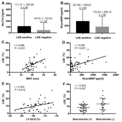 |
Myocardial fibrosis in preserved ejection fraction hypertrophic cardiomyopathy: predictive values of non-invasive markers
Yumin Li1, Xiaoyue Zhou2, Jia Liu1, Yukun Cao1, Xiaoyu Han1, Guozhu Shao1, Jin Gu1, Tong Liu1, Na Li1, Yue Cui1, and Heshui Shi1
1Department of Radiology, Union Hospital, Tongji Medical College, Huazhong University of Science and Technology, Wuhan, China, 2MR Collaboration, Siemens Healthineers Ltd, ShangHai, China
This study aimed to determine the predictive value of non-invasive markers in patients with hypertrophic cardiomyopathy with preserved ejection fraction. The results demonstrated that an elevated level of N-terminal pro b-type natriuretic peptide (Nt-proBNP) was an independent predictor for the presence of late gadolinium enhancement (LGE), and combined with maximum wall thickness (MWT), had good diagnostic performance for the detection of LGE. Furthermore, decreased left ventricular global circumferential strain (GCS) was independently correlated with the LGE%, indicating its potential prognostic value for detecting myocardial fibrosis.
|
|||
0990.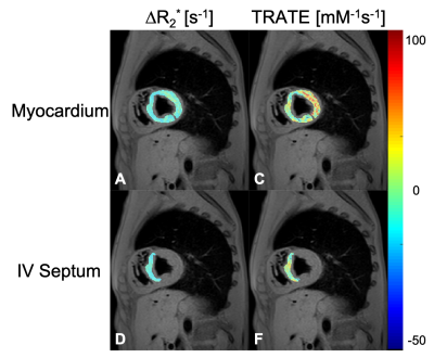 |
Relaxivity Contrast Imaging (RCI) of Myocardial Microstructure in Secondary Cardiomyopathy
Sudarshan Ragunathan1, Ryan K Robison2, Erik G Ellsworth3, and C Chad Quarles1
1Barrow Neurological Institute, Phoenix, AZ, United States, 2Philips, Nashville, TN, United States, 3Phoenix Children's Hospital, Phoenix, AZ, United States
Clinical cardiac MRI protocols can be insensitive to secondary cardiomyopathy induced myocardium thickening and altered cardiac function. In this work, we demonstrate the feasibility of acquiring cardiac relaxivity contrast imaging (RCI) data, and specifically the RCI-parameter termed transverse relaxivity at tracer equilibrium (TRATE). TRATE has previously been shown to detect changes in cellular microstructure. RCI acquisition comprises of 1) pre-/post-contrast T1 maps using a MOLLI protocol, and 2) pre-/post-contrast T2* maps using a multi-gradient echo protocol. Robust changes in post contrast R2*, and the subsequent TRATE values, support the potential role of using RCI to study myocardial microstructure changes.
|
|||
0991.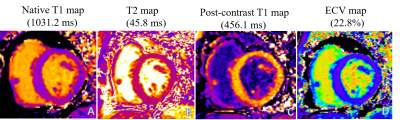 |
Cardiovascular MRI–derived myocardial strain vs. extracellular volume fractions in asymptomatic heart transplant patients
Xuehua Shen1, Yating Yuan2, Xiaoyue Zhou3, Kelvin Chow4, Jens Wetzl5, and Bo Liang2
1Department of Radiology, the Affiliated Hospital of Guizhou Medical University, Guiyang, China, China, 2Department of Radiology, Union Hospital, Tongji Medical College, Huazhong University of Science and Technology, Wuhan, China, Wuhan , China, China, 3MR Collaboration, Siemens Healthineers Ltd., Shanghai, China, Shanghai, China, China, 4MR R&D, Siemens Medical Solutions Inc., Chicago, USA, Chicago, USA, IL, United States, 5Siemens Healthcare GmbH, Erlangen, Germany, Erlangen, Germany, Germany
Cardiac magnetic resonance imaging has excellent tissue resolution and can quantitatively evaluate myocardial deformations and tissue characteristics. In this study, heart transplant (HT) patients had changed myocardial strain (LCGLS, LVGCS, and LVGRS) and tissue parameters (T1, T2, and ECV). There were also moderate correlations between these two different measurements. We suggest that myocardial movement is related to myocardial edema and fibrosis, and myocardial strain measurements could be an alternative to ECV with gadolinium injections.
|
|||
0992.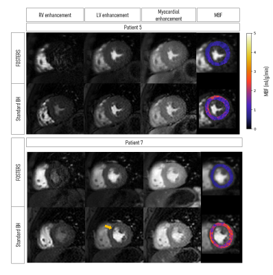 |
Free-Breathing High-Resolution Quantitative First-Pass Perfusion Cardiac MR using Dual-Echo Dixon
Cian Scannell1, Torben Schneider2, Ebraham Alskaf1, Filippo Bosio1, Joao Tourais3, Javier Sanchez-Gonzalez4, Mariya Doneva5, Christophe Schuelke5, Jakob Meineke5, Jochen Keupp5, Jouke Smink6, Marcel Breeuwer7, Amedeo Chiribiri1,
Markus Henningsson8,9, and Teresa M Correia1
1School of Biomedical Engineering and Imaging Sciences, King's College London, London, United Kingdom, 2Philips Healthcare, Guildford, United Kingdom, 3Magnetic Resonance Systems Lab, Delft University of Technology, Delft, Netherlands, 4Philips Healthcare Iberia, Madrid, Spain, 5Philips Research, Hamburg, Germany, 6MR Clinical Science, Philips Healthcare, Best, Netherlands, 7Department of Biomedical Engineering, Eindhoven University of Technology, Eindhoven, Netherlands, 8Department of Health, Medicine and Caring Sciences, Linköping University, Linköping, Sweden, 9Center for Medical Image Science and Visualization (CMIV), Linköping University, Linköping, Sweden
First-pass perfusion cardiac MR (FP-CMR) is an essential tool for characterizing ischemic heart disease. Moreover, automated quantitative methods enable a reliable and operator-independent assessment of myocardial blood flow. Conventional FP-CMR has limited spatial resolution and is performed under breath-hold. Therefore, diagnostic accuracy is compromised by respiratory induced motion artifacts and false-positives due to dark-rim artifacts. To overcome these challenges, free-breathing Fat-water separation for mOtion-corrected Spatio-TEmporally accelerated myocardial peRfuSion (FOSTERS) has been proposed. Here, we implement and evaluate a high-resolution dual-echo Dixon version of FOSTERS and compare its quantitative performance to standard-resolution FP-CMR in patients with suspected cardiovascular disease.
|
|||
|
0993.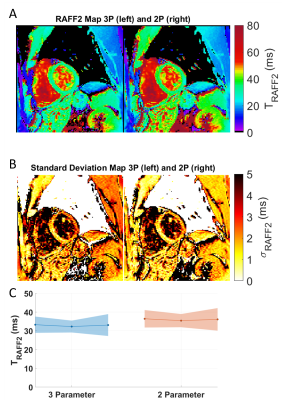 |
2D High Resolution Myocardial TRAFF2 Mapping: Reproducibility and Accuracy
Joao Tourais1, Ingo Hermann1,2, Rick Voncken3, Ömer Burak Demirel4, Sebastian Weingärtner1, and Mehmet Akcakaya4
1Magnetic Resonance Systems Lab, Department of Imaging Physics, TU Delft, Delft, Netherlands, 2Computer Assisted Clinical Medicine, Medical Faculty Mannheim, Heidelberg University, Mannheim, Germany, 3Scannexus BV, Maastricht, Netherlands, 4Department for Electrical and Computer Engineering, and Center for Magnetic Resonance Research, University of Minnesota, Minnesota, MN, United States
Relaxation Along a Fictitious Field (RAFF) times in the 2nd rotating frame (TRAFF2) have shown promise for the depiction of acute myocardial infarct in a mouse model. In this work, in vivo myocardial TRAFF2 maps are acquired in a single breath-hold at 3T. An additional image was acquired following saturation preparation to enable reconstruction with a three-parameter fit model. Visually high-quality map quality, homogeneous signal, artifact resilience and low inter-scan variability were achieved with TRAFF2 in vivo. This makes TRAFF2 an excellent candidate for myocardial tissue characterization in clinical use.
|
||
0994.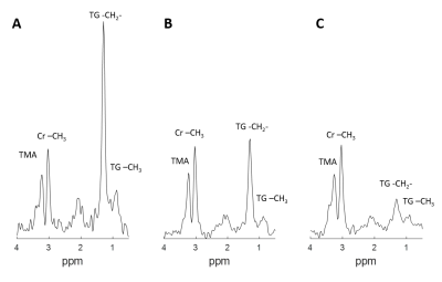 |
Cardiac 1H MRS detected reduced myocardial triglycerides after Doxorubicin injection: an in vivo animal study
Xiaoying Cai1,2, Selcuk Kucukseymen2, Maria-Alexandra Olaru3, Patrick Pierce2, Beth Goddu2, Jennifer Rodriguez2, and Reza Nezafat2
1Siemens Medical Solutions USA, Inc., Boston, MA, United States, 2Department of Medicine, Cardiovascular Division, Beth Israel Deaconess Medical Center and Harvard Medical School, Boston, MA, United States, 3Siemens Healthcare Limited GB&I, Frimley, United Kingdom
Cardiotoxicity remains a side effect of chemotherapy agents. Non-invasive imaging including cardiac MR has been applied to assess cardiac injury in patients receiving chemotherapy. Alterations in cardiac energetics have been reported in previous studies but have not been widely investigated in vivo. In this study, single voxel 1H spectroscopy was applied to detect lipid metabolism changes in a large animal model. The results showed reduced myocardial triglycerides content in the doxorubicin-treated group in comparison with the control group.
|
|||
0995.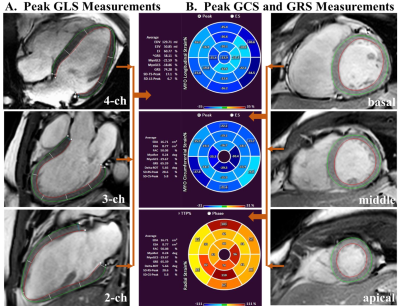 |
Myocardial T1 mapping and feature tracking in patients with OSA: potential novel insights into adaptive left ventricular remodeling phase
Xiaobin Zhou1, Xiaoyu Wei1, Yingjie Mei2, Qiong Ou3, Zhecheng Du4, and Hui Liu1
1Department of Radiology, Guangdong Provincial People’s Hospital, Guangdong Academy of Medical Sciences, Guangzhou, China, 2Philips Healthcare, Guangzhou, China, 3Sleep Center, Department of Pulmonary and Critical Care Medicine, Guangdong Provincial People’s Hospital, Guangdong Academy of Medical Sciences, Guangdong Provincial Geriatrics Institute, Guangzhou, China, 4Department of Medical Statistics, School of Public Health, Sun Yat-sen University, Guangzhou, China
The comprehensive assessment of left ventricular (LV) remodeling by cardiac magnetic resonance (CMR) imaging with T1 mapping and feature tracking (FT) technology in obstructive sleep apnea (OSA) was conducted in this study. Indexed extracellular volume (iECV) and indexed cellular volume (iCV) of myocardial tissue construction were derived from T1 mapping. The results show that elevated LV mass index (LVMi) in OSA was mainly from iCV, while not from iECV, with no LV ejection function (LVEF) or strain parameters impairment. Cellular hypertrophy accompanied with normal LV systolic function in patients with OSA may contribute to adaptive LV remodeling phase.
|
|||
0996.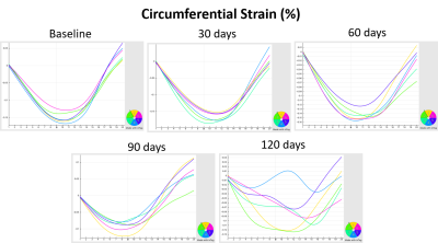 |
Assessment of Cardiovascular Characterization in Rat Model of non-Targeted Heart Radiation Using MRI
El-Sayed H Ibrahim1, Marek Lenarczyk1, and John Baker1
1Medical College of Wisconsin, Milwaukee, WI, United States
The effect of exposure outside of the geometric radiation field on non-targeted organs as it results in cardiac disease is uncertain. Characterization of myocardial tissue abnormalities and cardiac dysfunction with cardiac MRI represents a valuable tool to define the presence and stage of a disorder at given timepoint. In this study, cardiac MRI was performed in an established rat model of irradiation injury to the non-targeted heart at different timepoints post irradiation. The results showed the value of myocardial strain and T1 measurements as sensitive markers of cardiovascular changes due to non-direct irradiation effect on the heart.
|
|||
0997.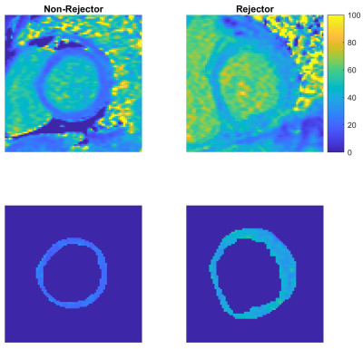 |
Extracellular Volume Fraction Mapping May Distinguish Acute Pediatric Heart Transplant Rejection from Non-Rejection
Bruce Damon1, Margaret M. Samyn2, Ke Yan2, Aryaz Sheybani3, Wubishet Belay3, Justin Godown3, Lazaro Hernandez4, Kristen George-Durrett3, and Jonathan Soslow3
1Radiology and Radiological Sciences, Vanderbilt University Medical Center, Nashville, TN, United States, 2Pediatrics, Medical College of Wisconsin, Milwaukee, WI, United States, 3Pediatric Cardiology, Vanderbilt University Medical Center, Nashville, TN, United States, 4Pediatric Cardiology, Joe DiMaggio Children’s Hospital, Hollywood, FL, United States
Acute rejection (AR) remains a significant cause of morbidity and mortality in pediatric patients after heart transplant. Endomyocardial biopsy (EMB) of the right ventricular septum is the standard method for diagnosing AR. However, EMB is prone to additional risk and sampling error as AR is a heterogeneous process. Here we show that extracellular volume fraction (ECV) mapping of the left ventricle (LV) analyzed with standard descriptive statistics and specialized texture analysis may differentiate pediatric heart transplant recipients as having AR or not. This CMR diagnostic approach offers diagnostic advantages including greater sampling volume and minimal invasiveness.
|
|||
0998.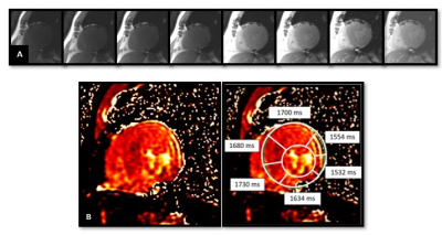 |
Relation of native T1 & ECV values to gadolinium enhancement & myocardial perfusion in hypertrophic cardiomyopathy in 3T MRI
Anoop Ayyappan1, Harshith Kramadhari1, Ajit Kumar1, and Kapilamoorthy T.R.1
1Department of IS & IR, Sree Chitra Tirunal Institute of Medical Sciences & Technology, Kerala, India
Hypertrophic cardiomyopathy (HCM) is a disorder of myocardium having a genetic basis, and high disease related mortality. Identification of areas of occult fibrosis along with overt fibrosis using T1 mapping will be useful in long term prognosis of patients along with other imaging parameters in MRI. This study was aimed at (a) determining the differences in native T1 values of controls and HCM patients, (b) to determine the relationship between early gadolinium enhancement (EGE)/Late gadolinium enhancement (LGE)/ perfusion abnormalities and native T1 and ECV values in patients of hypertrophic cardiomyopathy.
|
The International Society for Magnetic Resonance in Medicine is accredited by the Accreditation Council for Continuing Medical Education to provide continuing medical education for physicians.