Digital Posters
Susceptibility: Models & Mapping
ISMRM & SMRT Annual Meeting • 15-20 May 2021

Digital Posters
Susceptibility: Models & Mapping
| Concurrent 2 | 17:00 - 18:00 |
3965.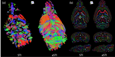 |
Asymmetric Susceptibility Tensor Imaging
Steven Cao1, Jingjia Chen1, Hongjiang Wei2, and Chunlei Liu1
1UC Berkeley, Berkeley, CA, United States, 2Shanghai Jiao Tong University, Shanghai, China
Susceptibility Tensor Imaging (STI) is a recently developed technique that uses phase data to solve for the underlying susceptibility tensor of the tissue. While STI has the potential for early diagnosis of many diseases including Parkinson’s and Alzheimer’s, it suffers from low image quality. From physics, the susceptibility tensor can be shown to be symmetric, so current approaches impose a symmetry constraint during inversion. We propose an inversion algorithm without this constraint, and instead enforce symmetry post-inversion by decomposing the result into symmetric and antisymmetric parts. We justify this approach empirically by comparing reconstructions of mouse brain and kidney data.
|
|||
 |
3966.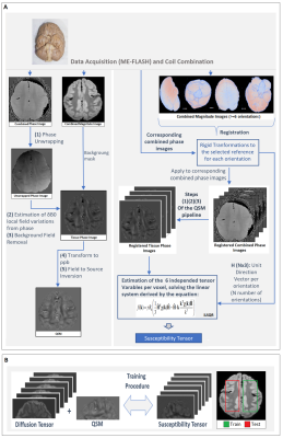 |
High angular resolution susceptibility and diffusion imaging in post mortem chimpanzee brain: Tensor characteristics and similarities
Dimitrios G. Gkotsoulias1, Riccardo Metere2, Yanzhu Su1, Cornelius Eichner1, Torsten Schlumm1, Roland Müller1, Alfred Anwander1, Toralf Mildner1, Carsten Jäger1, André Pampel1, Catherine Crockford3,4, Roman Wittig3,4, Liran Samuni 4,5,
Kamilla Pleh 6, Chunlei Liu7, and Harald E. Möller1
1Max Planck Institute for Human Cognitive and Brain Sciences, Leipzig, Germany, 2Donders Institute for Brain, Cognition and Behaviour, Nijmegen, Netherlands, 3Max Planck Institute for Evolutionary Anthropology, Leipzig, Germany, 4Tai Chimpanzee Project, Centre Suisse de Recherches Scientifiques en Cote d'Ivoire, Abidjan, Cote D'ivoire, 5Department of Human Evolutionary Biology, Harvard University, Cambridge, MA, United States, 6Project Group Epidemiology of Highly Pathogenic Microorganisms, Robert Koch Institute, Berlin, Germany, 7EECS, UC Berkeley, Berkeley, CA, United States
We present a ‘high angular resolution’ approach to susceptibility tensor imaging (STI) consisting of susceptibility-weighted acquisitions at 60 independent orientations in post- mortem chimpanzee brain. The derived susceptibility tensor and metrics are compared with the corresponding diffusion tensor-derived metrics and single-orientation quantitative susceptibility mapping (QSM) results. A preliminary approach to assess the voxel-wise relationship of the two tensors in white matter is presented: using machine learning strategies, an effort to estimate the susceptibility tensor from the diffusion tensor and minimum single-orientation QSM data in selected regions of interest was made.
|
||
|
3967.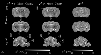 |
Rotation-Free Mapping of Magnetic Tissue Properties in White Matter
Anders Dyhr Sandgaard1, Valerij G. Kiselev2, Noam Shemesh3, and Sune Nørhøj Jespersen1,4
1Department of Clinical Medicine, Center for Functionally Integrative Neuroscience, Aarhus University, Aarhus, Denmark, 2Medical Physics, Department of Radiology, Faculty of Medicine, University of Freiburg, Freiburg, Germany, 3Champalimaud Neuroscience Programme, Champalimaud Centre for the Unknown, Lisbon, Portugal, 4Department of Physics and Astronomy, Aarhus University, Aarhus, Denmark
Mapping tissue magnetic properties may be a useful tool for understanding basic disease mechanism in neurodegenerative diseases. However, magnetic properties depend on the arrangement of magnetic susceptibility on all scales, including the microstructure, and typically require impractical sample rotations for robust estimation. By modeling white matter fibers as a system of cylinders, here we show that the first cumulant $$$\langle\Omega\rangle$$$ of the FID signal can be obtained without the need for sample rotation, by utilizing the fiber orientation distribution function estimated by diffusion imaging.
|
||
|
3968.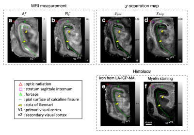 |
Magnetic susceptibility source separation: validation with Monte-Carlo simulation & application to human brain with histological comparison
Hyeong-Geol Shin1, Young Hyun Yun2, Seong Ho Yoo2, Sehoon Jung3, Sunhye Kim3, Hyung Jin Choi 2, and Jongho Lee1
1Seoul National University, Seoul, Korea, Republic of, 2Seoul National University College of Medicine, Seoul, Korea, Republic of, 3Research Institute of Industrial Science and Technology, Pohang, Korea, Republic of
We validate the magnetic susceptibility source separation method, which is recently proposed to separate paramagnetic (e.g., iron) and diamagnetic sources (e.g., myelin), by performing Monte-Carlo simulation. Then, the method is applied to ex-vivo and in-vivo human brains and compared to histology. In the simulation, we show the method successfully estimates the paramagnetic and diamagnetic susceptibility concentrations. When compared to histology, the ex-vivo and in-vivo results demonstrated that the positive and negative susceptibility maps from the method highly correspond to the iron and myelin distributions, respectively, proving its feasibility as a tool to map iron and myelin distributions in the brain.
|
||
3969.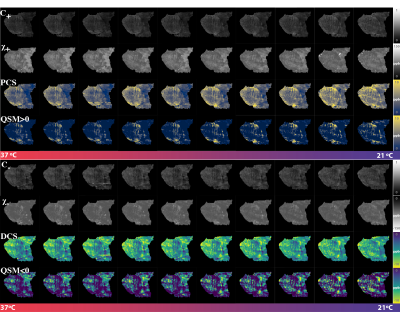 |
Validating DECOMPOSE QSM with temperature variant ex vivo brainstem imaging experiments
Jingjia Chen1, Khallil Taverna Chaim2, Maria Concepción García Otaduy2, and Chunlei Liu1,3
1Department of Electrical Engineering and Computer Sciences, University of California, Berkeley, Berkeley, CA, United States, 2LIM44, Instituto e Departamento de Radiologia, Faculdade de Medicina, Universidade de Sao Paulo, Sao Paulo, Brazil, 3Helen Wills Neuroscience Institute, University of California, Berkeley, Berkeley, CA, United States
A model and corresponding solver for estimating sub-voxel paramagnetic and diamagnetic susceptibility components are validated based on the temperature dependence of magnetic susceptibility. Two pieces of brainstem sample were scanned under various temperature conditions to test the properties of the separated paramagnetic and diamagnetic components.
|
|||
3970.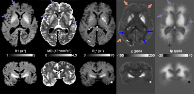 |
Studying magnetic susceptibility, microstructural compartmentalisation and chemical exchange in a formalin-fixed, whole-brain specimen
Kwok-Shing Chan1, Jeroen Mollink2, Jenni Schulz1, Anne-Marie van Cappellen van Walsum2, and José P. Marques1
1Donders Institute for Brain, Cognition and Behaviour, Radboud University, Nijmegen, Netherlands, 2Department of Medical Imaging, Anatomy, Donders Institute for Brain, Cognition and Behaviour, Radboud University Medical Center, Nijmegen, Netherlands
In this work, we study the feasibility of using formalin-fixed post-mortem specimen to investigate MRI phase contrast in brain tissue. Despite the magnetic susceptibility maps derived from in vivo and ex vivo imaging share similar contrasts, the non-susceptibility contributions on measured fields of ex vivo sample were clear affected by fixation process, and we found its microstructural compartmentalisation effect are also greatly reduced. Therefore, extra care has to be taken to directly translate ex vivo findings to in vivo applications.
|
|||
3971.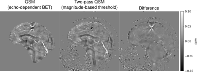 |
Robust Masking Techniques for Multi-Echo Quantitative Susceptibility Mapping
Ashley Stewart1,2, Simon Daniel Robinson2,3, Kieran O'Brien1,2,4, Jin Jin1,2,4, Georg Widhalm5, Gilbert Hangel3,5, Angela Walls6, Jonathan Goodwin7,8, Korbinian Eckstein3, Markus Barth1,2,9, and Steffen Bollmann1,2,9
1Centre for Innovation in Biomedical Imaging Technology, University of Queensland, Brisbane, Australia, 2Centre for Advanced Imaging, University of Queensland, Brisbane, Australia, 3High Field MR Center, Department of Biomedical Imaging and Image-Guided Therapy, Medical University of Vienna, Vienna, Austria, 4Siemens Healthcare Pty Ltd, Brisbane, Australia, 5Department of Neurosurgery, Medical University of Vienna, Vienna, Austria, 6Clinical & Research Imaging Centre, South Australian Health and Medical Research Institute, Adelaide, Australia, 7Department of Radiation Oncology, Calvary Mater Hospital, Newcastle, Australia, 8School of Mathematical and Physical Science, University of Newcastle, Newcastle, Australia, 9School of Information Technology and Electrical Engineering, University of Queensland, Brisbane, Australia
Quantitative Susceptibility Mapping (QSM) is a post-processing technique applied to gradient-echo phase data. QSM generally requires a signal mask to identify reliable phase values before reconstruction. Most QSM pipelines do not include masking procedures, and often suggest masking techniques that introduce artefacts, work only in the human brain, and lose critical information, especially near strong susceptibility sources. We propose two novel echo-dependent masking strategies and find that they significantly reduce streaking artefacts, particularly surrounding strong sources and tissue boundaries in multi-echo data. Our techniques are open-source and implemented in a new framework for automated, scalable, and robust QSM processing.
|
|||
3972.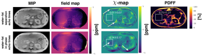 |
Quantitative susceptibility mapping in water–fat regions using in-phase echoes introduces significant quantification bias
Christof Boehm1, Maximilian N. Diefenbach1,2, Sophia Kronthaler1, Jakob Meineke3, Kilian Weiss4, Marcus R. Makowski1, and Dimitrios C. Karampinos1
1Department of Diagnostic and Interventional Radiology, School of Medicine, Klinikum rechts der Isar, Technical University of Munich, Munich, Germany, 2Division of Infectious Diseases and Tropical Medicine, University Hospital, LMU Munich, Munich, Germany, 3Philips Research Lab, Hamburg, Germany, 4Philips Healthcare, Hamburg, Germany
Gradient echo imaging using in-phase echoes has been proposed to reduce the field-map estimation in water–fat regions to a convex nonlinear least squares problem. However, it is known that the fat spectrum is complex and the definition of in-phase echo times remains problematic. In this work, the in-phase assumption is compared to standard water–fat imaging with respect to field- and quantitative susceptibility-mapping. The in-phase assumption is shown to be associated with quantification bias when subsequently estimating the field map and magnetic susceptibility in the spine, the liver and the breast.
|
|||
3973.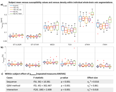 |
Investigating the Effect of Flow Compensation Schemes and Processing Pipelines on the Accuracy of Venous Quantitative Susceptibility Mapping
Ronja C. Berg1, Christine Preibisch1, Claus Zimmer1, David L. Thomas2,3, Karin Shmueli4, and Emma Biondetti5
1School of Medicine, Department of Neuroradiology, Technical University of Munich, Munich, Germany, 2Dementia Research Centre, UCL Queen Square Institute of Neurology, University College London, London, United Kingdom, 3Wellcome Centre for Human Neuroimaging, UCL Queen Square Institute of Neurology, University College London, London, United Kingdom, 4Department of Medical Physics and Biomedical Engineering, University College London, London, United Kingdom, 5Institut du Cerveau – ICM, INSERM U 1127, CNRS UMR 7225, Sorbonne Université, Paris, France
Venous quantitative susceptibility mapping (QSM) enables quantification of venous oxygenation. Flow-compensated acquisition is generally recommended for venous QSM, although its effect on the accuracy of venous susceptibility values has not been systematically evaluated. Moreover, QSM processing methods tend to be optimized for brain parenchyma tissues rather than veins. Here, we compared five different acquisition protocols (incorporating distinct flow compensation schemes) and six QSM processing methods in ten healthy volunteers. We found that venous susceptibility values depend strongly on the QSM pipeline (effect size ηp2=0.861) and much less on acquisition parameters including flow compensation (effect size ηp2=0.016).
|
|||
3974.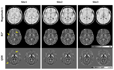 |
Reproducibility of R2* and quantitative susceptibility mapping in deep grey matter at 3T: Cross-vendor non-harmonized protocol study
Nashwan Naji1, M. Louis Lauzon2,3, Peter Seres1, Emily Stolz1, Richard Frayne2,3, Catherine Lebel4, Christian Beaulieu1, and Alan H. Wilman1
1Department of Biomedical Engineering, University of Alberta, Edmonton, AB, Canada, 2Departments of Radiology and Clinical Neurosciences, Hotchkiss Brain Institute, University of Calgary, Calgary, AB, Canada, 3Seaman Family MR Research Centre, Foothills Medical Centre, Calgary, AB, Canada, 4Department of Radiology, Alberta Children’s Hospital Research Institute and Hotchkiss Brain Institute, University of Calgary, Calgary, AB, Canada
R2* and QSM provide noninvasive ways to measure iron concentration in human brain. Performing multi-center studies help exploring wider demographical and pathological conditions. However, data pooled from multiple sites typically contain local acquisition sequence variations. In this study, the reproducibility of R2* and QSM at 3T are evaluated in 21 healthy adults (“traveling phantoms”) scanned 2x per site using independently site-optimized sequences from three sites and two scanner vendors. Mean R2* and susceptibility measurements in four deep grey matter structures were found to be highly correlated (r2 ≥ 0.98) and reproducible with SD of 1.2 s-1 and 4.1 ppb, respectively.
|
|||
3975.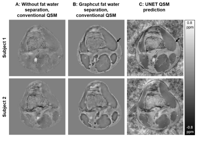 |
Deep learning based quantitative susceptibility mapping (QSM) in the presence of fat by using synthetically generated multi-echo phase data
Jannis Hanspach1, Aurel Jolla1, Michael Uder1, Bernhard Hensel2, Steffen Bollmann3, and Frederik Bernd Laun1
1Institute of Radiology, University Hospital Erlangen, Friedrich‐Alexander‐Universität Erlangen‐Nürnberg (FAU), Erlangen, Germany, 2Friedrich-Alexander-Universität Erlangen-Nürnberg (FAU), Erlangen, Center for Medical Physics and Engineering, Erlangen, Germany, 3University of Queensland, Brisbane, Australia, School of Information Technology and Electrical Engineering, Brisbane, Australia
Deep Learning reconstruction methods are increasingly investigated in Quantitative Susceptibility Mapping (QSM). In this work, we applied a UNET to reconstruct susceptibility maps in the presence of fat from unwrapped phase maps. The network was trained using synthetically generated multi-echo phase data and does not require explicit masking for the background field correction. Our results show that the proposed approach is well-suited to rapidly reconstruct high quality susceptibility maps in the presence of fat (e.g., outside the central nervous system) in in vivo data.
|
|||
|
3976.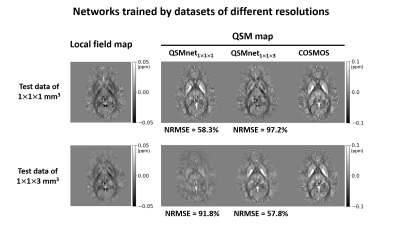 |
Necessity for a common dataset for a fair comparison between deep neural networks for QSM
Chungseok Oh1, Woojin Jung1, Hwihun Jeong1, and Jongho Lee1
1Seoul National University, Seoul, Korea, Republic of
We demonstrated that at least two conditions are required for a fair comparison between deep neural networks for dipole inversion: First, test data need to have the same characteristics as training data. Second, hyperparameter tuning should be performed if training dataset is changed. Our study implies that a common dataset is necessary for a fair comparison of deep neural networks for QSM.
|
||
3977.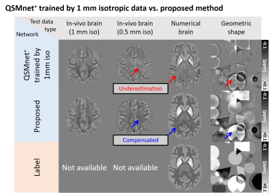 |
Generalization of deep learning-based QSM by expanding the diversity of spatial gradient in training data
Woojin Jung1, Steffen Bollmann2, Se-Hong Oh3, Hyeong-geol Shin1, Sooyeon Ji1, and Jongho Lee1
1Department of Electrical and Computer Engineering, Seoul National University, Seoul, Korea, Republic of, 2The University of Queensland, Brisbane, Australia, 3Biomedical Engineering, Hankuk University of Foreign Studies, Yongin, Korea, Republic of
In this work, the effect of spatial gradients in the training data on deep learning-based QSM is explored. We observe that deep learning-based QSM underestimates the susceptibility values when spatial gradients differ between training and test data. For demonstration, three types of networks were trained by using different spatial gradients of training images and evaluated on test data with varying spatial gradients. The results indicate that expanding the spatial gradient distribution of training data improves the performance of deep learning-based QSM. Furthermore, we demonstrate that augmenting spatial gradients may improve deep-learning based QSM to work for various image resolutions.
|
|||
3978.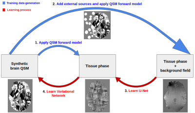 |
NeXtQSM - A complete deep learning pipeline for data-consistent quantitative susceptibility mapping trained with synthetic data
Francesco Cognolato1,2, Kieran O’Brien2,3, Jin Jin2,3, Simon Robinson4,5, Markus Barth1,2,6, and Steffen Bollmann1,2,6
1Centre for Advanced Imaging, The University of Queensland, Brisbane, Australia, 2ARC Training Centre for Innovation in Biomedical Imaging Technology, The University of Queensland, Brisbane, Australia, 3Siemens Healthcare Pty Ltd, Brisbane, Australia, 4High Field MR Center, Department of Biomedical Imaging and Image-Guided Therapy, Medical University of Vienna, Vienna, Austria, 5Department of Neurology, Medical University of Graz, Graz, Austria, 6School of Information Technology and Electrical Engineering, The University of Queensland, Brisbane, Australia
Deep learning based quantitative susceptibility mapping has shown great potential in recent years, outperforming traditional non-learning approaches in speed and accuracy in many applications. Here we aim to overcome the limitations of in vivo training data and model-agnostic deep learning approaches commonly used in the field. We developed a new synthetic training data generation method that enables the background field correction and a data-consistent solution of the dipole inversion to be learned using a variational network in one pipeline. NeXtQSM is a complete deep learning based pipeline for computing robust, fast and accurate quantitative susceptibility maps.
|
|||
3979. |
Total Deep Variation Regularization for Improved Iterative Quantitative Susceptibility Mapping (TDV-QSM)
Carlos Milovic1, Jose Manuel Larrain2,3, and Karin Shmueli1
1Department of Medical Physics and Biomedical Engineering, University College London, London, United Kingdom, 2Department of Electrical Engineering, Pontificia Universidad Catolica de Chile, Santiago, Chile, 3Biomedical Imaging Center, Pontificia Universidad Catolica de Chile, Santiago, Chile
Quantitative Susceptibility Mapping (QSM) is an ill-posed inverse problem. Traditionally, it is solved by minimization of a functional. Regularization terms may be interpreted as a denoising process. Many state-of-the-art methods are based on Total Variation regularization terms, with great success. With the advent of Deep Learning, new regularization strategies have been derived from training datasets. Total Deep Variation (TDV) is a recently proposed technique, showing impressive results. We applied a pre-trained TDV network as a denoising step in an iterative QSM solver. Results show improved error metrics for synthetic brain phantoms and enhanced in-vivo reconstructions, compared to Total-Variation-based algorithms.
|
|||
3980.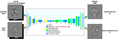 |
Exploring domain adaption for deep neural network trained QSM
JUAN LIU1 and Kevin Koch2
1Yale University, New Haven, CT, United States, 2Medical College of Wisconsin, Milwaukee, WI, United States
We aim to address the domain adaption problem of neural networks for QSM reconstruction which are learned from synthetic data while applied on real data. To address the unsupervised domain adaption, we apply domain-specific batch normalization layers in convolutional neural networks while allowing them to share all other model parameters. The proposed method is evaluated on multiple orientation datasets and single-orientation QSM datasets. Compared withTKD, MEDI, and DL-based method first training on synthetic datasets then model-based fine-tuning on real datasets, the proposed method achieved better performance.
|
|||
3981.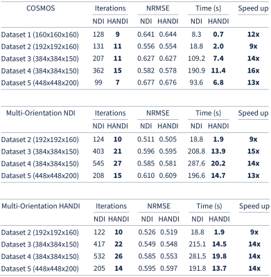 |
HANDI: Hessian Accelerated Nonlinear Dipole Inversion for rapid QSM
Christian Kames1,2, Jonathan Doucette1,2, and Alexander Rauscher1,2,3
1UBC MRI Research Centre, The University of British Columbia, Vancouver, BC, Canada, 2Department of Physics and Astronomy, The University of British Columbia, Vancouver, BC, Canada, 3Department of Pediatrics, The University of British Columbia, Vancouver, BC, Canada
We propose an extension of nonlinear dipole inversion (NDI), a first order method for solving the nonlinear formulation of the dipole inversion in quantitative susceptibility mapping (QSM), by using a second order method to increase the convergence rate of the minimization. The proposed method, Hessian Accelerated Nonlinear Dipole Inversion (HANDI), is shown to require fewer iterations than NDI, resulting in reconstruction times of a few seconds, more than 10x faster than NDI, without sacrificing accuracy. We further propose a learned proximal Newton method (HANDINet) and show that it outperforms learned variational networks based on NDI and standard dipole deconvolution minimizations.
|
|||
3982.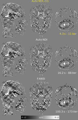 |
Automatic, Non-Regularized Nonlinear Dipole Inversion for Fast and Robust Quantitative Susceptibility Mapping
Carlos Milovic1 and Karin Shmueli1
1Department of Medical Physics and Biomedical Engineering, University College London, London, United Kingdom
Quantitative Susceptibility Mapping (QSM) is an ill-posed inverse problem often solved by regularization: minimizing a functional until it converges. This is usually time-consuming, requiring fine-tuning of several parameters by many repetitions of the whole optimization solver. Nonlinear Dipole Inversion is a QSM method that solves a nonlinear Tikhonov-regularized functional with a gradient descent solver. We show that stopping this method early provides optimal results, largely independent of the regularization weight. Here, we propose a non-regularized nonlinear conjugate gradient solver with a new stopping criterion based on analysing susceptibility map spatial frequency coefficients to achieve fast, parameter-free and automatic QSM.
|
|||
3983.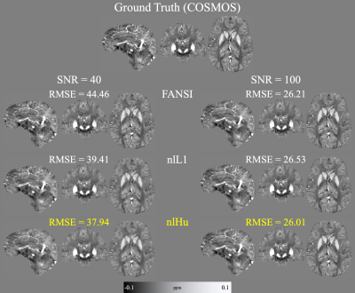 |
Improving Quantitative Susceptibility Mapping reconstructions via non-linear Huber loss data fidelity term (Huber-QSM)
Mathias Gabriel Lambert1,2,3, Carlos Milovic4, and Cristian Tejos1,2,3
1Department of Electrical Engineering, Pontificia Universidad Católica de Chile, Santiago, Chile, 2Biomedical Imaging Center, Pontificia Universidad Católica de Chile, Santiago, Chile, 3Millennium Nucleus for Cardiovascular Magnetic Resonance, Santiago, Chile, 4Department of Medical Physics and Biomedical Engineering, University College London, London, United Kingdom
Compared to L2-norm based QSM reconstructions, methods based on L1-norm data consistency are less prone to artifact generation caused by phase inconsistencies (e.g. unwrapping artifacts, intravoxel dephasing). However, L2-norm methods present better denoising performance in high SNR regions. Here, we present a QSM algorithm that combines the strengths of the L1 and L2 norms, using Huber's loss function as the data consistency term. Simulations and in vivo reconstructions showed enhanced performance, with superior artifact suppression capabilities of our proposed method.
|
|||
3984.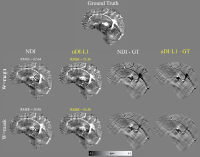 |
Non-regularized Dipole Inversion with streaking suppression via L1-norm optimization
Mathias Gabriel Lambert1,2,3, Cristian Tejos1,2,3, and Carlos Milovic4
1Department of Electrical Engineering, Pontificia Universidad Católica de Chile, Santiago, Chile, 2Biomedical Imaging Center, Pontificia Universidad Católica de Chile, Santiago, Chile, 3Millennium Nucleus for Cardiovascular Magnetic Resonance, Santiago, Chile, 4Department of Medical Physics and Biomedical Engineering, University College London, London, United Kingdom
Non-regularized QSM reconstructions are feasible by stopping gradient descent methods before they diverge. These methods consume less computation time, while avoiding the time required for parameter selection. In this work we present a linear dipole inversion method without regularization that uses the L1-norm in a proximal step to prevent streaking propagation and present more robustness against phase outliers. Compared to the Nonlinear Dipole Inversion method, our implementation achieved lower RMSE scores in phantom experiments and in vivo reconstructions with fewer artifacts.
|
The International Society for Magnetic Resonance in Medicine is accredited by the Accreditation Council for Continuing Medical Education to provide continuing medical education for physicians.