Digital Posters
QSM Clinical Applications
ISMRM & SMRT Annual Meeting • 15-20 May 2021

| Concurrent 2 | 17:00 - 18:00 |
3985.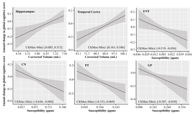 |
Quantitative susceptibility mapping MRI of brain iron and PET of β-amyloid predict cognitive decline during aging
Lin Chen1,2, Anja Soldan3, Kenichi Oishi1, Andreia Faria1, Marilyn Albert3, Peter van Zijl1,2, and Xu Li1,2
1Department of Radiology and Radiological Sciences, Johns Hopkins University, Baltimore, MD, United States, 2F.M. Kirby Research Center for Functional Brain Imaging, Kennedy Krieger Institute, Baltimore, MD, United States, 3Department of Neurology, Johns Hopkins University, Baltimore, MD, United States
We investigated associations of brain volume, iron levels as measured by QSM-MRI, and β-amyloid plaque load as measured by 11C-PiB PET imaging on prospective cognitive trajectories, measured using a global and domain-specific cognitive composite scores (e.g., episodic memory, executive function, visuospatial processing and language) in cognitively normal older adults with maximum follow-up of 5 years. Greater volume of multiple cortical and subcortical brain regions was strongly associated with the rate of cognitive decline. Associations between brain iron and β-amyloid and longitudinal cognitive decline were weaker, with brain iron in the basal ganglia and entorhinal cortex predicting global decline.
|
|||
3986.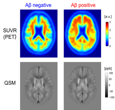 |
Prediction of Amyloid-β Deposition Using Multiple Regression Analysis of Quantitative Susceptibility Mapping
Ryota Sato1, Kohsuke Kudo2, Niki Udo3, Masaaki Matsushima4, Ichiro Yabe4, Akinori Yamaguchi2, Makoto Sasaki5, Masafumi Harada6, Noriyuki Matsukawa7, Tomoki Amemiya1, Yasuo Kawata1, Yoshitaka Bito1, Hisaaki Ochi1, and Toru
Shirai1
1Healthcare Business Unit, Hitachi, Ltd., Tokyo, Japan, 2Department of Diagnostic Imaging, Hokkaido University Graduate School of Medicine, Hokkaido, Japan, 3Department of Psychiatry, Hokkaido University Graduate School of Medicine, Hokkaido, Japan, 4Department of Neurology, Faculty of Medicine and Graduate School of Medicine, Hokkaido University, Hokkaido, Japan, 5Institute for Biomedical Sciences, Iwate Medical University, Iwate, Japan, 6Department of Radiology, Tokushima University, Tokushima, Japan, 7Department of Neurology, Nagoya City University, Aichi, Japan
For early diagnosis of Alzheimer’s disease, we created and evaluated a prediction method of amyloid β deposition based on multiple regression analysis of quantitative susceptibility mapping. A multiple regression model to predict standard uptake values (SUVs) of amyloid PET was constructed based on susceptibilities in 47 brain regions with the constraint Aβ deposition and susceptibility being positively correlated. The correlation coefficients between true and predicted SUVs were increased by incorporating the constraint, and the area under the receiver operating characteristics curve to predict Aβ positivity was 70%. The results suggest that the model could predict Aβ positivity at moderate accuracy.
|
|||
3987.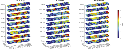 |
Quantifying iron deposition in Multiple System Atrophy via multi-echo Quantitative Susceptibility Mapping
Marta Lancione1,2, Matteo Cencini2,3, Mauro Costagli3,4, Graziella Donatelli2,5, Michela Tosetti2,3, Claudio Pacchetti6, Pietro Cortelli7,8, and Mirco Cosottini5
1IMT School for Advanced Studies Lucca, Lucca, Italy, 2IMAGO7 Foundation, Pisa, Italy, 3IRCCS Stella Maris, Pisa, Italy, 4Department of Neuroscience, Rehabilitation, Ophtalmology, Genetics, Maternal and Child Sciences (DINOGMI), University of Genova, Genova, Italy, 5Azienda Ospedaliero-Universitaria Pisana, Pisa, Italy, 6Parkinson and Movement Disorder Unit, IRCCS Mondino Foundation, Pavia, Italy, 7Department of Biomedical and NeuroMotor Sciences, University of Bologna, Bologna, Italy, 8Clinica Neurologica, IRCCS Istituto delle Scienze Neurologiche di Bologna, Bologna, Italy
In this work we quantified iron deposition in gray matter nuclei in a cohort of MSA patients with parkinsonian and cerebellar phenotypes using Quantitative Susceptibility Mapping (QSM). As it captures different tissues contribution depending on TE, we performed an ROI-based histogram analysis on susceptibility maps computed at each TE of a multi-echo GRE sequence. We observed significant differences among groups in several ROIs, such as putamen, globus pallidus, caudate nucleus, substantia nigra and dentate nucleus. The area under the ROC curve was higher for short TEs suggesting that short-TE QSM enhances diagnostic performances in the presence of strong susceptibility sources.
|
|||
3988.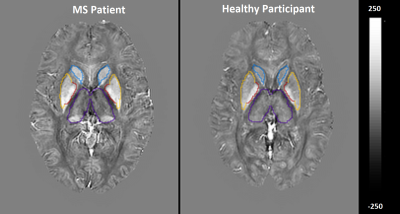 |
Clinical correlations of iron-rich deep grey matter of MS patients
Ibrahim Khormi1,2, Oun Al-iedani1,2, Amir Fazlollahi2,3, Bryan Paton2,4, Jeannette Lechner-Scott2,4,5, Abdulaziz Alshehri1,2, Kieran O'Brien6,7, Steffen Bollmann8, Rishma Vidyasagar9, Scott Ayton9, Anne-Louise Ponsonby9,10, and Saadallah Ramadan1,2
1School of Health Sciences, University of Newcastle, Newcastle, Australia, 2Hunter Medical Research Institute, Newcastle, Australia, 3CSIRO Health and Biosecurity, Brisbane, Australia, 4University of Newcastle, Newcastle, Australia, 5John Hunter Hospital, Newcastle, Australia, 6Siemens Healthcare Pty Ltd, Brisbane, Austria, 7ARC Training Centre for Innovation in Biomedical Imaging Technology, The University of Queensland, Brisbane, Australia, 8The University of Queensland, Brisbane, Australia, 9The Florey Institute of Neuroscience & Mental Health, Parkville, Australia, 10Murdoch Children's Research Institute, Royal Children's Hospital, University of Melbourne, Melbourne, Australia
This novel study compared quantitative magnetic susceptibility signal of deep grey matter (DGM) structures in the MS brain. We evaluated the QSM metrics in selected deep grey matter regions in 5 RRMS patients and 9 matched HCs. The STI suite software package was used for QSM image reconstruction. Compared to a reference region in HCs, a significant susceptibility change was detected in most DGM regions showing statistically significant RRMS cohort differences. QSM metrics in caudate showed strong correlations with depression scores, while pallidum and thalamus correlated significantly with anxiety. While results are limited due to small numbers, they provide the opportunity for further investigation in larger cohorts and strengthen these preliminary results.
|
|||
3989.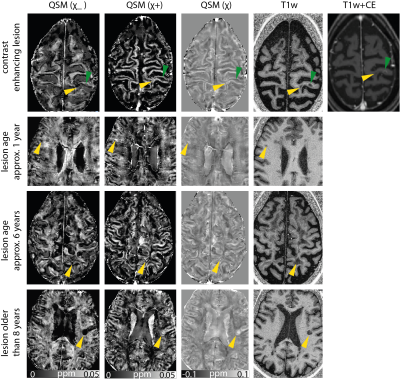 |
Separation of positive and negative susceptibility contrast at 7 Tesla allows for a more detailed characterization of multiple sclerosis lesions
Julian Emmerich1,2, Frederik L. Sandig3, and Sina Straub1
1Division of Medical Physics in Radiology, German Cancer Research Center (DKFZ), Heidelberg, Germany, 2Faculty of Physics and Astronomy, University of Heidelberg, Heidelberg, Germany, 3Division Radiology, German Cancer Research Center (DKFZ), Heidelberg, Germany
Quantitative susceptibility mapping (QSM) is known for its usefulness in imaging multiple sclerosis lesions. However, the co-occurrence of demyelination and iron accumulation due to the underlying inflammatory processes limit QSM from truthfully representing histology-like information as phase effects from positive and negative susceptibility sources within the same voxel cancel out in QSM. Here, it is shown that the separation of positive and negative susceptibility sources provides additional information to characterize susceptibility lesions.
|
|||
3990.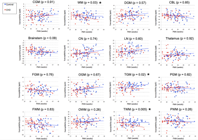 |
Quantitative susceptibility mapping in the infant brain diagnosed with congenital heart disease
Zungho Zun1,2,3,4, Kushal Kapse1, Nicole Andersen1, Scott Barnett1,2,3,4, Anushree Kapse1, Kristina Espinosa1, Jessica Quistorff1, Catherine Lopez1, Jonathan Murnick1,3,4, Mary T. Donofrio2,3,5, and Catherine Limperopoulos1,2,3,4
1Division of Diagnostic Imaging and Radiology, Children's National Hospital, Washington, DC, United States, 2Division of Fetal and Transitional Medicine, Children's National Hospital, Washington, DC, United States, 3Department of Pediatrics, George Washington University, Washington, DC, United States, 4Department of Radiology, George Washington University, Washington, DC, United States, 5Division of Cardiology, Children’s National Hospital, Washington, DC, United States
Quantitative susceptibility mapping may be used to assess brain development in infants based on its ability to evaluate iron and myelin contents. We measured magnetic susceptibilities in healthy infants and those diagnosed with congenital heart disease (CHD) in the early postnatal period and investigated the associations with neurodevelopmental outcomes. We report significantly lower magnetic susceptibilities in white matter and temporal lobe of the CHD cohort and significant associations between magnetic susceptibility of the temporal lobe and Bayley-III language scores at 18 months. Our findings may suggest disturbed iron deposition in the temporal lobe can lead to delays in language development.
|
|||
3991.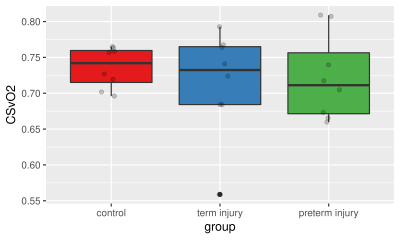 |
Quantitative Susceptibility Mapping of Venous Vessels in Neonates With Perinatal Asphyxia
Alexander Mark Weber1, Yuting Zhang2, Christian Kames3, and Alexander Rauscher1
1Pediatrics, UBC, Vancouver, BC, Canada, 2Radiology, Children’s Hospital of Chongqing Medical University, Chongqing, China, 3Physics, UBC, Vancouver, BC, Canada We aimed to test whether Cerebral venous oxygen saturation (CSVO2) could be measured using quantitative susceptibility mapping (QSM) in three distinct groups: healthy term neonates, and asphyxia injured term and preterm neonates. We acquired multi-echo gradient echo MRI data in 16 neonates with perinatal asphyxia and moderate or severe hypoxic-ischemic encephalopathy (HIE) (eight term-age, eight preterm, in eight healthy term-age controls. QSM derived oxygen saturation values in preterm and term neonates agreed well with past literature. CSVO2 in preterm and term neonates with HIE, however, were not found to be significantly different from each other or healthy controls. |
|||
3992.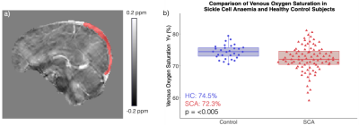 |
Investigating the Effect of Positive Airways Pressure on Venous Oxygenation in Sickle Cell Anemia with Quantitative Susceptibility Mapping
Russell Murdoch1, Hanne Stotesbury2, Jamie Kawadler2, Dawn Saunders2, Fenella Kirkham2, and Karin Shmueli1
1Department of Medical Physics and Biomedical Engineering, University College London, London, United Kingdom, 2Imaging and Biophysics, Developmental Neurosciences, UCL Great Ormond Street Institute of Child Health, London, United Kingdom
In 88 sickle cell anaemia (SCA) patients and 30 healthy controls, quantitative susceptibility mapping (QSM) showed significantly lower venous oxygen saturation (Yv) in the superior sagittal sinus of SCA patients. SCA subjects with silent cerebral infarcts (SCI) showed significantly lower Yv relative to those without SCI. 39 SCA subjects participated in a clinical study investigating the effect of auto-adjusting positive airways pressure (APAP) over a period of six months. No significant Yv differences were observed between 21 patients receiving APAP and 18 receiving standard care. In the APAP group, treatment compliance was positively correlated with increases in Yv.
|
|||
3993.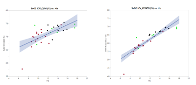 |
Validation Study of Venous Oxygenation in Internal Cerebral Vein in Patients with Sickle Cell Disease by QSM and CISSCO Method
Jian Shen1, Aart Nederveen2, and John Wood1,3
1Biomedical Engineering, University of Southern California, Los Angeles, CA, United States, 2Radiology, Academic Medical Center, Amsterdam, Netherlands, 3Children's Hospital Los Angeles, Los Angeles, CA, United States
Brain oxygenation can be measured using either T2-based methods or magnetic susceptibility-based methods. This study validates QSM and CISSCO methods in measuring the oxygenation in the internal cerebral vein and compares the results in sickle cell disease patients, anemia subjects with normal hemoglobin and healthy controls. Both methods reveal the group difference in the oxygen saturation in the deep structures. The limitations for both methods are discussed and the explanation for the bias between the two methods is provided.
|
|||
3994. |
Investigating the Magnetic Susceptibility of Silent Cerebral Infarcts in Sickle Cell Anaemia Using Two Different Gradient Echo Acquisitions
Russell Murdoch1, Hanne Stotesbury2, Jamie Kawadler2, Dawn Saunders2, Fenella Kirkham2, and Karin Shmueli1
1Department of Medical Physics and Biomedical Engineering, University College London, London, United Kingdom, 2Imaging and Biophysics, Developmental Neurosciences, UCL Great Ormond Street Institute of Child Health, London, United Kingdom
Little is known about the histopathology of silent cerebral infarcts (SCI) in sickle cell anaemia (SCA). Therefore, we measured the magnetic susceptibility (χ) of SCI in 45 SCA subjects and 10 healthy controls using quantitative susceptibility mapping (QSM) calculated from two different gradient-echo (GRE) acquisitions. Significantly lower χ was measured in lesions located in the parietal lobes relative to the frontal lobe. No significant relationships with mean lesion χ were observed with subject age, lesion size or depth in white matter. Strong correlations and minimal biases were observed between mean lesion χ measured using the two GRE acquisitions.
|
|||
3995.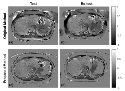 |
Improved Regularization for Quantitative Susceptibility Mapping of Liver Iron Overload
Julia V Velikina1, Ruiyang Zhao1,2, Collin Buelo2, Alexey A Samsonov1, Scott Reeder1,2,3,4,5, and Diego Hernando1,2
1Radiology, University of Wisconsin-Madison, Madison, WI, United States, 2Medical Physics, University of Wisconsin - Madison, Madison, WI, United States, 3Biomedical Engineering, University of Wisconsin - Madison, Madison, WI, United States, 4Emergency Medicine, University of Wisconsin - Madison, Madison, WI, United States, 5Medicine, University of Wisconsin - Madison, Madison, WI, United States
QSM may enable accurate quantification of liver iron overload. However, QSM of the abdomen faces a number of unique challenges due to large variations in susceptibility, presence of fat, and motion, which further confound the ill-posed inverse problem. We propose an approach to optimize the use of additional information provided by CSE imaging to regularize the QSM inversion problem. We validated this approach at 3T in patients with various levels of iron overload, including assessment of test-retest repeatability. This approach resulted in significantly reduced shading artifact, improved quality of susceptibility maps, and higher repeatability of measurements.
|
|||
3996.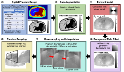 |
Quantitative Susceptibility Mapping of Liver Iron Overload using Deep Learning
Ruiyang Zhao1,2, Collin J Buelo2, Julia V Velikina1, Steffen Bollmann3, Ante Zhu4, Scott B Reeder1,2,5,6,7, and Diego Hernando1,2
1Radiology, University of Wisconsin-Madison, Madison, WI, United States, 2Medical Physics, University of Wisconsin-Madison, Madison, WI, United States, 3School of Information Technology and Electrical Engineering, University of Queensland, Brisbane, Australia, 4GE Global Research, Niskayuna, NY, United States, 5Biomedical Engineering, University of Wisconsin-Madison, Madison, WI, United States, 6Medicine, University of Wisconsin-Madison, Madison, WI, United States, 7Emergency Medicine, University of Wisconsin-Madison, Madison, WI, United States
A novel deep learning-based technique for quantitative susceptibility mapping (QSM) of liver iron overload was developed and validated. The proposed method relies on a 3D fully convolutional neural network, trained using synthetic dataset from a digital torso phantom that includes major organs. This study also included patients with iron overload who were imaged under 3T with using a single breath-hold multi-echo acquisition. Results showed promising performance and agreement with reference susceptibility measurements across a wide range of iron overload cases.
|
|||
3997.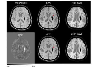 |
Comparison of True Susceptibility Weighted Imaging (tSWI) with SWI and QSM for Intracranial Hemorrhage
Ashmita De1, Derek J. Emery2, Kenneth S. Butcher3, and Alan H. Wilman1
1Department of Biomedical Engineering, University of Alberta, Edmonton, AB, Canada, 2Department of Radiology and Diagnostic Imaging, University of Alberta, Edmonton, AB, Canada, 3Division of Neurology, Department of Medicine, University of Alberta, Edmonton, AB, Canada
Quantitative Susceptibility Weighted Imaging or True SWI(tSWI) has been recently developed to overcome the shortcomings of SWI. SWI images are computed from filtered phase images, however, tSWI utilizes the susceptibility maps for its computation. Eight intracranial hemorrhage patients were scanned at 3T with 3D SWI sequence. Blooming effect and phase wrap artifacts that remain on SWI images of hemorrhage are removed in tSWI. tSWI provides better susceptibility weighting than magnitude within the hemorrhage and improved the texture visualization as compared Quantitative Susceptibility Mapping(QSM). Thus, tSWI can be a useful addition for hemorrhage visualization when SWI cannot provide a clear depiction.
|
|||
3998.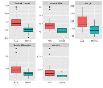 |
Radiomic Features on Quantitative Susceptibility Mapping Classify Amyotrophic Lateral Sclerosis Patients from Mimics
Anja Samardzija1, Thanh Nguyen2, Elizabeth Sweeney2, Kailyn Lee2, Ilhami Kovanlikaya2, Yi Wang 2, Andrew Schweitzer2, and Apostolos Tsiouris2
1Electrical and Computer Engineering, Cornell University, Highlands, NJ, United States, 2Weill Cornell Medicine, New York City, NY, United States
We trained a Random Forest classification model to classify amyotrophic lateral sclerosis (ALS) patients from those with mimicking clinical presentations based on QSM radiomic features extracted from the primary motor cortex. In a validation set, the model has 0.8 accuracy, 0.75 specificity of 0.75 and 0.84 sensitivity, which is superior to models using the mean QSM value as cutoff with 0.59 accuracy, 0.94 specificity and 0.14 sensitivity.
|
|||
3999.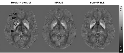 |
Quantitative susceptibility mapping in the basal ganglia of systemic lupus erythematosus patients with neuropsychiatric complaints
Marjolein Bulk1, Thijs van Harten1, Boyd Kenkhuis1, Francesca Inglese1, Ingrid Hegeman1, Sjoerd van Duinen1, Ece Ercan1, Cesar Magro-Checa1,2, Jelle Goeman1, Christian Mawrin3, Mark van Buchem1, Gerda Steup-Beekman1, Tom Huizinga1,
Louise van der Weerd1, and Itamar Ronen1
1Leiden University Medical Center, Leiden, Netherlands, 2Zuyderland Medical Center, Heerlen, Netherlands, 3Otto-von-Guericke University, Magdeburg, Germany
We explored the link between iron accumulation and neuroinflammation in basal ganglia in SLE using quantitative susceptibility mapping. We hypothesized that SLE patients, and in particular NPSLE patients would have increased numbers of activated microglia co-localizing with iron, which in turn would be reflected in increased local susceptibility. Based on QSM results in patients, as well as on iron staining of post-mortem SLE brain tissue, our results suggest that neuroinflammation in NPSLE is not necessarily associated with iron accumulation, and that the inflammatory pathomechanism in SLE may differ from the one observed in neurodegenerative diseases and in multiple sclerosis.
|
|||
4000.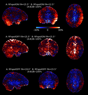 |
Scan Efficiency Optimisation for Quantitative Susceptibility Mapping of White Matter at 7T.
Jan Sedlacik1,2, Raphael Tomi-Tricot1,3, Pip Bridgen1,2, Tom Wilkinson1,2, Sharon Giles1,2, Karin Shmueli4, Jo V Hajnal1,2, and Shaihan J Malik1,2
1Centre for the Developing Brain, School of Biomedical Engineering & Imaging Sciences, King’s College London, London, United Kingdom, 2Biomedical Engineering Department, School of Biomedical Engineering & Imaging Sciences, King’s College London, London, United Kingdom, 3MR Research Collaborations, Siemens Healthcare Limited, Frimley, United Kingdom, 4MRI Group, Department of Medical Physics and Biomedical Engineering, University College London, London, United Kingdom
Quantitative Susceptibility Mapping (QSM) used for microstructural assessment of white matter (WM) is very attractive at ultra high magnetic field strengths, due to the increased signal-to-noise ratio (SNR) and phase sensitivity. This allows shortening echo and repetition times and, therefore, acquisition time. Further advantages of QSM are the low flip angles used for scanning, which results in low specific absorption rates, and the B1 insensitivity of the signal phase. However, suboptimal choice of imaging parameters will result in suboptimal SNR. The purpose of this work is to find and test optimal scanning parameters for QSM of the WM at 7T.
|
|||
4001.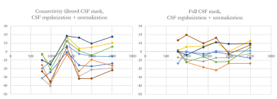 |
Whole brain CSF segmentation for consistent zero-referencing and longitudinal study applicability of MEDI+0
Alexey Dimov1, Thanh Nguyen1, Susan Gauthier2, and Yi Wang3
1Radiology, Weill Cornell Medicine, New York, NY, United States, 2Neurology, Weill Cornell Medicine, New York, NY, United States, 3Weill Cornell Medicine, New York, NY, United States
The role of CSF segmentation for zero-referenced MEDI+0 is demonstrated. In the present study we show that it is essential to enforce consistency in the CSF mask between different timepoints to ensure value reproducibility and to minimize effects of random shifts in the solution of MEDI+0 minimization
|
|||
4002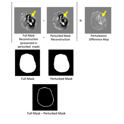 |
Artifact Evaluation of Quantitative Susceptibility Mapping Reconstructions Using Mask Parameter Perturbation Video Permission Withheld
Priya S Balasubramanian1,2, Alexandra Grace Roberts1,2, Pascal Spincemaille2, Thanh Nguyen 2, and Yi Wang1,2
1Cornell University, New York City, NY, United States, 2Weill Cornell Medical College, New York City, NY, United States
QSM uses a mask to separate tissue of interest from background to avoid the strong susceptibility sources and poor signals that are known to cause artifacts in QSM. In brain QSM, the strongly paramagnetic superior sagittal sinus (SSS) lies at the border of brain mask. Perturbing the brain mask by reducing the SSS inclusion in QSM reconstruction also affects the convergence of strong susceptibility sources inside the brain. This mask perturbation propagates into QSM perturbation, which may be used as a measure of artifacts associated with strong susceptibility sources as validated in numerical phantom and COSMOS data.
|
|||
4003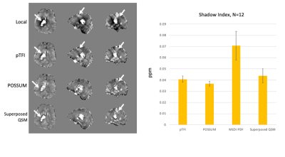 |
Regional Susceptibility Reconstruction Improves Artifact Incidence and Error in Quantitative Susceptibility Mapping through POSSUM Video Permission Withheld
Priya S Balasubramanian1,2, Pascal Spincemaille2, Thanh Nguyen2, and Yi Wang1,2
1Cornell University, New York City, NY, United States, 2Weill Cornell Medical College, New York City, NY, United States
Quantitative susceptibility mapping methods that selectively mask based on susceptibility thresholds and do regional reconstructions have been introduced to reduce shadowing artifacts when dealing with a large dynamic susceptibility range within the image. Here, we combine this technique with total field reconstructions, which have been shown to reduce errors resulting from incomplete background field removal. Reduced errors and shadow artifacts are demonstrated when compared to prior techniques.
|
|||
4004.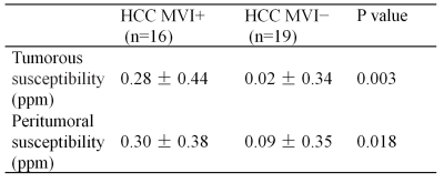 |
Quantitative Susceptibility Mapping in Preoperative Assessment of Microvascular Invasion of Hepatocellular Carcinoma: a Preliminary Study
Chang Liu1, Hongru Jia1, Weiqiang Dou2, Jing Ye1, and Xianfu Luo1
1Northern Jiangsu People’s Hospital, Yang zhou, China, 2GE Healthcare,MR Research China, Bei jing, China
In this study, we aimed to investigate susceptibility changes related to microvascular invasion (MVI) of hepatocellular carcinoma. We applied liver quantitative susceptibility mapping to measure lesions’ susceptibility. Significant differences in both tumorous and peritumoral susceptibility values were observed for MVI positive and negative lesions. With these findings, QSM imaging can be considered a potential technique for noninvasive preoperational assessment of MVI in hepatocellular carcinoma.
|
The International Society for Magnetic Resonance in Medicine is accredited by the Accreditation Council for Continuing Medical Education to provide continuing medical education for physicians.