Digital Posters
Hyperpolarization: Gas
ISMRM & SMRT Annual Meeting • 15-20 May 2021

| Concurrent 2 | 13:00 - 14:00 |
3557.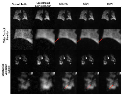 |
Convolutional Neural Networks for Super-resolution of Hyperpolarized 129Xe MR Images of the Lung
Junlan Lu1, Suphachart Leewiwatwong2, David Mummy3, Elianna Bier2, and Bastiaan Driehuys3
1Medical Physics, Duke University, Durham, NC, United States, 2Biomedical Engineering, Duke University, Durham, NC, United States, 3Radiology, Duke University, Durham, NC, United States
Although hyperpolarized 129Xe gas exchange MRI enables imaging ventilation, barrier, and RBC components in a single breath-hold, the necessary under-sampling imposed by limited imaging time constrains image resolution. Therefore, it is common to acquire an additional dedicated ventilation scan, which increases cost and imaging time. Instead, we demonstrate that deep convolutional neural networks with template-based augmentation can be trained to transform under-sampled low-resolution 129Xe ventilation images to a level of detail comparable to that of a dedicated ventilation scan. We evaluate the performance of multiple super-resolution models based on signal-to-noise ratio and structural similarity.
|
|||
3558.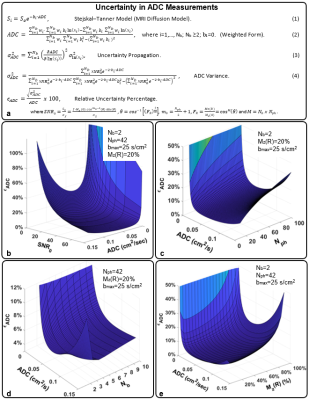 |
Optimizing Acquisition and Analysis for Diffusion Weighted Hyperpolarized 129Xe MRI of Pediatric and Adult Lungs
Abdullah S. Bdaiwi1,2, Peter J. Nedbalski1, Md M. Hossain3, Matthew M. Willmering1, Laura L. Walkup1,2,4, Hui Wang5, Robert P. Thomen1, Kai Ruppert1, Jason C. Woods1,4,6, and Zackary I. Cleveland1,2,4,6
1Center for Pulmonary Imaging Research, Cincinnati Children's Hospital Medical Center, Cincinnati, OH, United States, 2Biomedical Engineering Department, University of Cincinnati, Cincinnati, OH, United States, 3Division of Biostatistics and Epidemiology, Cincinnati Children's Hospital Medical Center, Cincinnati, OH, United States, 4Division of Pulmonary Medicine, Cincinnati Children’s Hospital Medical Center, Cincinnati, OH, United States, 5Philips, Cincinnati, OH, United States, 6Department of Pediatrics, University of Cincinnati, Cincinnati, OH, United States
Hyperpolarized (HP) 129Xe MRI can non-invasively quantify lung microstructure through measuring the gas-phase apparent diffusion coefficient (ADC), providing insights into disease pathophysiology—e.g., emphysema severity. To maximize the utility of this method, we developed an analytical model, based on HP-specific Bloch equations and error propagation, to optimize ADC measurements in terms of acquisition parameters (b-value, flip angle, phase encodes, etc.) and the ADC itself and validated this model via Monte Carlo and phantom studies. Finally, a lower bound on the expected 129Xe ADC was obtained by measuring ADC as a function of age in human subjects as young as six years.
|
|||
3559.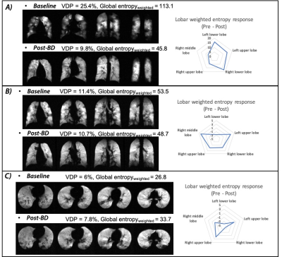 |
Assessment of dynamic airflow heterogeneity after bronchodilator in asthma using hyperpolarized helium-3 MRI
Mu He1, Lindsay A. Somerville1, Nicolas J. Tustison1, James Patrie1, Jaime F. Mata1, Joanne M. Cassani2, Roselove Nunoo-Asare1, Alan Ropp1, Wilson G. Miller1, Yun Michael Shim1, Talissa A. Altes2, John P. Mulger1, and Eduard E. de Lange1
1University of Virginia, Charlottesville, VA, United States, 2University of Missouri, Columbia, MO, United States
With hyperpolarized helium-3 (3He) MRI regional differences in airflow heterogeneity can be assessed. In this study, we used weighted entropy to evaluate dynamic ventilation heterogeneity in asthmatics. Serial 3He/1H scans were co-registered and normalized for comparison. Weighted entropy was calculated based on entropy and Ventilation defect percentage (VDP) values derived from normalized 3He scans. Lobar analysis was performed to identify lobar weighted entropy. We found that asthmatics have more heterogeneous ventilation distribution in the lower lobes at baseline. Variable airflow heterogeneity changes were observed globally in asthmatics, with the most significant bronchodilator response in the left lung.
|
|||
3560.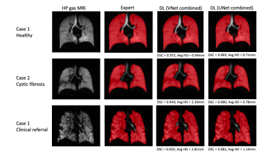 |
Comparison of 3D convolutional neural networks and loss functions for ventilated lung segmentation using multi-nuclear hyperpolarized gas MRI
Joshua R Astley1,2, Alberto M Biancardi1, Paul J Hughes1, Laurie J Smith1, Helen Marshall1, Guilhem J Collier1, James Eaden1, Nicholas D Weatherley1, Jim M Wild1, and Bilal A Tahir1,2
1POLARIS, University of Sheffield, Sheffield, United Kingdom, 2Oncology and Metabolism, University of Sheffield, Sheffield, United Kingdom
Deep learning has shown great promise for numerous medical image segmentation tasks, including delineation of ventilated lung volumes from hyperpolarized gas MRI. We previously demonstrated the utility of a VNet convolutional neural network (CNN), trained on a combination of 3He and 129Xe scans, in producing accurate segmentations that outperform conventional methods. In this work, we compared the performance of several 3D CNNs and loss functions for segmentation of ventilated lungs on a significantly larger and more diverse multi-nuclear hyperpolarised gas MRI dataset using several training strategies. We observe that the UNet CNN provides the best performing model for our dataset.
|
|||
3561. |
Hyperpolarized 3He MRI ADC and Ventilation Features Predict Rapidly Worsening Emphysema Using Machine-learning
Maksym Sharma1, Alexander M Matheson1, David G McCormack2, David A Palma1,3, and Grace Parraga1,2,3
1Medical Biophysics, Western University, London, ON, Canada, 2Division of Respirology, Department of Medicine, Western University, London, ON, Canada, 3Department of Oncology, Western University, London, ON, Canada Pulmonary hyperpolarized 3He MRI provides a way to measure lung ventilation heterogeneity in patients with COPD, including terminal airspace enlargement or emphysema that is typically quantified using CT densitometry. Unfortunately, MRI-derived biomarkers of emphysema progression remain unconfirmed, and also likely because of radiation dose considerations, CT follow-up of emphysema is rarely performed, and hence its longitudinal progression is not well-understood. Here we developed a machine-learning pipeline that identified hyperpolarized 3He MRI texture features that independently and uniquely correlated and predicted rapidly-worsening emphysema nearly 3 years later, measured as CT RA950, using a Decision Tree algorithm that achieved 82% prediction accuracy. |
|||
3562.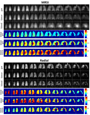 |
Imaging Gas-Exchange Lung Function using Density-Weighted MRSI and Hyperpolarised 129Xe Gas
Rolf F Schulte1, Guilhem J Collier2, James Ball2, Graham Norquay2, Madhwesha Rao2, and Jim M Wild2
1GE Healthcare, Munich, Germany, 2POLARIS, Department of Infection Immunity & Cardiovascular Disease, University of Sheffield, Sheffield, United Kingdom
3D density-weighted MRSI in combination with a frequency-tailored RF excitation pulse was designed, implemented and used to detect xenon gas in the lungs and xenon dissolved in lung tissue and blood. These images were used to calculate quantitative ratio maps of tissue-to-gas, blood-to-gas, and blood-to-tissue with good SNR.
|
|||
3563.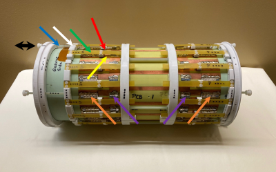 |
A 129Xe/1H Switched Frequency High Pass Birdcage Coil for Hyperpolarized 129Xe Gas Lung Imaging in Neonates at 1.5 T
Ronald Pratt1, Randy Giaquinto1, Wolfgang Loew1, Christopher Ireland1, Neil Stewart2,3, Nara Higano2, Jason Woods2, and Charles Dumoulin1
1Imaging Research Center/Radiology, Cincinnati Children's Hospital Medical Center, Cincinnati, OH, United States, 2Center for Pulmonary Imaging Research/Radiology, Cincinnati Children's Hospital Medical Center, Cincinnati, OH, United States, 3POLARIS, Imaging Sciences, Dept of Infection, Immunity & Cardiovascular Disease, University of Sheffield, Sheffield, United Kingdom Production of hyperpolarized 129Xe gas has opened new frontiers for imaging lung anatomy and function using MRI. Our institution has the goal of using hyperpolarized 129Xe gas for MR imaging of neonatal lungs at 1.5 T. To realize this goal, it is essential that the RF coil used for imaging operate at both the 129Xe and 1H frequencies. This report provides fabrication details of a novel 129Xe/1H switched frequency transmit/receive 16 rung high-pass birdcage RF coil. Images collected with hyperpolarized 129Xe gas and water in phantoms demonstrate that the coil provides excellent image quality at both frequencies. |
|||
3564.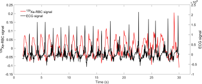 |
Temporal correlation of alveolar-capillary 129Xe signal dynamics with the cardiac cycle
Graham Norquay1, Guilhem J Collier1, and Jim M Wild1
1Infection, Immunity and Cardiovascular Disease, University of Sheffield, Sheffield, United Kingdom
Global 129Xe MRS was acquired in the lungs synchronously with an ECG recording to enable association of dissolved-phase 129Xe signal dynamics with the cardiac cycle. Time-domain Voigt fitting was used to calculate resonance parameters corresponding to 129Xe in red blood cells and tissue/blood plasma in the lungs. Positive and negative signal changes of 129Xe in the blood were found to be associated with ventricular systole and diastole in a healthy volunteer.
|
|||
3565.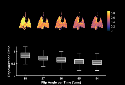 |
Feasibility of Xenon Polarization Transfer Contrast Imaging using Continuous RF Irradiation
Faraz Amzajerdian1, Tahmina Achekzai1, Luis Loza1, Hooman Hamedani1, Yi Xin1, Harilla Profka1, Ian Duncan1, Stephen Kadlecek1, Kai Ruppert1, and Rahim Rizi1
1University of Pennsylvania, Philadelphia, PA, United States
Xenon-polarization Transfer Contrast (XTC) imaging is a powerful technique for quantifying exchange rates between gas- and dissolved-phase xenon compartments and involves a series of radiofrequency (RF) saturation pulses applied to the targeted dissolved-phase resonance. Although increasing the number of pulses and their spacing generates greater contrast, it also increases acquisition time. In this work, in an effort to reduce acquisition time, particularly for free-breathing imaging protocols, we explored the use of continuous RF irradiation to depolarize dissolved-phase xenon, with the goal of producing results similar to those achieved with pulsed saturations, but in a significantly shorter time period.
|
|||
3566.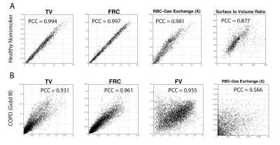 |
Reproducibility Study Measuring Ventilation, Gas Exchange and Surface-to-Volume Using Hyperpolarized Xenon in Free-breathing Human Subjects
Hooman Hamedani1, Stephen Kadlecek1, Faraz Amzajerdian1, Ryan Baron1, Kai Ruppert1, Ian Duncan1, Yi Xin1, Luis Loza1, Tahmina Achekzai1, Maurizio Cereda1, Kevin Ma1, David DiBardino1, and Rahim Rizi1
1University of Pennsylvania, Philadelphia, PA, United States
We have previously shown the advantages of multi- over single-breath imaging of lung ventilation using HP gas MRI, and have recently introduced our approach for comprehensively assessing the lung function during a tidal breathing scheme to quantify ventilation and dissolved parameters of xenon distribution. Here, we presented the repeatability of the imaging markers in a healthy subject and a patient with severe chronic obstructive pulmonary disease (COPD) with persisting cough. We have shown that the technical back-to-back reproducibility of measuring lung function with our scheme is excellent in a healthy subject and satisfactory in a COPD subject with persisting cough.
|
|||
3567.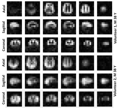 |
3D isotropic spectroscopic imaging of hyperpolarized 129Xe in the human brain
Madhwesha R Rao1, Guilhem J Collier1, Graham Norquay1, Rolf F Schulte2, and Jim M Wild1
1University of Sheffield, Sheffield, United Kingdom, 2GE Healthcare, Munich, Germany
3D isotropic images of hyperpolarized 129Xe dissolved in the brain were acquired with 1 L of inhaled xenon gas dose using a density-weighted MR spectroscopic imaging method. Images were acquired from 2 healthy male volunteers. The data was acquired with a voxel size of (2cm)3 and reconstructed to a voxel size of (0.625 cm)3 through zero filling. The isotropic voxel size and the high spectral resolution surpass previous reports using conventional 2D and echo planar spectroscopic imaging methods.
|
|||
3568.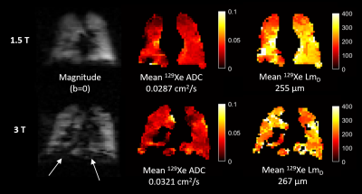 |
Background field inhomogeneity effects on hyperpolarized 129Xe diffusion-weighted MRI at 1.5T and 3T
Ho-Fung Chan1, Guilhem J Collier1, Madhwesha Rao1, and Jim M Wild1
1Infection, Immunity and Cardiovascular Disease, University of Sheffield, Sheffield, United Kingdom
Background field inhomogeneities due to air-tissue interface magnetic susceptibility differences in the lungs increases derived ADC measurements from 3He PGSE diffusion-weighted (DW) lung MRI at higher field strengths. To determine if the same effects are observed with 129Xe DW-MRI, 129Xe ADC and mean diffusive length scale (LmD) from healthy volunteers imaged at 1.5T and 3T were compared. A small bias towards increased 129Xe ADC (6.3%) and LmD (2.2%) values at 3T was obtained, which is smaller than the reported bias of 3He ADC (15.5%) and 3He mean chord length (10.5%), and similar to the reported 129Xe DW-MRI inter-scan repeatability differences.
|
|||
3569.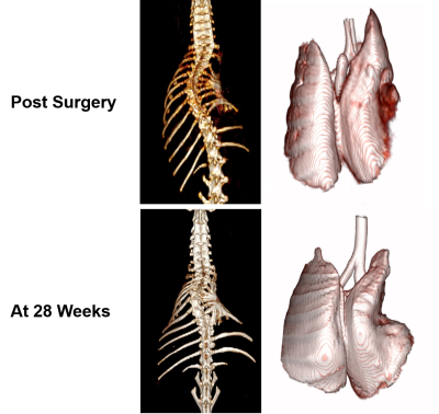 |
Detection of pulmonary abnormalities in a rabbit thoracic insufficiency syndrome model using hyperpolarized xenon-129 MRI
Kai Ruppert1, Faraz Amzajerdian1, Yi Xin1, Hooman Hamedani1, Luis Loza1, Tahmina S Achekzai1, Ryan J Baron1, Ian F Duncan1, Harrilla Profka1, Yiwen Qian1, Stephen Kadlecek1, Alessandra Fusco2, Benjamin Sinder3, Patrick J
Cahill3, Brian Snyder3,4, Thomas P Schaer2, and Rahim R Rizi1
1University of Pennsylvania, Philadelphia, PA, United States, 2School of Veterinary Medicine, University of Pennsylvania, Kennett Square, PA, United States, 3Children's Hospital of Philadelphia, Philadelphia, PA, United States, 4Boston Children's Hospital, Boston, MA, United States
Thoracic insufficiency syndrome (TIS) progresses to the development of restrictive lung disease and is commonly treated through surgical intervention. In this work, we used a rib-tether rabbit model to investigate the sensitivity of dynamic 1D simultaneous dissolved- and gas-phase hyperpolarized xenon-129 MRI imaging to pulmonary abnormalities secondary to TIS. We found asymmetric lung ventilation patterns and increases in alveolar septal wall thickness in both lungs of a rib-tethered rabbit compared to an age-matched control animal. These findings could help identify the optimal timepoint at which to conduct chest expansion surgery so as to maximize the resulting improvements in lung maturation.
|
|||
3570.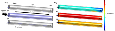 |
Modelling realistic Rb density and temperature distributions in a high throughput xenon-129 polariser
James Ball1, Jim M. Wild1, and Graham Norquay1
1POLARIS, Department of Infection, Immunity and Cardiovascular Disease, The University of Sheffield, Sheffield, United Kingdom
Hyperpolarised xenon-129 (129Xe) production via continuous-flow spin-exchange optical pumping often produces lower than predicted 129Xe polarisation. Frequently, thermodynamics within polariser systems is not considered, as well as spatially variant changes in rubidium (Rb) source distributions and changes in 129Xe relaxation over time. This work models realistic Rb and temperature distributions within a large-scale 129Xe-Rb polariser cell. Results show that Rb density varies significantly depending upon incident photon flux and Rb source distribution in the cell. Modelling allows estimation of the Rb presaturator length required at different gas flow rates in order to reach optimal Rb density and high 129Xe polarisation.
|
|||
3571.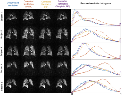 |
Template-based bias field correction of Hyperpolarized 129Xe Gas Ventilation MRI
Junlan Lu1, David Mummy2, Suphachart Leewiwatwong3, Elianna Bier3, and Bastiaan Driehuys2
1Medical Physics, Duke University, Durham, NC, United States, 2Radiology, Duke University, Durham, NC, United States, 3Biomedical Engineering, Duke University, Durham, NC, United States
Accurately correcting hyperpolarized 129Xe ventilation MRI for coil-induced bias field remains the most significant obstacle to precise, repeatable quantitative image analysis. Estimates using B1 maps from RF-depletion in radially acquired images may provide an improvement on the standard N4ITK solution, which recent works suggests may perform an overly aggressive correction. Here, we develop a template-based approach to bias field correction using B1 maps derived from multiple subjects. This paradigm is then evaluated by applying it to a set of test images reflective of a range of disease types and levels of ventilation obstruction.
|
|||
3572.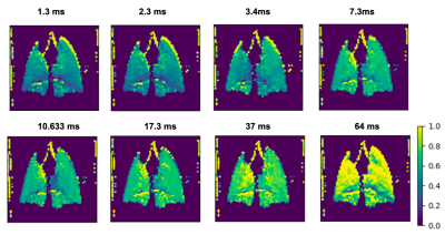 |
Performance of XTC imaging in a free-breathing mouse model exploring variable saturation delay times
Tahmina Susan Achekzai1, Luis Loza2, Stephen Kadlecek2, Kai Ruppert2, Faraz Amzajerdian2, and Rahim R. Rizi1
1Radiology, University of Pennsylvania, Philadelphia, PA, United States, 2University of Pennsylvania, Philadelphia, PA, United States
XTC imaging takes advantage of the exchange between gas and dissolved phase Xe to provide information on lung function, as direct dissolved phase imaging is limited in signal. In this study, we demonstrate XTC imaging in a regular and transgenic mouse model. We aim to optimize depolarization effectiveness by varying delay time and number of saturation pulses applied in the imaging scheme and demonstrate the dependence of depolarization to these parameters.
|
|||
3573.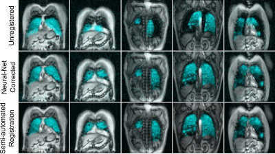 |
Fully-automated Multi-spectral Pulmonary Registration for Hyperpolarized Noble Gas MRI Using Neural Networks
Alexander M Matheson1, Rachel L Eddy1, Jonathan L MacNeil2, Marrissa J McIntosh1, and Grace M Parraga1,2
1Medical Biophysics, Robarts Research Institute, Western University, London, ON, Canada, 2School of Biomedical Engineering, Robarts Research Institute, Western University, London, ON, Canada
Co-registered hyperpolarized gas and proton MRI are required to calculate functional lung biomarkers using semi-automated pipelines. Automated registration between different spectral acquisitions is difficult due to differences in contrast and imaging features between different spectral images. Convolutional neural networks create abstract representations of images that may overcome these feature differences. We retrospectively pooled data sets previously registered using a semi-automated pipeline and applied random two-dimensional affine transformations and noise. Neural networks generated inverse transformation matrices from these data to correct the applied transformations. The trained network successfully corrected mis-alignment with an average error of less than one pixel.
|
|||
3574.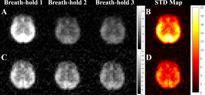 |
The effects of an initial depolarization pulse on the dissolved phase hyperpolarized 129Xe images
Yurii Shepelytskyi1,2, Vira Grynko1,2, Tao Li3, Ayman Hassan4,5, Karl Granberg4, and Mitchell S Albert2,3,5
1Chemistry and Materials Science Program, Lakehead University, Thunder Bay, ON, Canada, 2Thunder Bay Regional Health Research Institute, Thunder Bay, ON, Canada, 3Chemistry, Lakehead University, Thunder Bay, ON, Canada, 4Thunder Bay Regional Health Sciences Centre, Thunder Bay, ON, Canada, 5Northern Ontario School of Medicine, Thunder Bay, ON, Canada
Hyperpolarized (HP) xenon-129 (129Xe) freely dissolves in pulmonary blood and travels to highly perfused organs. Dissolved phase HP 129Xe imaging is commonly used for evaluating gas-blood exchange in lungs, imaging cerebral perfusion, detecting hemodynamic response, and kidney perfusion. However, the signal-to-noise ratio (SNR) of HP 129Xe dissolved phase images varies between breath-holds, especially for brain imaging. In this work, we demonstrated a significant reduction in the variability of MRI image SNR by implementing an additional depolarization pulse prior to image acquisition.
|
|||
 |
3575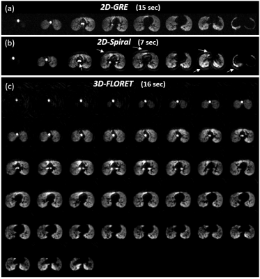 |
2D and 3D Spiral for Diffusion Weighted MRI with Hyperpolarized 129Xe Video Permission Withheld
Abdullah S. Bdaiwi1,2, Matthew M. Willmering1, Hui Wang3, and Zackary I. Cleveland1,2,4,5
1Center for Pulmonary Imaging Research, Cincinnati Children's Hospital Medical Center, Cincinnati, OH, United States, 2Biomedical Engineering Department, University of Cincinnati, Cincinnati, OH, United States, 3Philips, Cincinnati, OH, United States, 4Department of Pediatrics, University of Cincinnati, Cincinnati, OH, United States, 5Division of Pulmonary Medicine, Cincinnati Children’s Hospital Medical Center, Cincinnati, OH, United States
Diffusion-weighted hyperpolarized 129Xe MRI is a validated measure of lung microstructure and can assess changes in alveolar dimensions. These images are commonly acquired via 2D gradient recalled echo (GRE), but 129Xe GRE images suffer from coarse resolution in the slice dimension and breath-hold durations (≤16 s) that may be difficult for pediatric and severely ill subjects. To overcome these limitations, we implemented 2D- and 3D-spiral (FLORET, Fermat looped, orthogonally encoded trajectories) sequences for 129Xe diffusion imaging. These sequences enable either rapid acquisition or high-resolution, isotropic lung coverage and display image quality and ADC accuracy comparable to that of conventional 2D-GRE.
|
||
|
3576.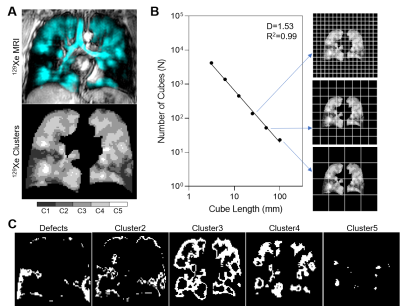 |
Evaluating the Fractal Nature of 129Xe MRI Ventilation Heterogeneity
Rachel L Eddy1,2, Alexander M Matheson3,4, Marrissa J McIntosh3,4, and Grace Parraga3,4
1St. Paul's Hospital, UBC Centre for Heart Lung Innovation, Vancouver, BC, Canada, 2Division of Respiratory Medicine, Department of Medicine, University of British Columbia, Vancouver, BC, Canada, 3Robarts Research Institute, London, ON, Canada, 4Department of Medical Biophysics, Western University, London, ON, Canada
Pulmonary ventilation has been shown to follow a fractal distribution using fluorescence imaging. 129Xe MRI provides high spatial-temporal resolution images of pulmonary ventilation so here, we aimed to determine the fractal properties of 129Xe MRI ventilation heterogeneity using the box-counting method. In 25 patients with asthma, MRI ventilation heterogeneity followed a power law and mean fractal dimension for MRI signal ranged from 1.39-1.82. Fractal analysis can provide a new tool to measure regional MRI ventilation heterogeneity and investigate pulmonary structure-function relationships in patients with lung disease.
|
The International Society for Magnetic Resonance in Medicine is accredited by the Accreditation Council for Continuing Medical Education to provide continuing medical education for physicians.