Digital Posters
Elastography: Applications
ISMRM & SMRT Annual Meeting • 15-20 May 2021

| Concurrent 4 | 13:00 - 14:00 |
0999.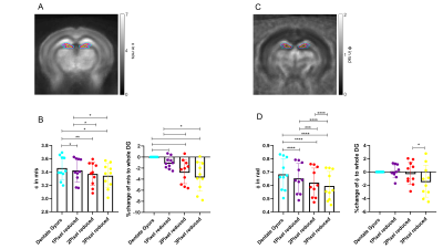 |
In vivo MR elastography of the murine hippocampus is sensitive to the microscopic mechanical properties of dentate gyrus subzones
Anna Morr1, Marcin Nowicki2, Gergely Bertalan1, Rafaela Vieira da Silva1, Carmen Infante Duarte1, Stefan Paul Koch1, Philipp Boehm-Sturm1, Ute Krügel2, Jürgen Braun1, Barbara Steiner1, Josef Käs2, Thomas Fuhs2, and Ingolf Sack1
1Charité - Universitätsmedizin Berlin, Berlin, Germany, 2Universität Leipzig, Leipzig, Germany
In this study, we investigated the mechanical properties of subregions of the murine dentate gyrus in vivo using MR elastography (MRE) and ex vivo with atomic force microscopy (AFM). Consistent with AFM, subregional analysis of the dentate gyrus by MRE revealed that the subgranular zone (SGZ), in which adult neurogenesis takes place, is characterized by marked soft-solid properties. We conclude that MRE is sensitive to micromechanical properties beyond the image resolution. The observed soft-solid material properties of the SGZ quantified by AFM and MRE might be of functional relevance for neurogenesis in the mouse brain.
|
|||
1000.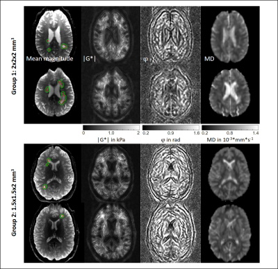 |
Are multiple sclerosis lesions stiffer or softer than surrounding brain tissue?
Helge Herthum1, Stefan Hetzer2, Michael Scheel1, Friedemann Paul3, Heiko Tzschätzsch1, Tom Meyer1, Mehrgan Shahryari1, Jürgen Braun4, and Ingolf Sack1
1Experimentelle Radiologie, Charité Universitätsmedizin Berlin, Berlin, Germany, 2Berlin Center for Advanced Neuroimaging (BCAN), Berlin, Germany, 3NeuroCure Clinical Research Center and Clinical and Experimental Multiple Sclerosis Research Center, Berlin, Germany, 4Institut für medizinische Informatik, Charité Universitätsmedizin Berlin, Berlin, Germany
Using multifrequency MR elastography with 2- and 1.5mm resolutions in a cohort of 19 patients with multiple sclerosis and a total 206 analyzed lesions we found no evidence for systematic differences in the viscoelastic properties between MS lesions and surrounding brain tissue. Although future technical improvements in MRE resolution and parameter reconstruction methods might reveal significant differences between lesion and brain properties, our results indicate that those differences are minor compared to other published changes in brain viscoelasticity of the human brain.
|
|||
1001.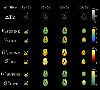 |
Brain MR-Elastography in microgravity analogous conditions
Fatiha Andoh1, Claire Pellot-Barakat1, and Xavier Maître1
1Université Paris-Saclay, CEA, CNRS, Inserm, BioMaps, Orsay, France
The head down tilt (HDT) position is commonly used to simulate vascular and tissue fluid dynamics during spaceflights. In HDT position, the cerebral autoregulation faces difficulties to adjust the vascular tone while the cephalad fluid shifts may yield increased intracranial pressures and altered mechanical properties. MRI T2 mapping in HDT position have shown fluid overpressure in the brain and resulting loss of water contents in the CSF and orbital compartments. Whole brain MRE was performed here in similar HDT conditions. It was sensitive enough to provide new insights on the overall mechanical response of brain tissues in microgravity analogous conditions.
|
|||
1002.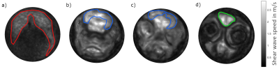 |
Micro MR elastography of the zebrafish brain
Jakob Jordan1, Gergely Bertalan1, Heiko Tzschätzsch1, Tom Meyer1, Anton Gauert2, Anja Heeren-Hagemann2, Jürgen Braun3, and Ingolf Sack1
1Department of Radiology, Charité - Universitätsmedizin Berlin, Berlin, Germany, 2Department of Hematology/Oncology, Charité - Universitätsmedizin Berlin, Berlin, Germany, 3Institute of Medical Informatic, Charité - Universitätsmedizin Berlin, Berlin, Germany
Zebrafish have emerged as a versatile, cost-efficient and easy-to-handle animal model in many areas of biomedical research. Using multifrequency MR elastography at 7-Tesla and 1000 to 1400 Hz excitation frequency, we analyzed the viscoelastic properties of the post-mortem zebrafish brain. Shear wave speed as a surrogate marker of stiffness was mapped with an in-plane resolution of 40 microns showing stiffer properties (1.2±0.3 m/s) of the brain than skeletal muscle (0.89±0.09 m/s). Furthermore, sub-regions showed distinct stiffness properties, which, however, lacked significance in this preliminary feasibility study.
|
|||
1003.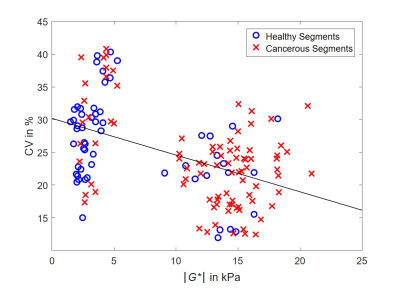 |
Heterogeneity of Viscoelastic Properties in Prostate Cancer Assessed by Ex Vivo MR Elastography
Rolf Reiter1,2, Shreyan Majumdar2, Steven Kearney2, Andre Kajdacsy‐Balla3, Virgilia Macias3, Michael Abern4, Simone Crivellaro4, Thomas J. Royston2, and Dieter Klatt2
1Radiology, Charité - Universitätsmedizin Berlin, Berlin, Germany, 2Bioengineering, University of Illinois at Chicago, Chicago, IL, United States, 3Pathology, University of Illinois at Chicago, Chicago, IL, United States, 4Urology, University of Illinois at Chicago, Chicago, IL, United States
We aimed to evaluate the heterogeneity of viscoelastic tissue properties of fresh prostatectomy specimens of men with prostate cancer using MR elastography and histopathology as a reference. Our results suggest that prostate cancer is characterized by a stiff yet homogeneous biomechanical signature. A possible explanation for this finding might be differences of healthy vs. cancerous tissue based on anatomical zones and their specific nonmalignant pathologies (prostatitis in the peripheral zone and benign prostatic hyperplasia in the central gland), and the unique nondestructive growth pattern of PC with intervening stroma which might provide a rigid scaffold.
|
|||
1004.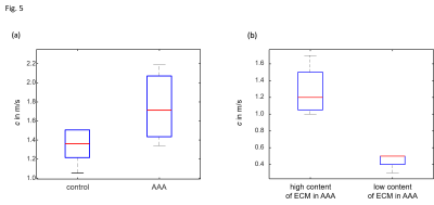 |
MR elastography of abdominal aortic aneurysm specimens
Dilyana B. Mangarova1,2, Julia Brangsch1, Marcus R. Makowski1,3, Bernd Hamm1, Ingolf Sack1, Jürgen Braun 4, and Gergely Bertalan1
1Department of Radiology, Charité - Universitätsmedizin Berlin, Berlin, Germany, 2Department of Veterinary Pathology, Free University of Berlin, Berlin, Germany, 3Department of Diagnostic and Interventional Radiology, Technical University of Munich, Berlin, Germany, 4Institute for Medical Informatics, Charité - Universitätsmedizin Berlin, Berlin, Germany
Abdominal aortic aneurysms (AAA) are among the worldwide leading causes of death. Standard MRI and ultrasonography cannot predict the risk of life-threatening AAA rupture. The ECM composition and therefore the stiffness of the aortic wall changes during AAA development and progression. Therefore, we investigated AAA stiffness in murine tissue samples using MR elastography with an in-plane resolution of 40 microns. Compared to healthy aorta, we found an increase of vessel wall stiffness in AAA of approximately 20% and an excellent correlation of histology-quantified ECM accumulation with thrombus stiffness.
|
|||
1005.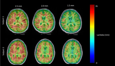 |
Viscoelastic Differences in Human Gray and White Matter at Different Spatial Resolutions
Shruti Mishra1,2, Bin Deng2,3,4, W. Scott Hoge1,2,3, Giacomo Annio5, Ralph Sinkus5, and Samuel Patz1,2
1Department of Radiology, Brigham & Women's Hospital, Boston, MA, United States, 2Harvard Medical School, Boston, MA, United States, 3Athinoula A. Marginos Center for Biomedical Imaging, Charlestown, MA, United States, 4Radiology, Massachusetts General Hospital, Boston, MA, United States, 5Laboratory for Vascular Translational Science (LVTS), Institut National de la Santé et de la Recherche Médicale (INSERM), Paris, France
Magnetic resonance elastography (MRE) was utilized to demonstrate differences in shear wavelength between human cortical gray matter and subcortical white matter and evaluate the effect of spatial resolution on wavelength estimation. Much greater brain architectural detail was discerned in the viscoelastic maps when going from 2.5 mm to 1.5 mm isotropic spatial resolution. We also demonstrate in individual healthy subjects, through two adjacent multi-voxel regions of interest, that cortical gray matter is stiffer than adjacent subcortical white matter, and that this difference is accentuated at higher resolutions. Furthermore, we show that the mean estimated wavelength increases with decreasing spatial resolution.
|
|||
1006. |
Assessment of Obesity-Related Inflammation in Subcutaneous Adipose Tissue with MR Elastography (MRE)
Jiahui Li1, Prachi Singh2, Marzanna Obrzut1, Xin Lu1, Kevin J. Glaser1, Alina Allen3, Sudhakar K. Venkatesh1, Taofic Mounajjed4, Jun Chen1, Armando Manduca1, Vijay Shah3, Richard L. Ehman1, and Meng Yin1
1Radiology, Mayo Clinic, Rochester, MN, United States, 2Sleep and Cardiometabolic Health, Pennington Biomedical Research Center, Baton Rouge, LA, United States, 3Gastroenterology, Mayo Clinic, Rochester, MN, United States, 4Anatomic Pathology, Mayo Clinic, Rochester, MN, United States
Multiparametric MRI and MRE was performed in 27 obese patients who had biopsies of liver and subcutaneous adipose tissues. We found significant correlations between the mechanical properties of liver and subcutaneous fat and their histological and biochemistry results. A model combining liver proton density fat fraction and subcutaneous fat stiffness had a slightly higher AUC for diagnosing nonalcoholic steatohepatitis than liver stiffness (AUC: 0.87 vs. 0.84, p=0.74). The results indicate that obesity-induced systemic inflammation affects both adipose and liver tissue mechanical properties and, therefore, models utilizing mechanical biomarkers from adipose tissue may improve the diagnosis of steatohepatitis in obese patients.
|
|||
1007. |
Anisotropic stiffness of the supraspinatus muscle estimated via MR elastography and transversely isotropic nonlinear inversion.
Elijah Van Houten1, Cyril Tous2,3, Alexandre Jodoin4, Matthew McGarry5, Philip Bayly6, Keith Paulsen5,7, Curtis Johnson8, and Nathalie Bureau2,4
1Mechanical Engineering, Université de Sherbroke, Sherbrooke, QC, Canada, 2Centre de recherche du Centre hospitalier de l’Université de Montréal, Montréal, QC, Canada, 3Université de Montréal, Montréal, QC, Canada, 4Department of Radiology, Centre hospitalier de l’Université de Montréal, Montréal, QC, Canada, 5Thayer School of Engineering, Dartmouth College, Hanover, NH, United States, 6Department of Mechanical Engineering & Materials Science, Washington University in St. Louis, St. Louis, MO, United States, 7Dartmouth-Hitchcock Medical Center, Lebanon, NH, United States, 8Department of Biomedical Engineering, University of Delaware, Newark, DE, United States
Surgical planning for rotator cuff repair would benefit significantly from quantitative measures of the muscle stiffness to avoid re-tearing of the muscle after surgery. MR Elastography (MRE) can provide quantitative measures of soft tissue stiffness in-vivo, but repeatable, stable results require accurate tissue models capable of treating the specific geometry and highly anisotropic structure of muscle tissue. Here, MRE based on a nearly incompressible, transversely isotropic continuum model is developed and applied to the case of the supraspinatus muscle. Overall, results show good agreement between reconstructed anisotropic stiffness values and independently published results based on wavelength analysis.
|
|||
1008.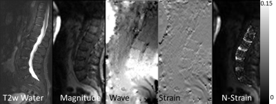 |
Assessment of Shear Strain in Intervertebral Discs In Vivo with Magnetic Resonance Elastography
Yuan Le1, Jun Chen1, Phillip J. Rossman1, Ziying Yin1, Kevin J. Glaser1, Joshua D. Trzasko1, Yi Sui1, Stephan Kannengiesser2, Bradley D. Bolster, Jr.3, Joel P. Felmlee1, and Richard L. Ehman1
1Radiology, Mayo Clinic, Rochester, MN, United States, 2Siemens Healthcare GmbH, Erlangen, Germany, 3Siemens Medical Solutions USA, Inc., Salt Lake City, UT, United States
This study is to test the feasibility of visualizing waves propagating in the spine and measuring the strain in the intervertebral discs (IVDs) in vivo with Magnetic Resonance Elastography (MRE). Our results show that waves can be visualized, and that IVD shear strain can be quantitatively assessed. Degeneration of the IVD is thought to decrease disc shear stiffness. Preliminary results in a series of volunteers demonstrated increasing IVD shear strain with age, consistent with the known gradual degradation of IVD function with age.
|
|||
1009.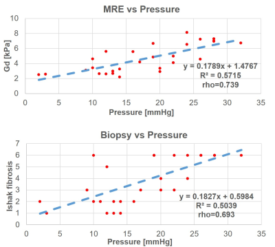 |
3D-MRE as independent predictor of direct portal venous pressure compared to Biopsy derived Fibrosis Measurements
Giacomo Annio1,2, Ahmed Hamimi3, Khaled Z. Abd-Elmoniem 3, Theo Heller3, Elliot Levy 3, David E. Kleiner4, Ohad Etzion5, Rabab Ali6, Ralph Sinkus1,2, and Ahmed M. Gharib3
1LVTS, INSERM U1148, Paris, France, 2Department of Biomedical Engineering, King's College London, London, United Kingdom, 3Biomedical and Metabolic Imaging Branch, National Institute of Diabetes and Digestive and Kidney Diseases, NIH, Bethesda, MD, United States, 4Center for Cancer Research, National Cancer Institute, Bethesda, MD, United States, 5Department of Gastroenterology and Liver Diseases, Ben-Gurion University of the Negev, Be'er Sheva, Israel, 6Department of Internal Medicine, Duke University, Durham, NC, United States
The assessment of liver fibrosis and inflammation is crucial to the clinical management of patients suffering from liver diseases. Portal venous pressure assessment has important prognostic implications for hepatic disease patients’ care. However, it can only be measured invasively. In this work we show that liver stiffness quantified via 3DMRE is a strong independent predictor of DPVP compared to invasive biopsy. Moreover stiffness as evaluated with 3DMRE is an operator independent and non-invasive biomarker which could hence constitute an optimal method for diagnosis and fibrosis staging in all patients and a non-invasive surrogate of DPVP.
|
|||
1010.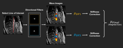 |
Estimating Aortic Stiffness Using Aortic MR Elastography Using an Inversion without Considering the Geometry
Huiming Dong1, Prateek Kalra1, Richard D White1, and Arunark Kolipaka1
1Radiology, The Ohio State University Wexner Medical Center, Columbus, OH, United States
Aortic stiffness is a valuable imaging marker because of its association with a variety of cardiovascular conditions such as abdominal aortic aneurysm (AAA). Calculating AAA stiffness from aortic MRE data presents a unique challenge due to the relatively small size of an AAA compared to the liver. In the present study, we propose a new inversion strategy that consists of directional filters that are designed to extract the propagating waves along the axial direction of the AAA, and uses the wave information along the axial direction for stiffness calculation to reduce the impact of AAA geometry on stiffness estimation.
|
|||
1011.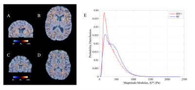 |
Preliminary MR elastography investigation on HIV+ cohort with cerebral small vessel disease
Abrar Faiyaz1, Irteza Enan Kabir1, Marvin M Doyley1, Ingolf Sack2, Md Nasir Uddin3, and Giovanni Schifitto3
1Department of ECE, University of Rochester, Rochester, NY, United States, 2Department of Radiology, Charité - Universitätsmedizin Berlin, Berlin, Germany, 3Department of Neurology, University of Rochester, Rochester, NY, United States
Neuroinflammation plays a crucial role in HIV-infected subjects at risk of cerebral small vessel disease. As MR-Elastography (MRE) has shown the potential to link inflammation with brain stiffness reduction, we investigated the brain viscoelastic properties of the HIV-infected and HIV-uninfected individuals. The preliminary results with MRE are promising, complementing other imaging modalities such as diffusion MRI (DTI, NODDI) to investigate brain microstructure integrity in HIV infection.
|
|||
1012.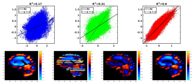 |
A Learned Phase Correction Algorithm Outperforms a Conventional Filter for 3D Vector MRE
Jonathan M Trevathan1, Jonathan M Scott1, Joshua D Trzasko1, Armando Manduca1, John Huston1, Richard L Ehman1, and Matthew C Murphy1
1Mayo Clinic, Rochester, MN, United States
Bulk motion during the motion encoding of multi-slice spin-echo EPI pulse sequences used to acquire 3D MRE data creates a slice-to-slice phase jitter. A 3D convolutional neural network was trained to estimate a phasor corresponding to noise-free displacements given noisy, complex-valued MRE data. The net outperformed its filtering counterpart in noisy and noise-free simulation data, and decreased test-retest repeatability error in vivo.
|
The International Society for Magnetic Resonance in Medicine is accredited by the Accreditation Council for Continuing Medical Education to provide continuing medical education for physicians.