Digital Posters
Elastography: Methods
ISMRM & SMRT Annual Meeting • 15-20 May 2021

| Concurrent 4 | 13:00 - 14:00 |
1013.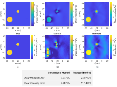 |
MR Elastography Reconstruction Based on a Linear Integral Equation Derived from the Helmholtz Identity
Motofumi Fushimi1, Naohiro Eda1, and Takaaki Nara1
1The University of Tokyo, Tokyo, Japan
This paper presents a novel reconstruction method for MR Elastography. Unlike most conventional methods that solve Navier's partial differential equation (PDE), the proposed approach solves a linear integral equation derived from Helmholtz’s identity. The proposed method can reconstruct the complex shear modulus without calculating higher-order derivatives of the measured displacement data, nor solving a nonlinear minimization problem that could result in local minima. Our preliminary study by numerical simulations demonstrated that the proposed method could robustly estimate the complex shear modulus. Future work is to compare the effectiveness of the proposed method with the PDE-based conventional methods.
|
|||
1014.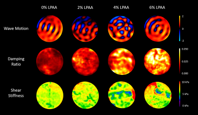 |
Viscoelastic MRE Phantoms with Tunable Damping Ratio Independent of Shear Stiffness
L. Tyler Williams1, Zheng Cao1, Matthew D. J. McGarry2, Elise A. Corbin1, and Curtis L. Johnson1
1Biomedical Engineering, University of Delaware, Newark, DE, United States, 2Thayer School of Engineering, Dartmouth College, Hanover, NH, United States
This study validates the use of LPAA/PAA gels as viscoelastic MRE phantoms with tunable damping ratio independent of shear stiffness. PAA concentration was 6wt% across all phantoms while LPAA concentration ranged from 0 to 6wt%. High-resolution MRE scans were conducted with 600, 700, 800, and 900 Hz vibrations. For each frequency, storage modulus and shear stiffness were constant while loss modulus and damping ratio increased with increasing LPAA concentration. For each LPAA concentration, loss modulus increased with increasing frequency indicating viscoelastic behavior. These results verify the independent tunability of damping ratio and shear stiffness.
|
|||
1015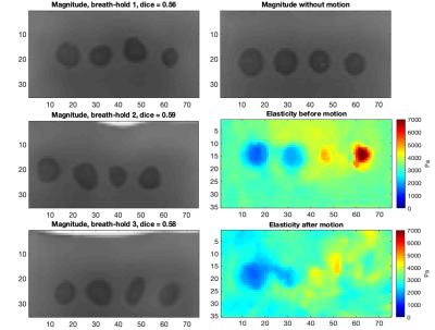 |
Investigation of Motion as a Source of Error in MR Elastography Video Permission Withheld
Emily Chan1, Daniel Fovargue1, Matt Kelly2, Ralph Sinkus1,3, and Julia A Schnabel1
1School of Biomedical Engineering & Imaging Sciences, King's College London, London, United Kingdom, 2Perspectum, Oxford, United Kingdom, 3INSERM U1148, Laboratory for Vascular Translational Science, University Paris Diderot, University Paris 13, Paris, France
Clinically, motion occurring between breath-hold acquisitions during a magnetic resonance elastography (MRE) scan is assumed negligible and therefore not corrected. In this work, we demonstrate through simulations on a phantom that motion of this degree does impact elasticity reconstruction, which highlights the need for care to be taken to account for this motion.
|
|||
1016.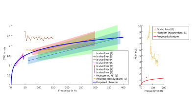 |
A viscoelastic phantom of the healthy human liver for elastography in MRI and ultrasound
Anna Morr1, Helge Herthum1, Felix Schrank1, Steffen Görner1, Jürgen Braun1, Ingolf Sack1, and Heiko Tzschätzsch1
1Charité - Universitätsmedizin Berlin, Berlin, Germany
Elastography in MRI (MRE) and ultrasound (USE) are the most reliable imaging techniques for liver fibrosis detection. However, there is no established, standardized material that mimics the elasticity and viscous behavior of liver tissue and can be used as a phantom for MRE and USE. We developed a phantom based on polyacrylamide which shows a viscoelastic dispersion from 5-3000 Hz similar to that observed in the healthy human liver, which is measurable by MRE and USE, stable over months and can be produced in a reproducible way. The novel phantom facilitates technical improvements and cross-modality comparisons in MRE and USE.
|
|||
1017.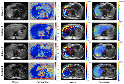 |
Simultaneous acquisition for magnetic resonance elastography and fat quantification of the liver
Shuo Li1, Suhao Qiu1, Chenfei Shen1, Yuan Feng1, and Yiping P. Du1
1School of Biomedical Engineering, Shanghai Jiao Tong University, Shanghai, China Magnetic resonance elastography (MRE) has been used for tumor detection and liver fibrosis evaluation. The existence of intratumoral fat suggests a lower risk of microvascular invasion of combined hepatocellular-cholangiocarcinoma. The combination of MRE and fat quantification can provide sufficient sensitivity to detect substantial macrovesicular hepatic steatosis or fibrosis. In clinical practice, MRE and fat quantification are scanned separately. However, this may cause slice mis-registration between MRE and fat quantification, and increase scan time. In this study, the feasibility of simultaneous acquisition for MRE and fat quantification was demonstrated. This technique can have several potential clinical applications without increasing scan time. |
|||
1018. |
Effect of different driver power amplitudes on the measurement of liver stiffness in pediatric liver MR elastography
Dong Kyu Kim1, Kyunghwa Han2, Haesung Yoon3, Mi-Jung Lee3, and Hyun Joo Shin3
1Radiology, Armed Forces Capital Hospital, Seongnam, Korea, Republic of, 2Center for Clinical Imaging Data Science, Severance Hospital, Seoul, Korea, Republic of, 3Radiology, Severance Hospital, Seoul, Korea, Republic of
The standard driver power of magnetic resonance elastography (MRE) in pediatric patients is recommended to be reduced by 20–50%, compared to adult patients, to keep the patients from any injury due to the vibrating driver, however, there is limited published information on driver power of MRE in pediatric applications. Furthermore, there is lack of study to evaluate whether different amplitudes of driver power may affect the measurement of liver stiffness on pediatric MRE. In this abstract, we assessed the effect of different driver power amplitudes on the measurement of liver stiffness in pediatric liver MRE.
|
|||
1019.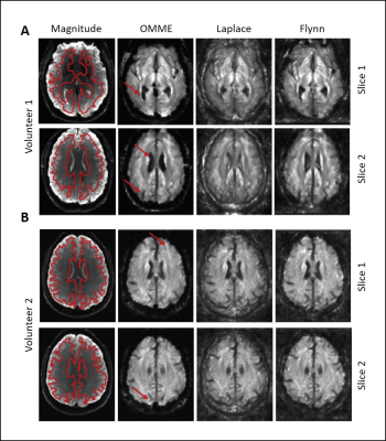 |
Optimal multiple motion encoding in phase contrast MRI
Helge Herthum1, Hugo Carrillo2, Axel Osses2, Sergio Uribe3, Ingolf Sack1, and Cristóbal Bertoglio4
1Experimentelle Radiologie, Charité Universitätsmedizin Berlin, Berlin, Germany, 2Center for Mathematical Modeling, Universidad de Chile, Santiago, Chile, 3Department of Radiology, School of Medicine, Pontificia Universidad Católica de Chile, Santiago, Chile, 4Bernoulli Institute, University of Groningen, Groningen, Netherlands
Optimal multiple motion encoding (OMME) for phase-contrast MRI was developed with application to magnetic resonance elastography for unwrapping motion images. OMME is formulated as a least-squares problem using multiple phase-contrast measurements with different motion encoding gradients (MEG). OMME is applied to phantom and in vivo human brain experiments. The wrap-free motion images are further used to reconstruct shear-wave-speed maps and compared to conventional phase unwrapping methods. OMME generates wrap free phase-contrast images with the wrap limit determined by the smallest MEG used, while maintaining the signal-to-noise ratio(SNR) of the largest MEG, which makes OMME especially suitable for high SNR applications.
|
|||
1020.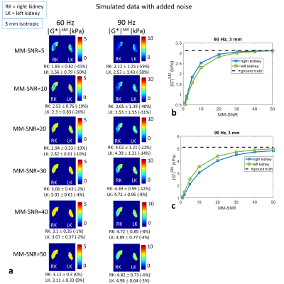 |
In Silico Evaluation of Magnetic Resonance Elastography of the Kidney
Deirdre McGrath1, Christopher Bradley1, and Susan Francis1
1Sir Peter Mansfield Imaging Centre, University of Nottingham, Nottingham, United Kingdom
The accuracy of clinical magnetic resonance elastography (MRE) is difficult to determine. Finite element modelling (FEM) simulation allows evaluation of the errors on acquired MRE measures, and also informs methodological optimisation. In this study kidney MRE is simulated using an anthropomorphic personalised model of the kidneys, and simulated data is compared with a 2D GRE MRE acquisition of the same volunteer on whom the model was based. The optimum MRE imaging resolution was identified, and through adding simulated noise to the FEM data, errors were estimated for the acquired MRE data, and recommendations made for kidney MRE optimisation.
|
|||
1021.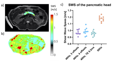 |
Optimization of GRE-MRE in the pancreas
Anne-Sophie van Schelt1, Nienke Wassenaar1, Eric M Schrauben1, Marian Troelstra1,2, Jules L. Nelissen1, Ralph Sinkus2,3, Jaap Stoker4, Aart J Nederveen1, and Jurgen H Runge1
1Radiology and Nuclear Medicine, Amsterdam UMC location AMC, Amsterdam, Netherlands, 2Department of Imaging Sciences and Biomedical Engineering, King's College London, London, United Kingdom, 3Inserm U1148, LVTS, University Paris Diderot, Paris, France, 4Radiology and Nuclear medicine, Amsterdam UMC location AMC, Amsterdam, Netherlands
Pancreatic Magnetic Resonance Elastography (MRE) allows non-invasive estimation of pancreatic tissue visco-elastic properties for multiple pathophysiological diseases. The aim of this study is to optimize pancreatic GRE-MRE to provide an in-depth analysis of the propagation and registration of shear waves. Optimization was performed using four combinations of frequency, the number of measured wave-phase offsets, and TE. Pancreas GRE-MRE at 40Hz, TE 6.9ms and 4 or 5 wave-phase offsets can estimate the SWS of the pancreas head with optimal encoding and propagation and has an acceptable breath-hold duration for clinical purposes.
|
|||
1022.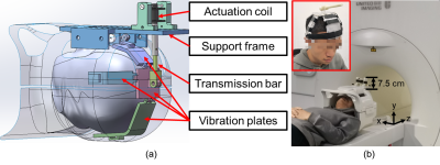 |
An Electromagnetic Actuator for Brain Magnetic Resonance Elastography
Suhao Qiu1, Zhao He1, Runke Wang1, Ruokun Li2, Fuhua Yan2, and Yuan Feng1
1the Institute for Medical Imaging Technology, Shanghai Jiao Tong University, Shanghai, China, 2the Department of Radiology, Ruijin Hospital, Shanghai, China
Effective actuators are crucial for brain magnetic resonance elastography (MRE). In this study, we designed, tested, and verified an electromagnetic actuator. With a grappler-shaped design, the actuator was easy to use and comfortable to wear on head. Phantom and brain experiments indicated that the proposed actuator did not interference with routine imaging sequences. It generated stable shear waves with a full width at half maximum of 0.3 Hz in the frequency spectrum. Phantom and brain MRE demonstrated that the actuator could carry out multi-frequency MRE with a high frequency accuracy.
|
|||
1023.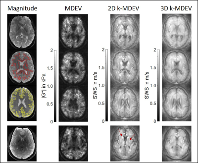 |
Magnetic resonance elastography of the in vivo human brain using multifrequency wavenumber analysis in 2D and 3D.
Helge Herthum1, Heiko Tzschätzsch1, Tom Meyer1, Mehrgan Shahryari1, Lisa Stencel1, Jing Guo1, Jürgen Braun2, and Ingolf Sack1
1Experimentelle Radiologie, Charité Universitätsmedizin Berlin, Berlin, Germany, 2Institut für medizinische Informatik, Charité Universitätsmedizin Berlin, Berlin, Germany
Wavenumber analyses in MR elastography (MRE), which use first-order finite difference operators, are known to be more stable against noise than second-order finite derivative methods. However, wavenumber analyses for the human brain normally suffer from abundant heterogeneities and solid-fluid interfaces. We here present multifrequency wavenumber analysis for MRE of the human brain in 2D and 3D based on adapted bandpass filters. We show that both approaches provide better repeatability in test-retest experiments compared to standard analyses. Moreover, wavenumber analyses yield stable values and rich detail in regions of lower signal-to-noise-ratio such as deep gray matter.
|
|||
1024.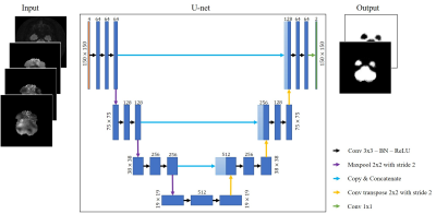 |
Deep Learning Automation for Human Brain Masking of Multi-Band MR Elastography
Aaron Thomas Anderson1,2, Siddharth Muralidaran3, Alexander M Cerjanic1,4, Bradley P Sutton1,4, Mark A Anastasio1,4, and Frank J Brooks1,4
1Beckman Institute for Advanced Science & Technology, University of Illinois at Urbana-Champaign, Urbana, IL, United States, 2Stephens Family Clinical Research Institute, Carle Foundation Hospital, Urbana, IL, United States, 3Electrical & Computer Engineering, University of Illinois at Urbana-Champaign, Urbana, IL, United States, 4Bioengineering, University of Illinois at Urbana-Champaign, Urbana, IL, United States
The processing steps involved in MR elastography include custom imaging sequences, image reconstruction, and material property estimation, but the most person-hours are spent on manual image masking. Manual corrections are needed because automated brain segmentation often fails near temporal lobe artifacts, which are unique for each subject. A deep learning method, specifically, a U-net architecture, is trained to map input MRE-image intensity data to corresponding manually corrected masks (N=44) with the goal of automating the masking of future MRE brain datasets. We observed the U-net-based masking maintained data quality (OSS-SNR) for all subjects.
|
|||
1025.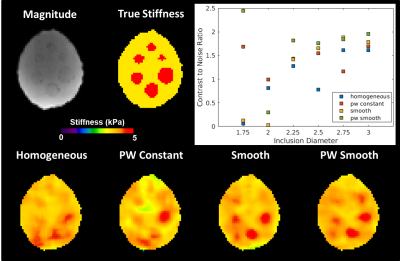 |
Effect of Spatial Inhomogeneity Models on Performance of Machine Learning Based Inversion Algorithms for Brain Magnetic Resonance Elastography
Jonathan M Scott1, Joshua D Trzasko1, Armando Manduca1, Matthew L Senjem1, Clifford R Jack1, John Huston III1, Richard L Ehman1, and Matthew C Murphy1
1Mayo Clinic, Rochester, MN, United States
Four machine learning inversion algorithms with different material spatial property assumptions (trained on simulated data with homogeneous, piecewise constant, smooth, or piecewise smoothly varying material properties) were evaluated in a brain simulating phantom with stiff inclusions, a test-retest repeatability dataset, and an Alzheimer’s disease dataset. The piecewise smooth inversion produced the highest contrast to noise ratio and allowed the best visualization of inclusions in the phantom study. All four inversions produced stiffness estimates that were repeatable and sensitive to stiffness changes in Alzheimer’s disease.
|
|||
1026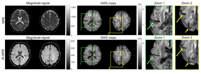 |
Cerebral inversion recovery MR elastography for cortical stiffness quantification Video Permission Withheld
Ledia Lilaj1, Helge Herthum2, Tom Meyer2, Mehrgan Shahryari2, Gergely Bertalan2, Alfonso Caiazzo2, Jürgen Braun3, Thomas Fischer2, Sebastian Hirsch4,5, and Ingolf Sack2
1Radiology, Charité – Universitätsmedizin Berlin, Berlin, Germany, 2Charité – Universitätsmedizin Berlin, Berlin, Germany, 3Institute of Medical Informatics, Charité – Universitätsmedizin Berlin, Berlin, Germany, 4Berlin Center for Advanced Neuroimaging, Charité – Universitätsmedizin Berlin, Berlin, Germany, 5Bernstein Center for Computational Neuroscience, Berlin, Germany
Inversion recovery magnetic resonance elastography (IR-MRE) is a new acquisition technique that performs MRE while selectively nulling the signal generated by one compartment with a specific T1. In phantom and in vivo experiments, free fluid, water or cerebrospinal fluid, was nulled. The shear wave speed (SWS) maps showed no overall change in average values. However, in cortical areas, IR-MRE revealed cortical SWS values 10% higher than in standard MRE, resulting in sharper tissue-CSF interfaces. Besides, ventricles are 39% narrower in IR-MRE than in MRE. In conclusion, IR-MRE allows a more precise quantification of cortical stiffness.
|
|||
1027.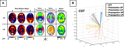 |
Multi-Excitation Actuator Design for Anisotropic Brain MRE
Diego A. Caban-Rivera1, Daniel R. Smith1, Keshav A. Kailash2, Ruth J. Okamoto2, Matthew D.J. McGarry 3, Lance T. Williams1, Charlotte Guertler2, Grace Mcilvain1, Damian Sowinski3, Elijah E.W. Van Houten 4, Keith D. Paulsen3,5, Philip V. Bayly2, and Curtis L. Johnson1
1Biomedical Engineering, University of Delaware, Newark, DE, United States, 2Mechanical Engineering and Material Science, Washington University in St. Louis, St. Louis, MO, United States, 3Thayer School of Engineering, Dartmouth College, Hanover, NH, United States, 4Mechanical Engineering, Université de Sherbrooke, Sherbrooke, QC, Canada, 5Dartmouth-Hitchcock Medical Center, Lebanon, NH, United States
The goal of this study is to thoroughly examine a novel customized multi-excitation MRE actuator combining both left-right (LR) and anterior-posterior (AP) drivers for use in estimating anisotropic mechanical properties of the brain. We demonstrate the key differences in wave patterns and how these lead to distinct stiffness estimations. The displacement fields from both excitations were combined to recover repeatable anisotropic properties in the corticospinal tract. The improved ME-MRE actuator can be used for future studies of white matter mechanical properties in health and disease.
|
|||
1028. |
Intra-patient comparison of 3D and 2D MRE techniques for assessment of liver fibrosis
Roberta Catania1, Amir A. Borhani1, Camila Lopes Vendrami1, Roger C Grimm2, Bradley D. Bolster3, and Frank Miller1
1Northwestern University Feinberg School of Medicine, Chicago, IL, United States, 2Mayo Clinic, Rochester, MN, United States, 3Siemens Medical Solutions USA, Inc., Salt Lake City, UT, United States
3D MRE allows volumetric assessment of liver stiffness. Performance of this method as compared to 2D techniques (GRE and seEPI), was evaluated, with respect to the ROI areas per slice, stiffness values, and presence of artifacts. The 3D MRE provided larger area of liver and was less prone to artifacts. Liver Stiffness Measures (LSM) based on 3D technique were lower than 2D technique. 3D MRE exhibits less susceptibility to artifacts and provides larger measurable areas of liver. LSM based on 3D MRE however were lower than 2D techniques.
|
|||
1029.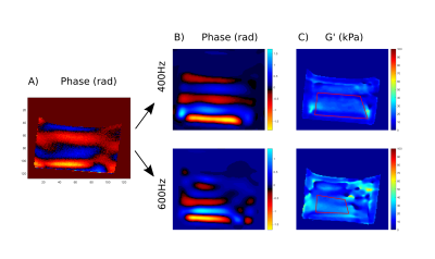 |
Short echo time dual-frequency MR Elastography with Optimal Control RF pulses
Pilar Sango Solanas1, Kevin Tse Ve Koon1, Eric Van Reeth1,2, Cyrielle Caussy3,4, and Olivier Beuf1
1Univ Lyon, INSA‐Lyon, Université Claude Bernard Lyon 1, UJM-Saint Etienne, CNRS, Inserm, CREATIS UMR 5220, U1206, F‐69616, Lyon, France, 2CPE Lyon, Département Sciences du Numérique, Lyon, France, 3Univ Lyon, Laboratoire CarMen, Inserm, INRAe, INSA Lyon, Université Claude Bernard Lyon 1, Pierre-Bénite, France, 4Hospices Civils de Lyon, Département Endocrinologie, Diabète et Nutrition, Hôpital Lyon Sud, Pierre-Bénite, France
Magnetic Resonance Elastography (MRE) quantifies the mechanical properties of tissues, typically applying motion encoding gradients (MEG). High frequencies are difficult to reach due to slew rate limitations and low frequencies induce too long TEs, yielding magnitude images with low SNR. Multifrequency results allow better characterizations of tissues using data usually acquired through sequential monofrequency experiments. In this study, we generate optimal control-based RF pulses to outperform simultaneous multifrequency MRE. The pulse is applied with a constant gradient during the mechanical excitation to simultaneously achieve spatially selective excitation and motion encoding. Results on phantom demonstrated the feasibility of the proposed method.
|
|||
1030.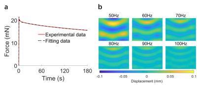 |
Comparative analysis of indentation and magnetic resonance elastography for measuring viscoelastic properties
Yu Chen1, Suhao Qiu1, Zhao He1, Fuhua Yan2, Ruokun Li2, and Yuan Feng1
1Institute for Medical Imaging Technology, School of Biomedical Engineering, Shanghai Jiao Tog University, Shanghai, China, 2Department of Radiology, Ruijin Hospital, Shanghai Jiao Tong University School of Medicine, Shanghai, China
Comparing viscoelastic properties of soft tissues measured in vivo and ex vivo are of interests for basic science and clinical translation. Magnetic resonance elastography (MRE) and indentation are two commonly used techniques for measurement of tissue viscoelasticity. In this study, we used a gelatin phantom to compare measurements by MRE and indentation. Using 2nd-order Prony series, frequency response measured from indentation was estimated. We observed apparent frequency-dependent behaviors from MRE in the range of 50-100 Hz, but not from indentation. Results implied that MRE and indentation measures at different frequency ranges.
|
|||
1031.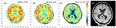 |
Development of robust MRE for measurement of high-speed functionally mediated changes in human brain viscoelasticity
Bin Deng1,2,3, W. Scott Hoge3,4, Shruti Mishra3,4, Giacomo Annio5, Ralph Sinkus5, and Samuel Patz3,4
1Athinoula A. Martinos Center for Biomedical Imaging, Massachusetts General Hospital, Charlestown, MA, United States, 2Radiology, Massachusetts General Hospital, Boston, MA, United States, 3Harvard Medical School, Boston, MA, United States, 4Radiology, Brigham and Women’s Hospital, Boston, MA, United States, 55. Laboratory for Vascular Translational Science (LVTS, Institut National de la Santé et de la Recherche Médicale (INSERM), Paris, France
Our aim is to develop a robust, high-spatial resolution and highly reproducible MRE methodology for the measurement of rapid functionally mediated changes in human brain viscoelasticity. Based on prior work in mice, we expect to observe changes in shear wavelength of ~2mm due to functional activation. Using a modified Ristretto fMRE pulse sequence to acquire control data in healthy volunteers, we evaluated the reproducibility of MRE measurements at 2.5-, 2- and 1.5-mm isotropic spatial resolution. Excellent intra-scan consistency was observed between two interleaved paradigm acquisitions at all resolutions with comparable data quality. Robust inter-scan repeatability was also observed.
|
|||
1032.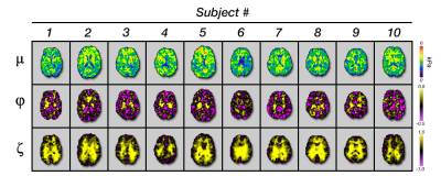 |
Anisotropic Mechanical Properties of White Matter Tracts Estimated with Multi-Excitation MRE and TI-NLI
Daniel R. Smith1, Diego A. Caban-Rivera1, Matthew D. J. McGarry2, L. Tyler Williams1, Grace McIlvain1, Charlotte Guertler3, Ruth J. Okamoto3, Damian Sowinski2, Elijah Van Houten4, Phil V. Bayly3, Keith D. Paulsen2, and Curtis L. Johnson1
1Biomedical Engineering, University of Delaware, Newark, DE, United States, 2Thayer School of Engineering, Dartmouth College, Hanover, NH, United States, 3Mechanical Engineering and Materials Science, Washington University in St. Lous, St. Louis, MO, United States, 4Mechanical Engineering, Universite de Sherbrooke, Sherbrooke, QC, Canada
In this study, we used multi-excitation MR elastography (MRE) in conjunction with transversely isotropic NLI (TI-NLI) to estimate the anisotropic viscoelastic parameters of the human brain. We collected data on ten subjects and took averages of the parameters within individual regions of the brain. Through comparison of regions of interest, we found significant differences between the parameter estimate of gray matter and white matter and between individual white matter tracts. The results of this study indicate that multi-excitation MRE and TI-NLI can generate consistent anisotropic parameter estimates for WM that capture innate differences in individual tract structure.
|
The International Society for Magnetic Resonance in Medicine is accredited by the Accreditation Council for Continuing Medical Education to provide continuing medical education for physicians.