Digital Posters
Multicontrast Methods
ISMRM & SMRT Annual Meeting • 15-20 May 2021

| Concurrent 4 | 15:00 - 16:00 |
 |
1232.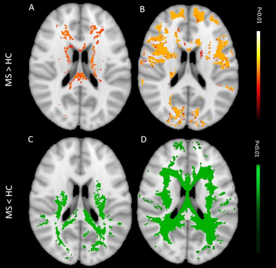 |
Quantitative myelin-sensitive MRIs exhibit differential sensitivity to multiple sclerosis pathology in distinct brain lesions and regions
Reza Rahmanzadeh1,2,3, Po-Jui Lu1,2,3, Muhamed Barakovic1,2,3, Matthias Weigel1,2,3, Laura Gaetano4, Riccardo Galbusera1,2,3, Thanh D. Nguyen5, Francesco La Rosa 6,7, Daniel S. Reich8, Pascal Sati8,9, Yi Wang5, Meritxell Bach Cuadra6,7, Ernst-Wilhelm Radue1,2,
Jens Kuhle1,3, Ludwig Kappos1,3, Stefano Magon10, and Cristina Granziera1,2,3
1Neurologic Clinic and Policlinic, Departments of Medicine, Clinical Research and Biomedical Engineering, University Hospital Basel and University of Basel, Basel, Switzerland, 2Translational Imaging in Neurology (ThINk) Basel, Department of Biomedical Engineering, University Hospital Basel and University of Basel, Basel, Switzerland, 3Research Center for Clinical Neuroimmunology and Neuroscience (RC2NB) Basel, University Hospital Basel and University of Basel, Basel, Switzerland, 4Hoffmann-La Roche Ltd., Basel, Switzerland, 5Department of Radiology, Weill Cornell Medical College, New York, NY, United States, 6Signal Processing Laboratory (LTS5), Ecole Polytechnique Fédérale de Lausanne (EPFL), Laussane, Switzerland, 7Radiology Department, Center for Biomedical Imaging (CIBM), Lausanne University and University Hospital, Laussane, Switzerland, 8Translational Neuroradiology Section, National Institute of Neurological Disorders and Stroke, NIH, 10 Center Drive MSC 1400, Building 10 Room 5C103, Bethesda, MD, United States, 9Department of Neurology, Cedars-Sinai Medical Center, Los Angeles, CA, United States, 10Pharmaceutical Research and Early Development, Roche Innovation Center Basel, F. Hoffmann-La Roche Ltd., Basel, Switzerland
The differential sensitivity of myelin-sensitive advanced MRIs (aMRIs) to the pathology in various brain lesions and regions in multiple sclerosis (MS) is currently debated. This study aimed to address this issue using myelin water fraction maps (MWF), quantitative susceptibility mapping (QSM) and T1 relaxometry (qT1). Our results show that (i) qT1 is the most sensitive in differentiating white matter and cortical MS lesions from normal-appearing tissue (ii) QSM is best differentiating lesions with various extent of damage (lesions with vs without paramagnetic rim & periventricular vs juxta-cortical lesions) and (iii) MWF outperforms the other aMRI methods in identifying occult normal appearing pathology.
|
||
1233.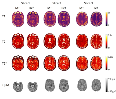 |
Three-dimensional whole-brain simultaneous quantitative mapping of T1, T2, T2*, and susceptibility with MR Multitasking
Tianle Cao1,2, Sen Ma1, Nan Wang1, Sara Gharabaghi3, Yibin Xie1, Zhaoyang Fan1,4,5, Elliot Hogg6, E. Mark Haacke 3,7,8, Michele Tagliati6, Anthony G. Christodoulou1,2, and Debiao Li1,2
1Biomedical Imaging Research Institute, Cedars Sinai Medical Center, Los Angeles, CA, United States, 2Department of Bioengineering, University of California, Los Angeles, Los Angeles, CA, United States, 3Magnetic Resonance Innovations, Inc., Bingham Farms, MI, United States, 4Department of Radiation Oncology, Keck School of Medicine, University of Southern California, Los Angeles, CA, United States, 5Department of Radiology, University of Southern California, Los Angeles, CA, United States, 6Department of Neurology, Cedars Sinai Medical Center, Los Angeles, CA, United States, 7Department of Radiology, Wayne State University School of Medicine, Detroit, MI, United States, 8The MRI Institute for Biomedical Research, Bingham Farms, MI, United States
A new approach for simultaneous quantitative mapping of T1, T2, T2*, and susceptibility was developed. This technique employs hybrid T2 preparation/inversion pulse modules followed by fully flow-compensated multi-echo FLASH readouts. Our method reconstructs images with different T2 preparation times, echo times, and inversion times in the MR Multitasking framework and use them for parameter quantification. Results of both visual comparison and statistical analysis showed that our proposed method agreed well with reference methods while being more time efficient.
|
|||
1234.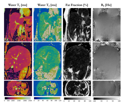 |
3D Isotropic-resolution Non-rigid Motion Compensated Liver T1, T2 and fat fraction mapping
Giorgia Milotta1, Gastao Cruz2, Radhouene Neji2, Claudia Prieto2, and Rene Botnar2
1University College London, London, United Kingdom, 2King's College London, London, United Kingdom
Quantitative T1 and T2 mapping has shown promising results in the quantification of liver fibrosis and inflammation, whereas proton density fat fraction mapping has been used to quantify the hepatic lipid content in fatty liver diseases. Conventionally multiple sequential 2D breath-held scans are performed to acquire sequential 2D T1, T2 and fat fraction maps. However, this approach suffers from limited spatial resolution and coverage and potential misregistration errors. In this work, we sought to develop a free-breathing non-rigid motion corrected 3D sequence for simultaneous and co-registered acquisition of joint liver T1, T2 and fat fraction maps for quantitative tissue characterization
|
|||
1235.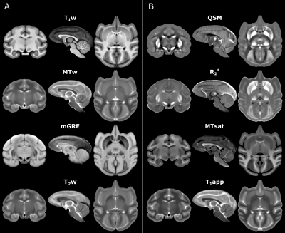 |
Multi-contrast MRI Atlas of the Cynomolgus Macaque Brain
Rakshit Dadarwal1,2 and Susann Boretius1,2
1Functional Imaging Laboratory, German Primate Center, Göttingen, Germany, 2Georg August Universität Göttingen, Göttingen, Germany
Neuroimaging studies require a species-specific template to promote an exquisite spatial normalization. In this work, we provide a high-resolution multi-contrast MRI template of the cynomolgus macaque (Macaca fascicularis) brain. This template may serve as a standard neuroanatomical template to normalize single subject scans in the stereotaxic space. The incorporation of multiple MRI contrast mechanisms provides excellent contrast between gray matter, white matter, and subcortical structures with fine neuroanatomical details.
|
|||
1236.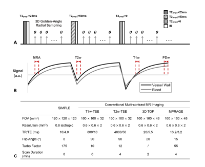 |
Generation of co-registered multi-contrast MR images for carotid atherosclerosis evaluation based on a single SIMPLE sequence
Jiaqi Dou1, Yajie Wang1, Huiyu Qiao1, Zhensen Chen1, Yuze Li1, Haikun Qi2, Jie Sun3, Dongxiang Xu3, Xihai Zhao1, and Huijun Chen1
1Center for Biomedical Imaging Research, School of Medicine, Tsinghua University, Beijing, China, 2School of Biomedical Engineering and Imaging Sciences, King's College London, London, United Kingdom, 3Department of Radiology, University of Washington, Seattle, WA, United States
Carotid atherosclerosis is a leading cause of ischemic stroke worldwide. Multi-contrast magnetic resonance imaging (MRI) is an ideal non-invasive technique to assess carotid plaque. However, its clinical application is limited by long scan time and non-rigid registration between different contrasts. In this study, we generated a set of co-registered T1w, T2w, PDw images, and MR angiogram (MRA) in one scan based on the SIMPLE (SImultaneous T1 and T2 Mapping of carotid PLaquE) sequence. Preliminary experiments on patients validated the feasibility of the generated multi-contrast images for carotid plaque assessment and comparable performance with conventional sequences.
|
|||
1237.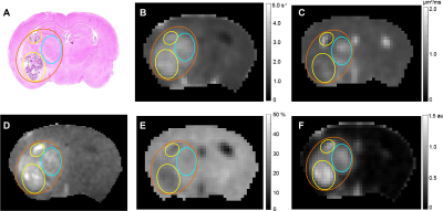 |
Multiparametric MRI Distinguishes Cerebral Radiation Necrosis vs. Recurrent Glioma in Mouse Models
Xia Ge1, John A Engelbach1, Liya Yuan2, Sonika Dahiya3, Feng Gao4, Keith M Rich2, Joseph JH Ackerman1,5,6,7, and Joel R Garbow1,5
1Radiology, Washington University in St Louis, St Louis, MO, United States, 2Neurosurgery, Washington University in St Louis, St Louis, MO, United States, 3Neuropathology, Washington University in St Louis, St Louis, MO, United States, 4Surgery, Washington University in St Louis, St Louis, MO, United States, 5Alvin J Siteman Cancer Center, Washington University in St Louis, St Louis, MO, United States, 6Internal Medicine, Washington University in St Louis, St Louis, MO, United States, 7Chemistry Department, Washington University in St Louis, St Louis, MO, United States
Malignant brain tumor patients treated with radiation are at risk of developing subsequent treatment effects, including radiation necrosis (RN), which cannot be differentiated from recurrent tumor. We have developed mouse models of RN and of admixed tumor growing in previously irradiated brain (“mixed lesion”) that recapitulate the major histologic characteristics of human RN and recurrent glioma, respectively. These models provide a platform for the development of imaging biomarkers capable of differentiating RN vs. tumor. We demonstrate a multiparametric, clinically translatable 1H MRI pipeline (R1, R1-post-contrast, R2, ADC, MTR, and DCEAUC) that shows significant promise for differentiating RN vs. recurrent tumor.
|
|||
1238.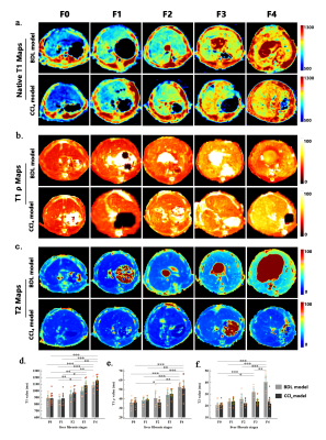 |
Changes of native T1, T1ρ, and T2 values during liver fibrosis in rats at 11.7T MRI
Yimei Lu1, Qianfeng Wang2, and Dengbin Wang1
1Department of Radiology, Xinhua Hospital, Shanghai Jiao Tong University School of Medicine, shanghai, China, 2Fudan University, Institute of Science and Technology for Brain-Inspired Intelligence, shanghai, China
The relaxation times (including T1, T2, and T1ρ) are tissue-specific parameters , and depend on the physical, chemical and biological characteristics of the tissue. In liver fibrosis, the deposition of macromolecules (such as collagen fibers, proteoglycans, etc.) in the extracellular matrix may affect the movement of free protons, resulting in tissue relaxation times change. Many studies have shown that native T1 and T2 values are related to the degree of liver fibrosis. We investigate the influence of different pathological changes on T1 mapping, T1ρ, and T2 mapping in two vivo animal models of liver fibrosis, with a focus on liver fibrosis.
|
|||
1239.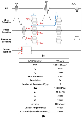 |
Multi-Physics Multi-Contrast Magnetic Resonance Imaging
Mehdi Sadighi1, Mert Şişman1, and B. Murat Eyüboğlu1
1Electrical and Electronics Eng., Middle East Technical University (METU), Ankara, Turkey
In this study, a Diffusion-Weighted (DW) Spin Echo (SE) based pulse sequence with current injection is proposed to combine data acquisitions of the Diffusion Tensor ($$$\overline{\overline{D}}$$$), current-induced magnetic flux density $$$B_z$$$ and the magnetohydrodynamic (MHD) flow imaging. The current density distribution ($$$\overline{J}$$$) can be estimated from the measured $$$B_z$$$ using the MRCDI method and the conductivity tensor can be reconstructed from the acquired $$$\overline{\overline{D}}$$$ and the estimated $$$\overline{J}$$$ using the DT-MREIT method. The acquired results of an experimental phantom with anisotropic diffusion using DW-SE pulse sequence shows that the proposed method could provide multi-contrast imaging based on multiple physical properties.
|
|||
1240.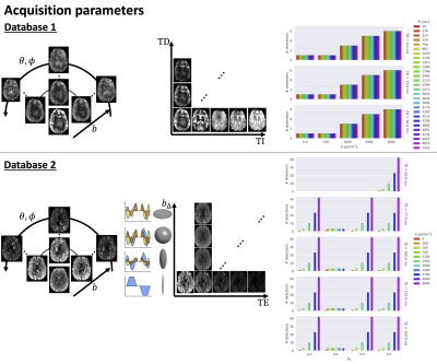 |
Optimising multi-contrast MRI experiment design using concrete autoencoders
Chantal Tax1,2, Hugo Larochelle3, Joao P. De Almeida Martins4, Jana Hutter5, Derek K. Jones2, Maxime Chamberland2, and Maxime Descoteaux6
1Image Sciences Institute, University Medical Center Utrecht, Utrecht, Netherlands, 2CUBRIC, Cardiff University, Cardiff, United Kingdom, 3Google Brain, Montreal, QC, Canada, 4Department of Radiology and Nuclear Medicine, St. Olav's University Hospital, Trondheim, Norway, 5Centre for Medical Engineering, King's College London, London, United Kingdom, 6SCIL, University of Sherbrooke, Sherbrooke, QC, Canada
Multi-contrast MRI provides a comprehensive picture of tissue microstructure, but the high dimensionality of the parameter space increases scan time. In this work, we present a data-driven approach to multi-contrast MRI experiment design using concrete autoencoders. Concrete autoencoders simultaneously perform measurement subset-selection and learn a prediction of the full set of measurements. This approach was evaluated on two multi-contrast databases encoding diffusion, relaxation, and susceptibility. The results showed similar patterns of measurement-subset selection and mean-squared errors across different training sets. The increasing availability of public multi-contrast MRI databases can further push data-driven approaches in providing recommendations for experiment design.
|
|||
1241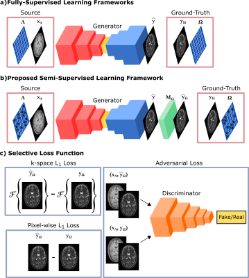 |
A Semi-Supervised Learning Framework for Jointly Accelerated Multi-Contrast MRI Synthesis without Fully-Sampled Ground-Truths Video Permission Withheld
Mahmut Yurt1,2, Salman Ul Hassan Dar1,2, Berk Tinaz1,2,3, Muzaffer Ozbey1,2, Yilmaz Korkmaz1,2, and Tolga Çukur1,2,4
1Department of Electrical and Electronics Engineering, Bilkent University, Ankara, Turkey, 2National Magnetic Resonance Research Center, Bilkent University, Ankara, Turkey, 3Department of Electrical and Computer Engineering, University of Southern California, Los Angeles, CA, United States, 4Neuroscience Program, Aysel Sabuncu Brain Research Center, Bilkent University, Ankara, Turkey
Current approaches for synthetic multi-contrast MRI involve deep networks trained to synthesize target-contrast images from source-contrast images in fully-supervised protocols. Yet, their performance is undesirably circumscribed to training sets of costly fully-sampled source-target images. For practically advanced multi-contrast MRI synthesis accelerated across the k-space and contrast sets, we propose a semi-supervised generative model that can be trained to synthesize fully-sampled images using only undersampled ground-truths by introducing a selective loss function expressed only on the acquired k-space coefficients randomized across training subjects. Demonstrations on multi-contrast brain images indicate that the proposed model maintains equivalent performance to the gold-standard fully-supervised model.
|
|||
1242.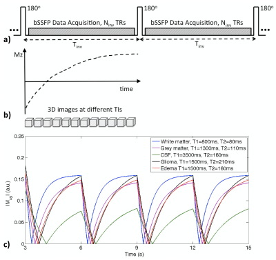 |
Multi-Contrast Whole Brain MRI: Optimization of Imaging Parameters and Motion Compensation
Jing Liu1, Angela Jakary1, Duan Xu1, and Janine Lupo1
1Radiology and Biomedical Imaging, University of California San Francisco, San Francisco, CA, United States
Multi-contrast anatomical brain MR images are widely used for tissue characterization and lesion detection, by applying multiple sequences. We developed a single continuous data acquisition applied with multiple inversion pulses, allowing multi-contrast imaging with high data acquisition efficiency. In order to achieve accurate tissue segmentation based on the multi-contrast images, imaging parameters are optimized based on simulated signal evolutions for achieving good image quality and accurate tissue characterization. A motion compensation approach for 3D whole-brain, multi-contrast MRI, was developed by exploiting the flexible data sampling strategy that allows for arbitrary combination of data collected throughout acquisition time and inversion times.
|
|||
1243.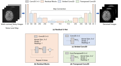 |
Adaptive Multi-contrast MR Image Denoising based on a Residual U-Net using Noise Level Map
Jiahao Hu1,2,3, Yilong Liu1,2, Zheyuan Yi1,2,3, Yujiao Zhao1,2, Fei Chen3, and Ed X. Wu1,2
1Laboratory of Biomedical Imaging and Signal Processing, The University of Hong Kong, Hong Kong, China, 2Department of Electrical and Electronic Engineering, The University of Hong Kong, Hong Kong, China, 3Department of Electrical and Electronic Engineering, Southern University of Science and Technology, Shenzhen, China
Multi-contrast MRI offers us images with complementary diagnostic information. Despite the dramatic difference in contrast, multi-contrast images often share highly correlated structure information. A deep learning (DL) based strategy is proposed to denoise multi-contrast MR images with flexible noise-levels using residual U-Net. This method utilizes the structural similarities across contrasts by simultaneously denoising multiple contrasts while existing single-contrast MRI denoising methods neglect the analogous structure information. The proposed method outperforms BM3D in terms of better noise reduction and details preservation. More importantly, we introduce a noise-level map that can be manually set to fit the different noise levels.
|
|||
1244.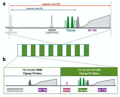 |
Simultaneous morphological and quantitative lumbar MRI with 3D isotropic high-resolution using MIXTURE T2
Daichi Murayama1, Takayuki sakai1, Masami Yoneyama2, and Shigehiro Ochi1
1Radiology, Eastern Chiba Medical Center, Chiba, Japan, 2Philips Japan, Tokyo, Japan
We propose a new sequence called MIXTURE (Multi-Interleaved X-prepared TSE with inTUitive RElaxometry). MIXTURE based is a 3D TSE that can set arbitrary echo times using the T2 preparation pulses, and enables several image contrasts (such as PDW, T2W) and T2 mapping by acquiring at least two echo time images. MIXTURE could simultaneously provide morphology, pathology and T2 quantitative lumbar imaging in one single scan. This sequence is very promising to evaluate the degeneration of nerve roots and intervertebral disc for 3D T2 mapping and improve the ability of diagnostic imaging around the lumbar for 3D multi contrast MRI.
|
|||
1245.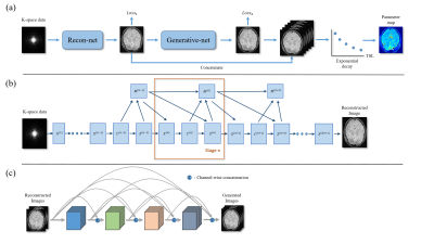 |
Accelerated MR parametric mapping with a hybrid deep learning model
Haoxiang Li1,2, Jing Chen1, Yuanyuan Liu1, Hairong Zheng1, Dong Liang1, and Yanjie Zhu1
1Shenzhen Institutes of Advanced Technology, Chinese Academy of Sciences, Shenzhen, China, 2Shenzhen College of Advanced Technology, University of Chinese Academy of Sciences, Shenzhen, China
Magnetic Resonance (MR) parametric mapping like $$$T_1$$$ , $$$T_2$$$, proton density is a powerful tool for biology tissue characterization, which is useful for clinical application such as diagnosis of pathologies including Alzheimer’s disease and multiple sclerosis1, evaluation of myocardial fibrosis2 and assessment of knee cartilage damage3. However the long scan time makes it challenging for practical clinical application. The purpose of this study was to develop a deep learning based method for accelerated MR parametric mapping with good performance at high acceleration rate both by reducing the contrast number and undersampling the k-space data.
|
|||
1246.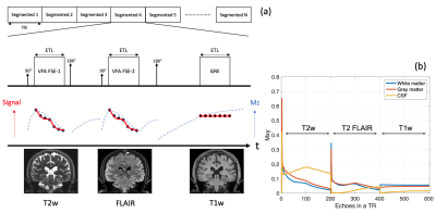 |
Simultaneous 3D T1 weighted, T2 weighted and FLAIR imaging using Serial Connection of Echo Train Acquisitions (SCETA) for Multi-contrast imaging
Naoyuki Takei1, Shohei Fujita2,3, Issei Fukunaga2, Mitsuharu Miyoshi1, Shigeki Aoki2, Suchandrima Banerjee4, and Tetsuya Wakayama1
1MR Applications and Workflow, GE Heatlcare, Tokyo, Japan, 2Juntendo University School of Medicine, Tokyo, Japan, 3The University of Tokyo Graduate School of Medicine, Tokyo, Japan, 4MR Applications and Workflow, GE Heatlcare, Menlo Park, CA, United States
To aim for accelerated brain routine MRI, a novel 3D multi-contrast imaging technique using the hybrid acquisition of FSE and GRE was proposed. The basic image contrasts commonly used in clinical practice such as T1 weighted, T2 weighted and FLAIR were acquired simultaneously. The comparison with conventional 3D imagings was performed in term of image contrast and scan time to demonstrate the proof of concept. The proposed technique potentially gives a new perspective of the use of 3D acquisition strategy.
|
|||
1247.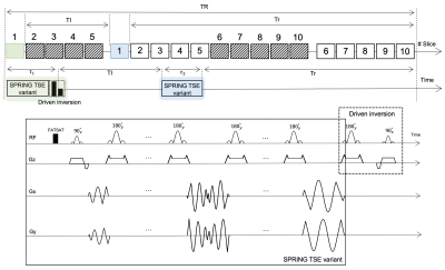 |
Rapid and Simultaneous Acquisition of T2-weighted and Fluid-attenuated Brain Images using a Spiral-ring Turbo Spin-echo Imaging
Zhixing Wang1, Xue Feng1, John Mugler2, and Craig Meyer1
1Biomedical Engineering, University of Virginia, Charlottesville, VA, United States, 2Radiology & Medical Imaging, University of Virginia, Charlottesville, VA, United States
This study describes a new approach to obtain T2-weighted and fluid-attenuated inversion recovery (FLAIR) images simultaneously in a short scan time. In this technique, the Cartesian readout is replaced by an annular spiral-ring segmentation, and a 180°(y)-90°(x) preparation RF pulse at the end of the echo train is used for driven inversion, rather than a single 180° inversion pulse as used for conventional FLAIR. Both the T2-weighted and FLAIR images can be acquired in a single scan, with higher scan efficiency and similar image quality, when compared with Cartesian TSE and Cartesian FLAIR.
|
|||
1248.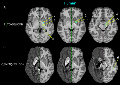 |
Merging T1 weighted images with QSM provides a unique contrast for brain tissue segmentation in humans and non-human primates
Rakshit Dadarwal1,2 and Susann Boretius1,2
1Functional Imaging Laboratory, German Primate Center, Göttingen, Germany, 2Georg August Universität Göttingen, Göttingen, Germany
Automatic brain tissue segmentations mostly rely on T1 weighted images (T1w) which provide an excellent grey matter – to -white matter contrast but T1w lacks sufficient contrast for major subcortical nuclei. Particularly in non-human primates, these segmentation approaches encounter their limits. By merging T1w with Quantitative Susceptibility Mapping, a unique brain tissue contrast could be generated. TQ-SILiCON maps allowed for an improved subcortical nuclei classification while retaining the excellent white matter delineation of T1w. The approach works equally well for humans and non-human primates.
|
|||
1249.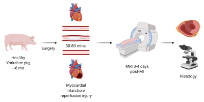 |
Comprehensive multiparametric cardiac MRI tissue phenotyping (LGE, T1, T2, DWI, BOLD & VAI) of acute myocardial infarction in swine
Holly Doig1, Maaike van den Boomen1,2,3, Erin Connors1, Joan Kim1, Jaume Coll-Font1,3, Robert A. Eder1, Shi Chen1, Yoshiko Iwamoto1, Kyrre E. Emblem4, Kawin Setsompop3, Niek H.J. Prakken2, Ronald J.H. Borra2,5, and Christopher T. Nguyen1,3
1Cardiovascular Research Center, Massachusetts General Hospital, Boston, MA, United States, 2Department of Radiology, University Medical Center Groningen, Groningen, Netherlands, 3A.A. Martinos Center for Biomedical Imaging, Massachusetts General Hospital, Boston, MA, United States, 4Department of Diagnostic Physics, Oslo University Hospital, Oslo, Norway, 5Department of Nuclear Medicine and Molecular Imaging, University Medical Center Groningen, Groningen, Netherlands
Myocardial infarction (MI) and ischemic reperfusion (IR) injury are a common western world problem and are aimed to be better understood by imaging techniques to enable non-invasive longitudinal follow up. Novel MRI based techniques, such as cardiac DWI, BOLD and VAI, are emerging and detailed assessment and comparison with current golden standard and histology is needed to determine their role in treatment planning and revalidation of MI patients. Here we show that specifically BOLD can be used as an early marker in the acute MI phase.
|
|||
1250.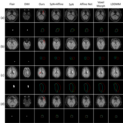 |
Stroke analysis with fully automatic multi-contrast MR image registration
Weijian Huang1, Yulon Qi2, Qiang He3, Ting Ma4, Xin Liu1, Guanxun Cheng2, Hairong Zheng1, and Shanshan Wang1
1Paul C Lauterbur Research Center, Shenzhen Institutes of Advanced Technology Chinese Academy of Sciences, Shenzhen, China, 2Radiology department, Peking University Shenzhen Hospital, shenzhen, China, 3United Imaging Research Institute of Innovative Medical Equipment, Shenzhen, China, 4Pengcheng Laboratory, Shenzhen, China
Stroke is a leading cause of death and disability worldwide. However, the misalignment between multi-contrast MR images bring difficulties in identifying the lesions. We propose an automatic framework including affine and deformation transformation for multi-contrast stroke images registration. In the framework, a new inverse operation is proposed to maintain the topology of images and a background suppression loss function is designed to optimize background predictions. The method achieves the best Dice score of 0.826 compared to 5 state-of-the-art methods. Moreover, our method is about 17 times faster than the most competitive method SyN when testing on a same CPU.
|
|||
1251.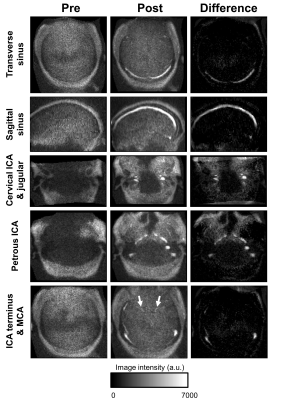 |
In-vivo ferumoxytol imaging and T1/T2 characterization at 64mT
Thomas Campbell Arnold1, Samantha By2, Hadrien Dyvorne2, Rafael O'Halloran2, Farzana Sayani3, Lisa M. Desiderio4, Brian Litt1,5, and Joel M. Stein4
1Bioengineering, University of Pennsylvania, Philadelphia, PA, United States, 2Hyperfine Research, Guilford, CT, United States, 3Medicine, Perelman School of Medicine, Philadelphia, PA, United States, 4Radiology, Perelman School of Medicine, Philadelphia, PA, United States, 5Neurology, Perelman School of Medicine, Philadelphia, PA, United States
MRI provides superior imaging for diverse clinical applications, but cost and other factors limit availability in various healthcare and lower resource settings. Low-field strength units promise to expand access but involve trade-offs including reduced signal, longer acquisitions, and uncertain benefit of contrast agents. Here we characterize T1 and T2 properties of ferumoxytol, an iron oxide agent with prolonged blood pool phase and higher R1 and R2 values than gadolinium, on a 64mT portable system. We demonstrate enhancement across a range of concentrations in phantoms and visualization of cerebral vasculature in patients receiving the agent for iron-deficiency anemia.
|
The International Society for Magnetic Resonance in Medicine is accredited by the Accreditation Council for Continuing Medical Education to provide continuing medical education for physicians.