Digital Posters
Novel Contrast Mechanisms
ISMRM & SMRT Annual Meeting • 15-20 May 2021

| Concurrent 4 | 15:00 - 16:00 |
1252. |
BOLD-free fMRI?
Tobias C Wood1 and Nikou Louise Damestani1
1Neuroimaging, King's College London, London, United Kingdom
We demonstrate a proof-of-concept fMRI experiment using a BOLD-insensitive MT-prepared ZTE pulse sequence, which exploits the recently proposed Arterial Blood Contrast mechanism. We show tightly localised responses to a visual checkerboard task in a small number of subjects.
|
|||
1253.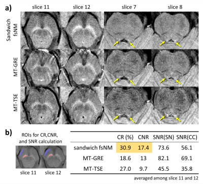 |
High neuromelanin contrast achieved using sandwiched flow saturation RF pulses: sandwich-fsNM imaging
Sooyeon Ji1, Eun-Jung Choi1, Eung Yeop Kim2, Dong Hoon Shin3, Hyeong-Geol Shin1, and Jongho Lee1
1Department of Electrical and Computer Engineering, Seoul National University, Seoul, Korea, Republic of, 2Department of Radiology, Gachon University Gil Medical Center, Incheon, Korea, Republic of, 3Department of Neurology, Gachon University Gil Medical Center, Incheon, Korea, Republic of
A neuromelanin-sensitive imaging protocol based on a GRE sequence, sandwich-fsNM, is developed using multiple flow saturation pulses instead of MT pulses. The flow saturation pulses are located both superior and inferior to the imaging volume, like a sandwich, to avoid asymmetric MT across slices. The proposed protocol yields higher contrast between crus cerebri and substantia nigra and better visualizes locus coeruleus, compared to MT prepared GRE or TSE sequences.
|
|||
1254.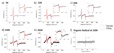 |
EPR measurements on human brain tissue at variable temperature
Fábio Seiji Otsuka1, Carlos Salmon1, Otaciro Nasimento2, and Maria Otaduy3
1Physics Department, University of São Paulo, Ribeirão Preto, Brazil, 2Physics Institute, University of São Paulo, São Carlos, Brazil, 3Medical School, University of São Paulo, São Paulo, Brazil
On this work it was evaluated the EPR measurements at variable temperature on tissue samples from different brain regions. For all samples, four different peaks were observed and their relationship with temperature were analyzed. High-spin rhombic iron(g = 4.3), copper(g = 2.06, g = 2.28) and organic radical (g = 1.989) peaks showed a Curie behavior with antiferromagnetic contributions. By other hand, a broad peak centered at g = 2.0 showed an antiferromagnetic behavior with Curie contribution. Additionally, a fifth peak was observed only for the Locus Coeruleus sample with a temperature dependent g-value (3.62 - 2.55).
|
|||
1255.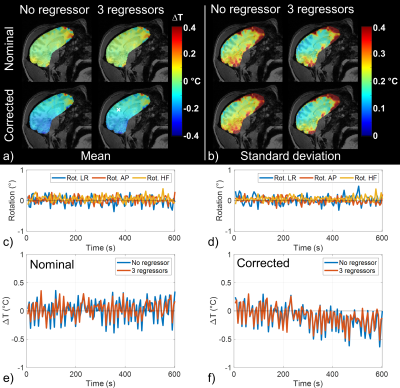 |
Radiofrequency heating measurement using MR thermometry and field monitoring: methodological considerations and first in vivo results.
Caroline Le Ster1, Franck Mauconduit1, Christian Mirkes2, Michel Bottlaender3,4, Fawzi Boumezbeur1, Boucif Djemai1, Alexandre Vignaud1, and Nicolas Boulant1
1Paris-Saclay University, CEA, CNRS, BAOBAB, Neurospin, Gif-sur-Yvette, France, 2Skope MRT, Zurich, Switzerland, 3Paris-Saclay University, CEA, CNRS, INSERM, BioMaps, Service hospitalier Joliot, Orsay, France, 4UNIACT, Neurospin, CEA, Gif-sur-Yvette, France
An MR thermometry (MRT) method with field monitoring is proposed to improve the measurement of small temperature variations occurring in head MRI exams. MRT experiments were performed at 7T with concurrent radiofrequency heating and field monitoring on an agar-gel phantom and on an anaesthetized macaque. Inclusion of field fluctuations in image reconstruction showed beneficial for the measurement of small temperature rises as encountered in standard brain exams.
|
|||
1256.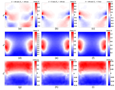 |
Experimental Evaluation of Spin Echo based Magnetic Resonance Magnetohydrodynamic Flow Velocimetry
Mert Şişman1, Mehdi Sadighi1, Hasan Hüseyin Eroğlu2,3, and B. Murat Eyüboğlu1
1Electrical and Electronics Engineering, Middle East Technical University (METU), Ankara, Turkey, 2Danish Research Centre for Magnetic Resonance, Centre for Functional and Diagnostic Imaging and Research, Copenhagen University Hospital, Amager and Hvidovre, Denmark, 3Center for Magnetic Resonance, DTU Health Tech, Technical University of Denmark, Kgs Lyngby, Denmark
Magnetohydrodynamic (MHD) flow occurs due to the Lorentz force formed by the interaction between the static magnetic field of the MR scanner and the externally injected electric current. The MR based imaging of MHD flow has become a research field of interest recently. The MHD flow can be extracted from the MR phase images obtained using a pulse sequence with flow-encoding gradients. In this study, the MHD flow distributions of a simulated model and a homogeneous experimental phantom are obtained. The MHD velocity contrast images reconstructed using simulated and experimental measurements are consistent.
|
|||
1257.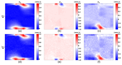 |
Simultaneous Magnetic Resonance Magnetohydrodynamic Flow Velocity and Diffusion Tensor Imaging
Mert Şişman1, Mehdi Sadighi1, and B. Murat Eyüboğlu1
1Electrical and Electronics Engineering, Middle East Technical University (METU), Ankara, Turkey
Recently, it is shown that the magnetohydrodynamic (MHD) flow has potential in clinical applications. In addition, diffusion tensor imaging (DTI) is widely used as an exclusive clinical diagnostic method. To obtain the MHD flow velocity distributions in three directions, twelve acquisitions with flow-encoding gradients and external current injection are needed. By choosing flow-encoding directions carefully, it is possible to obtain MHD flow velocity and DT distributions from the MR phase and magnitude images of the same acquisition. In this study, the MHD flow and DT data of an imaging phantom are acquired simultaneously using a spin echo based pulse sequence.
|
|||
1258.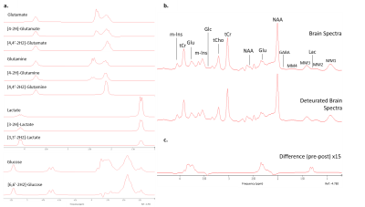 |
Theoretical evaluation of the feasibility to detect label-exchange by proton MRS at 7T in human brain after administration of deuterated glucose.
Simone Poli1, Lia Bally2, Roland Wiest3, and Roland Kreis1
1Department of Radiology and Biomedical Research, University of Bern, Bern, Switzerland, 2Department of Diabetes, Endocrinology, Nutritional Medicine and Metabolism, Inselspital, Bern University Hospital and University of Bern, Bern, CH, Bern, Switzerland, 3Institute of Diagnostic and Interventional Neuroradiology, Inselspital, Bern University Hospital and University of Bern, Bern, CH, Bern, Switzerland
This study aims to analyze the feasibility of indirect detection by 1H MRS of cerebral metabolism following intake of deuterated glucose in humans on a clinical 7T system. Brain spectra before and after [6,6’-2H2]-glucose administration were simulated with realistic SNR and linewidth to evaluate the best achievable precision in quantifying specific deuterated and non-deuterated metabolites. We expect labeling of glutamate, lactate and glutamine to be observable from single spectra of large VOIs, but also in MRSI at resolutions of 2-5 cm3. The simulations were corroborated with phantom data but eventual feasibility will remain to be confirmed in vivo.
|
|||
1259.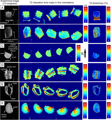 |
Relaxation Anisotropy in Biological Tissues
Nina Elina Hänninen1,2, Mikko Johannes Nissi1,2, Matti Hanni1,3,4, Olli Gröhn5, Miika Tapio Nieminen1,3,4, and Timo Liimatainen1,4
1Research Unit of Medical Imaging, Physics and Technology, University of Oulu, Oulu, Finland, 2Department of Applied Physics, University of Eastern Finland, Kuopio, Finland, 3Medical Research Center, University of Oulu and Oulu University Hospital, Oulu, Finland, 4Department of Diagnostic Radiology, Oulu University Hospital, Oulu, Finland, 5A.I. Virtanen Institute for Molecular Sciences, University of Eastern Finland, Kuopio, Finland
Relaxation times of tissues can depend on tissue orientation with respect to main magnetic field. However, these effects are still unknown in many biological tissues. Relaxation anisotropy provides a base to develop novel quantitative MRI contrasts and unwrap the theory behind different relaxation mechanisms. We investigated relaxation anisotropy of conventional and rotating frame relaxation parameters in brain, spinal cord, tendon, cartilage, kidney and cardiac muscle tissue. The findings show that relaxation anisotropy varies between relaxation parameters and between different tissue types.
|
|||
1260.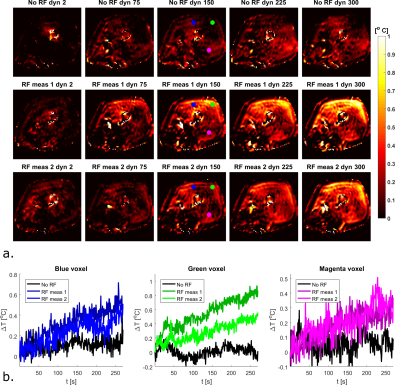 |
Towards high precision thermal based RF safety assessment with cardiac triggered MR thermometry
Bart R. Steensma1, Cornelis A.T. van den Berg1, and Alexander J.E. Raaijmakers2
1Center for Image Sciences - Computational Imaging Group, University Medical Center Utrecht, Utrecht, Netherlands, 2Biomedical Engineering - Medical Imaging Analysis, Eindhoven University of Technology, Eindhoven, Netherlands
To validate thermal simulations, high precision in vivo MR thermometry measurements are required. We demonstrate in vivo MR thermometry in the upper leg at 7T, where we improved the precision of our temperature measurements by using cardiac triggering with a PPU. The standard deviation in baseline measurements without RF heating decreased more than two-fold (0.1 ° C with PPU compared to 0.21 ° C without PPU). We were able to perform reproducible MR thermometry measurements in vivo, with local temperature increases of less than 1 °C.
|
|||
1261. |
Virtual non-contrast enhanced MRI (VNC-MRI)
Thomas Lindner1, Hanna Debus1, and Jens Fiehler1
1University Hospital Hamburg-Eppendorf, Hamburg, Germany
This study presents an approach to retrospectively remove contrast agent from the final images, denoted as "virtual non-contrast enhanced imaging"
|
|||
1262. |
Monte Carlo Simulation for Magnetic Nanoparticle Biosensors
Tristhal Parasram1, Rongsheng Lu2, Yi Chen2, and Dan Xiao1
1University of Windsor, Windsor, ON, Canada, 2Southeast University, Nanjing, China
Magnetic nanoparticles can bind to biomarkers. The particles dispersed in solution become clustered if the targets exist, leading to a different T2 relaxation time. The relaxation switch mechanism has been applied as a biosensor for various biomarkers. However, the change in relaxation rate is related to the nanoparticle size and concentration. Some theoretical models have been applied to predict this rate of change in search for the optimal system parameters. In this work, an open-source Monte Carlo algorithm has been developed to simulate water diffusion in microscopic environments with nanoparticles and the effective T2 relaxation times.
|
|||
1263.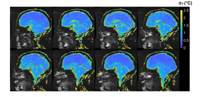 |
Feasibility of Magnetic Resonance Thermometry at 0.55T
Waqas Majeed1, Axel J. Krafft2, Sunil Patil1, Henrik Odéen3, John Roberts3, Florian Maier2, Dennis L. Parker3, and Himanshu Bhat1
1Siemens Medical Solutions USA Inc., Malvern, PA, United States, 2Siemens Healthcare GmbH, Erlangen, Germany, 3Department of Radiology and Imaging Sciences, University of Utah, Salt Lake City, UT, United States
Low field MRI offers advantages such as reduced cost and improved safety of implantable and interventional devices, and reduced RF energy deposition over high field alternatives. However, the accuracy of proton resonance frequency (PRF) thermometry suffers at low field due to reduced signal to noise ratio as well as lower temperature-sensitivity of MR phase. In this study we demonstrate that reduced off-resonance effects, increased T2* and shorter T1 associated with low field can be leveraged to achieve high quality MR thermometry in the brain and prostate at 0.55T.
|
|||
1264.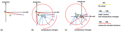 |
Improved PRF-based MR Thermometry for Tissues with Aqueous and Adipose
Chang-Sheng Mei1, Shenyan Zong2, and Guofeng Shen2
1Department of Physics, Soochow University, Taipei, Taiwan, 2Biomedical Instrument Institute, School of Biomedical Engineering, Shanghai Jiao Tong University, Shanghai, China
The proton resonance frequency (PRF) offset, which is linear to the temperature changes, has been developed and applied for thermal ablation therapies. However, the conventional PRF-based thermometry is limited in temperature measurements of fat due to the absence of hydrogen bonds. In this work, we proposed a revised and retrospective PRF method merged with the nonlinear circle fitting, to detect the accurate phase changes of water for achieving temperature measurements in aqueous and adipose tissues. The in vitro water-bathing heating for phantom and ex vivo focused ultrasound (FUS) heating for swine tissues were performed to verify the proposed method.
|
|||
1265.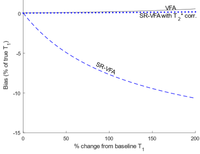 |
Effects of T2* on accuracy of single reference variable flip angle T1 – mapping for MR thermometry
Michael Malmberg1, Dennis L Parker2, and Henrik L Odéen2
1Biomedical Engineering, University of Utah, Salt Lake City, UT, United States, 2Radiology and Imaging Sciences, University of Utah, Salt Lake City, UT, United States
Neglecting the effects of T2* changes in the single reference variable flip angle (SR-VFA) method for T1-mapping produces a substantial systematic bias on T1 that increases with TE, T1 changes, and T2* changes. These effects were simulated using 1000 noisy signals of a single voxel using the SPGR equation. It was found that the bias can be corrected by measuring T2* changes dynamically, at the expense of noise, which noise could be mitigated through weighted T1-map combinations across echo times. Multi-echo T1-mapping with the SR-VFA method could be combined with PRFS thermometry to allow fast, accurate T1-mapping of heterogeneous tissue.
|
|||
1266.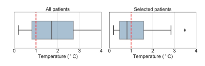 |
Accuracy of MR thermometry during deep radiofrequency hyperthermia treatments in the pelvic region
Iva VilasBoas-Ribeiro1, Sergio Curto1, Gerard C. van Rhoon1,2, and Margarethus M. Paulides1,3
1Department of Radiotherapy, Erasmus MC Cancer Institute, University Medical Center Rotterdam, Rotterdam, Netherlands, 2Department of Radiation Science and Technology, Faculty of Applied Sciences, Delft University of Technology, Delft, Netherlands, 3Center for Care and Cure Technologies Eindhoven (C3Te), Department of Electrical Engineering, Eindhoven University of Technology, Eindhoven, Netherlands
Proton resonant frequency shift is the most frequently used method for MR-thermometry. This method is sensitive to patient motion which poses issues that affect the MR-thermometry accuracy. Few studies have evaluated MR thermometry in the pelvis, but only in volumes far from the regions with air motion, and without an objective patient data exclusion criteria. In this study, we assessed accuracy of MR thermometry for a selected group of patients with cervical carcinoma. We showed that changing gastrointestinal air volume was an important confounder for MR thermometry accuracy and that this can be exploited for selection criteria prior to treatment.
|
|||
1267.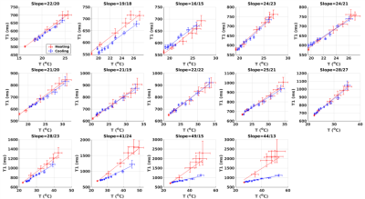 |
Towards correlating tissue status with dynamic PRF-T1 measurements using a single reference dual flip angle technique
Henrik Odéen1, Sara L Johnson1, Allison H Payne1, and Dennis L Parker1
1Radiology and Imaging Sciences, University of Utah, Salt Lake City, UT, United States
Simultaneous PRF/T1 measurements using a single reference variable flip angle method is used to investigate the utility of T1 as a complimentary measure to thermometry for treatment outcome in MR guided thermal interventions, such as focused ultrasound. Both temperature and thermal dose have been shown to be inaccurate indicators of treatment outcome, creating a potential need for alternative quantitative metrics. This can be of particular value in adipose tissues where PRF has very low sensitivity. It is shown that the slope of the T1 vs. temperature curve correlates with amount of delivered thermal dose.
|
|||
1268.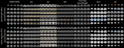 |
Analysis of neurodegeneration using diffusion and functional MRI in FTLD model marmoset
Mitsuki Rikitake1, Junichi Hata2, Fumiko Seki3, Shinsuke Ishigaki4, Kuniyuki Iwata-Endo4, Nobuyuki Iwade4, Takako Shirakawa1, Hirotaka James Okano2, Hideyuki Okano5, and Gen Sobue4
1Department of Redioligical Science, Human Health Science, Tokyo Metroplitan University, Tokyo, Japan, 2Jikei University Graduate School of Medicine, Tokyo, Japan, 3Central Institute for Experimental Animals, Kanagawa, Japan, 4Department of Neurology, Nagoya University Graduate School of Medicine, Nagoya, Japan, 5RIKEN Center of Brain Science, Saitama, Japan
Symptom of FTLD is known brain atrophy and neurodegeneration predominantly in frontal and temporal lobes.However, the regularity of neurodegeneration and image analysis methods have not been established.The purpose of this study was to conduct multiparametric MRI to analyze the neurodegeneration caused by FTLD using structural MRI and fMRI.We used marmosets, which have a relatively similar brain structure to humans, as the experimental subjects.Statistical imaging analysis using diffusion MRI showed gray matter degeneration was observed, mainly in the caudate nucleus.The fMRI showed decreased brain activity in the frontal lobe and basal ganglia.
|
|||
1269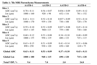 |
The Use of the 3He/129Xe MRI Lung Morphometry for a Longitudinal Observation of the Emphysema Progression in AATD Patients Video Permission Withheld
Elise Noelle Woodward1, Matthew S Fox1,2, Tingting Wu3, Hacene Serrai1, David G McCormack4, Grace Parraga3,4,5,6, and Alexei Ouriadov1,2,6
1Physics and Astronomy, Western University, London, ON, Canada, 2Lawson Health Research Centre, London, ON, Canada, 3Department of Medical Biophysics, Western University, London, ON, Canada, 4Department of Medicine, Respirology, Western University, London, ON, Canada, 5Robarts Research Institute, London, ON, Canada, 6School of Biomedical Engineering, Western University, London, ON, Canada
In this study, we demonstrated a possible solution to underestimation of the global mean that can mask the severity of emphysema progression. Four patients with Alpha-1 Anti-Trypsin Deficiency disorder (AATD) were measured in 2014 using hyperpolarized He to measure lung function, and again in 2018 to measure lung function. While it is evident looking at morphometry images that there is a significant reduction in pixels and therefore emphysema progression, it is not at first evident in morphometry estimates. By normalizing these morphometry estimates of ADC/Lm, we can better characterize emphysema progression in individuals with AATD
|
|||
1270.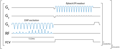 |
Metabolite-specific echo planar imaging of hyperpolarized 13C-pyruvate at 4.7T
Tyler Blazey1, Galen D Reed2, Joel R Garbow1, and Cornelius von Morze1
1Mallinckrodt Institute of Radiology, Washington University in St. Louis, St. Louis, MO, United States, 2GE Healthcare, Dallas, TX, United States
Hyperpolarization greatly increases the sensitivity of MRI studies of 13C-labeled substances. To expand our ability to investigate in vivo metabolism with hyperpolarized MRI, we developed a metabolite-specific 2D EPI pulse sequence with custom flyback spatially-selective RF pulses for [1-13C]pyruvate and [1-13C]lactate at 4.7T. Spectral profiles for each metabolite obtained using a [13C]urea phantom showed good correspondence with simulated profiles. In vivo testing was performed by imaging the liver and kidney of a rat following the injection of hyperpolarized [1-13C]pyruvate. Both the temporal and spatial resolution of [1-13C]pyruvate and [1-13C]lactate images was higher with our metabolite-specific EPI sequence than with CSI.
|
|||
1271. |
Feasibility of model-based omega-3 fatty acid fraction mapping using multi-echo gradient-echo imaging at 3T
Dominik Weidlich1, Julius Honecker2, Claudine Seeliger2, Daniela Junker1, Marcus R. Makowski1, Hans Hauner2, Dimitrios C. Karampinos1, and Stefan Ruschke1
1Department of Diagnostic and Interventional Radiology, School of Medicine, Technical University of Munich, Munich, Germany, 2Else Kröner Fresenius Center for Nutritional Medicine, Technical University of Munich, Freising, Germany
A non-invasive method allowing the probing of the relative ω-3 fatty acid content in adipose tissue has the potential to reveal unknown metabolic patterns in physiology and pathology. In the current study, a model-based triglyceride mapping scheme is extended to enable the differentiation between ω-3 and non-ω-3 fatty acids by exploiting additional knowledge about the chemical shift properties. This extended scheme is combined with a chemical shift encoding imaging sequence and evaluated in a phantom validation experiment against gas chromatography–mass spectrometry. Furthermore, feasibility is demonstrated in both in vitro and in vivo settings.
|
The International Society for Magnetic Resonance in Medicine is accredited by the Accreditation Council for Continuing Medical Education to provide continuing medical education for physicians.