Digital Posters
T1ρ, MT & CEST
ISMRM & SMRT Annual Meeting • 15-20 May 2021

| Concurrent 4 | 17:00 - 18:00 |
1471.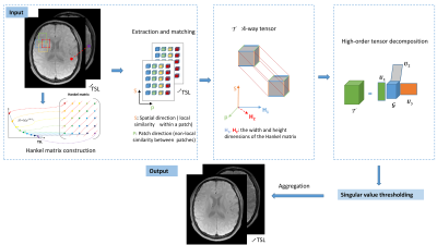 |
Accelerating T1ρ mapping using patch-based low-rank tensor
Yuanyuan Liu1, Zhuo-Xu Cui1, Xin Liu1, Dong Liang1, and Yanjie Zhu1
1Shenzhen Institutes of Advanced Technology, Chinese Academy of Sciences, Shenzhen, China
T1ρ mapping requires the acquisition of multiple T1ρ-weighted images with different spin lock times to obtain the T1ρ maps, resulting in a long scan time. Compressed sensing has shown good performance in fast quantitative T1ρ mapping. In this study, we used a patch-based low-rank tensor imaging method to reconstruct the T1ρ-weighted images from highly undersampled data. Preliminary results show that the proposed method achieves a 6-fold acceleration and obtains more accurate T1ρ maps than the existing methods.
|
|||
1472.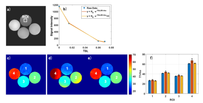 |
Robust Quantitative T1rho imaging
Huimin Zhang1, Baiyan Jiang1, Jian Hou1, Queenie Chan2, and Weitian Chen1
1Imaging and Interventional Radiology, The Chinese University of Hong Kong, Sha Tin, Hong Kong, 2Philips Healthcare, Hong Kong, Hong Kong
T1rho is a valuable biomarker to probe macromolecular components in tissue. However, conventional T1rho quantification techniques are susceptible to B1 RF and B0 field inhomogeneities, which cause failure of spin-lock and signal oscillation. Adiabatic pulses have been used before and after spin-lock for simultaneous B1 RF and B0 field inhomogeneities compensation and have achieved robust spin-lock, but it does not remove the effect of field inhomogeneities to the actual T1rho value. Therefore, robust T1rho quantification still remains a problem. In this work, we utilized AC-iTIP and combined it with a proposed correction method to address this problem.
|
|||
1473.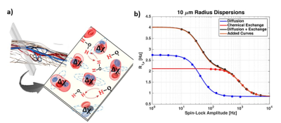 |
R1ρ Dispersion imaging in human skeletal muscle at 3 Tesla
Fatemeh Adelnia1, Zhongliang Zu1,2, Feng Wang1,2, Kevin D Harkins3, and John C Gore1,2,3,4,5
1Vanderbilt University Institute of Imaging Science, Nashville, TN, United States, 2Department of Radiology and Radiological Sciences, Vanderbilt University Medical Center, Nashville, TN, United States, 3Department of Biomedical Engineering, Vanderbilt University, Nashville, TN, United States, 4Department of Physics and Astronomy, Vanderbilt University, Nashville, TN, United States, 5Department of Molecular Physiology and Biophysics, Vanderbilt University, Nashville, TN, United States
R1ρ dispersion over a range of weak locking fields has the potential to reveal information on microvascular geometry and density such as microvascular spacing. This work presents in-vivo results supporting the application of R1ρ dispersion at low locking fields to the measurement of microvascular sizes and spacings in skeletal muscle. We present model fit parameters from measurements of R1ρ dispersion of human skeletal muscle that is close to ex-vivo data.
|
|||
1474.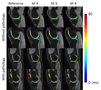 |
Highly accelerated T1ρ imaging using kernel-based low-rank compressed sensing reconstruction in knees with and without osteoarthritis
Jeehun Kim1,2, Chaoyi Zhang3, Mingrui Yang1, Hongyu Li3, Mei Li1, Richard Lartey1, Leslie Ying3,4, and Xiaojuan Li1
1Department of Biomedical Engineering, Program of Advanced Musculoskeletal Imaging (PAMI), Cleveland Clinic, Cleveland, OH, United States, 2Department of Electrical Engineering, Case Western Reserve University, Cleveland, OH, United States, 3Electrical Engineering, University at Buffalo, State University of New York, Buffalo, NY, United States, 4Biomedical Engineering, University at Buffalo, State University of New York, Buffalo, NY, United States
The T1ρ imaging is a promising biomarker for early diagnosis of osteoarthritis, but the application of the method is hindered by its long scan time. In this work, a novel compressed sensing algorithm based on kernel-based low-rank was proposed. The algorithm was evaluated with numerical simulation and volunteer scans, where the volunteers with and without osteoarthritis was scanned with prospective downsampling to evaluate the algorithm performance regarding the presence of pathology.
|
|||
1475.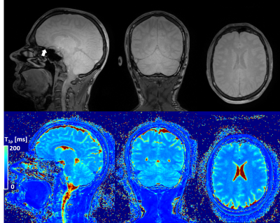 |
Reproducible high-resolution T1ρ maps of the brain in under seven minutes using compressed sensing
Gabriele Bonanno1,2,3, Tom Hilbert4,5,6, Patrick Leibig7, and Tobias Kober4,5,6
1Advanced Clinical Imaging Technology,Siemens Healthcare AG, Bern, Switzerland, 2Translational Imaging Center, sitem-insel AG, Bern, Switzerland, 3Departments of Radiology and Biomedical Research, University of Bern, Bern, Switzerland, 4Advanced Clinical Imaging Technology, Siemens Healthcare AG, Lausanne, Switzerland, 5Department of Radiology, University Hospital (CHUV) and University of Lausanne (UNIL), Lausanne, Switzerland, 6LTS5, École Polytechnique Fédérale de Lausanne, Lausanne, Switzerland, 7Siemens Healthcare GmbH, Erlangen, Germany
Relaxometry in the rotating frame can give unique insights into low frequency biological processes, and therefore may be a valuable quantitative MRI method for clinical research of neurodegenerative diseases. However, T1ρ maps require long scan times for whole-brain coverage with high isotropic resolution. We present a T1ρ mapping method based on an accelerated spiral-phyllotaxis 3D Cartesian gradient echo scan with compressed sensing reconstruction that allows for <7-min acquisition time and provides maps directly at the scanner to facilitate a fast workflow for clinical applications. Our method also demonstrated excellent repeatability and reproducibility.
|
|||
1476.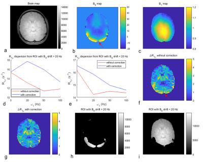 |
Correction of errors in estimates of T1ρ at low spin-lock amplitudes in the presence of B0 and B1 inhomogeneities
Zhongliang Zu1, Fatemeh Adelnia1, Kevin Harkins1, Feng Wang1, and John Gore1
1Vanderbilt University Medical Center, Nashville, TN, United States
Spin-lock imaging at low locking amplitudes are sensitive to the effects of water diffusion in intrinsic gradients and may provide information on tissue microvasculature. However, although composite pulse preparations have been used to reduce artifacts due to B0 and B1 inhomogeneities, there are still residual errors. Here, we analyze the source of these errors and developed an approximate theoretical analysis to correct these errors. Simulations and experiments on a healthy human subject show that these errors are mainly due to B0 shifts, and can be reduced by this correction approach when the B0 shift is relatively small compared with w1.
|
|||
1477.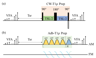 |
High Resolution Adiabatic T1ρ Mapping Using 3D MAPSS Sequence at 3T
Can Wu1,2 and Qi Peng3
1Department of Medical Physics, Memorial Sloan Kettering Cancer Center, New York, NY, United States, 2Philips Healthcare, Andover, MA, United States, 3Department of Radiology, Albert Einstein College of Medicine and Montefiore Medical Center, Bronx, NY, United States
The 3D MAPSS T1ρ sequence provides fast and accurate T1ρ quantification with high spatial fidelity using RF phase cycling and variable flip angle in a MP-GRE sequence. However, the conventional 3D MAPSS sequence uses continuous-wave RF pulses for spin locking and is thus sensitive to B0 and B1 field inhomogeneities. In this study, a 3D MAPSS T1ρ sequence with adiabatic RF pulses was implemented and was shown to be less sensitive to B0 frequency offset on phantom compared to conventional 3D MAPSS T1ρ mapping. It was also successfully applied to high-resolution 3D T1ρ mapping of knee cartilage and brain tissue.
|
|||
1478.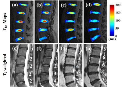 |
Comprehensive T1ρ Measurement of in vivo Lumbar Intervertebral Discs using a 3D Adiabatic T1ρ Prepared UTE (UTE-Adiab-T1ρ) Sequence
Zhao Wei1,2,3, Alecio F. Lombardi1, Zubiad Ibrahim1, Mohammadamin Cheraghi1, Koihi Masuda4, Jiang Du1, Eric Y. Chang1,5, Graeme M. Bydder1, Wenhui Yang2,3, and Ya-Jun Ma1
1Department of Radiology, UC San Diego, San Diego, CA, United States, 2Institute of Electrical Engineering, Chinese Academy of Sciences, Beijing, China, 3University of Chinese Academy of Sciences, Beijing, China, 4Department of Orthopedic Surgery, UC San Diego, San Diego, CA, United States, 5Radiology Service, Veterans Affairs, San Diego Healthcare System, San Diego, CA, United States
To assess the feasibility of using a 3D ultrashort echo time sequence with adiabatic T1ρ preparation (UTE-Adiab-T1ρ) to map the T1ρ of entire lumbar intervertebral discs (IVDs), 17 human subjects’ lumbar spines were scanned. Correlations between T1ρ values of all of the components of the IVD and disc degeneration grades and subjects' ages were calculated. T1ρ differences between subjects with and without low back pain were also assessed. The study showed that the UTE-Adiab-T1ρ sequence can measure T1ρ values of the whole IVD. These may be useful for assessment of IVD degeneration.
|
|||
1479.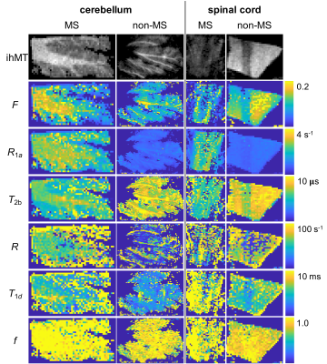 |
Quantitative inhomogeneous MT with Cramer Rao lower bound optimized protocol to distinguish tissue from donors with/without Multiple Sclerosis
Gopal Varma1, Aaron K Grant1, Olivier M Girard2, Guillaume Duhamel2, and David C Alsop1
1Radiology, Division of MR Research, Beth Israel Deaconess Medical Center, Harvard Medical School, Boston, MA, United States, 2CNRS, CRMBM, Aix-Marseille Univ, Marseille, France
Inhomogeneous magnetization transfer (ihMT) is predominantly associated with myelin imaging and shows promise in detection and monitoring myelin related disorders, including multiple sclerosis (MS). Extraction of quantitative parameters from ihMT data (qihMT) would ideally provide information independent of the ihMT sequence. We used Cramer Rao lower bound analysis to develop an optimized acquisition protocol for qihMT and simulations to test the fidelity of the protocol. Preliminary application to central nervous system samples showed differences in free pool longitudinal relaxation rate R1a, bound pool transverse relaxation time T2b, and dipolar relaxation time T1d between specimens from donors with and without MS.
|
|||
1480.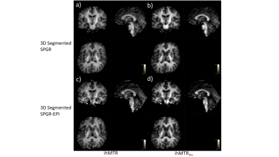 |
Fast 3D steady state inhomogeneous magnetization transfer imaging with Segmented Spoiled Gradient Echo - Echo Planar Imaging.
Masanori Ozaki1 and Masao Yui1
1Research and Development Center, Canon Medical Systems Corporation, Kawasaki, Japan
Inhomogeneous magnetization transfer is a promising technique to provide high sensitivity and specificity of detecting myelinated content in tissue. 3D steady-state ihMT imaging with 3D segmented Spoiled Gradient Echo (SPGR) sequence can provide myelin information of the whole brain within 5-10min of acquisition time. In this work, we demonstrate 3D steady-state ihMT imaging with a 3D segmented Spoiled Gradient Echo - Echo Planar Imaging (SPGR-EPI) sequence instead of 3D segmented SPGR to reduce acquisition time. We successfully reduced approximately 30% of the acquisition time for 3D steady-state ihMT imaging by applying 3D Segmented SPGR-EPI compared to 3D segmented SPGR.
|
|||
1481.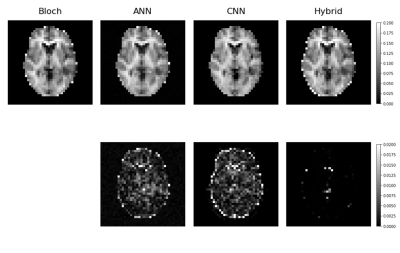 |
Accelerated Fitting for Quantitative Magnetization Transfer in Glioblastoma Multiforme Patients with Uncertainty using Deep Learning
Matt Hemsley1,2, Rachel W Chan2, Liam Lawrence1,2, Sten Myrehaug3,4, Arjun Sahgal2,3,4, and Angus Z Lau1,2
1Medical Biophysics, University of Toronto, Toronto, ON, Canada, 2Department of Physical Sciences, Sunnybrook Research Institute, Toronto, ON, Canada, 3Department of Radiation Oncology, Sunnybrook Health Sciences Centre, Toronto, ON, Canada, 4Department of Radiation Oncology, University of Toronto, Toronto, ON, Canada
qMT has been suggested as a biomarker in Glioblastoma patients. However, reconstruction involves a computationally expensive fitting procedure involving the Bloch-McConnell equations. In this work, the use of neural networks was investigated to perform the fit and to compute uncertainty heatmaps to identify regions of potential error. The dataset consisted of 164 scans from N=41 glioblastoma patients (33=training, 8=testing). Models were evaluated using MAE and correlation in the whole-head volumes and specific ROIs. The model output agreed with a conventional curve-fitting algorithm (r=0.93, and <1% error) with speed up factors of 240000x. Uncertainty predictions were correlated with prediction error (r=0.59).
|
|||
1482.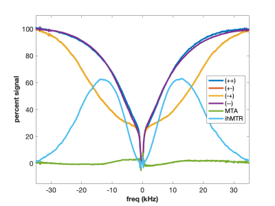 |
Super-Lorentzians and MT Asymmetries and Dipolar Order – Oh My!
Scott D. Swanson1
1Department of Radiology, University of Michigan, Ann Arbor, MI, United States
MT studies of model systems disentangle effects of MT asymmetry and dipolar oder. Choice of lipid materials controls amplitude of ihMT. These studies help outline the physical mechanisms of MT and provide guidance for design of MT, MTA, and ihMT phantoms.
|
|||
1483.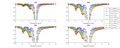 |
Toward more specific imaging of fibrosis: The z-spectrum of collagen
Nabeelah Jinnah1, Olivier Mougin1, Penny Gowland1, Caroline Hoad1, Gordon Moran1, Andrew Carradus1, and Hannah Williams1
1University of Nottingham, Nottingham, United Kingdom
We have characterized the z-spectrum of collagen at 3T and 7T. We found two peaks +1.9 ppm and at +3.5 ppm and fitting to the Bloch McConnell equations yielded pool sizes that increased monotonically with concentration.
|
|||
1484.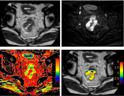 |
Differential diagnosis rectal cancer with and without lymph node metastasis using amide proton transfer-weighted imaging and T1 map
Anliang Chen1, Ailian Liu1, Jiazheng Wang2, Zhiwei Shen2, Deshuo Dong1, Wan Dong1, Yuhui Liu1, Qingwei Song1, and Renwang Pu1
1Radiology, The First Affiliated Hospital of Dalian Medical University, Dalian, China, 2Philips Healthcare, Beijing, China
Amide proton transfer-weighted (APTw) imaging can be used to assess changes of intracellular protein concentration and the pH value. Non-invasive visualization and quantification of tissue composition could be detected by T1 map. In this study, we aim to explore the value in difference of rectal cancer with and without lymph node metastasis using APTw imaging combined with T1 map. High diagnostic efficacy (AUC: 0.891; sensitivity: 81.8%; specificity: 100.0%) was achieved with combination of APT and T1 values.
|
|||
1485.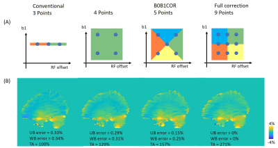 |
B0 and B1 correction anti-respectively for chemical exchange saturation transfer imaging
Ying-Hua Chu1, Yi-Cheng Hsu1, and Patrick Alexander Liebig2
1MR Collaboration, Siemens Healthcare Ltd., Shanghai, China, 2Siemens Healthcare GmbH, Erlangen, Germany
We propose a new simultaneous B0 and B1 correction for Chemical Exchange Saturation Transfer imaging. This method undersamples the number of offsets on the B1 and B0 plane. The CEST map's mean error almost halved at the upper brain region compared with the conventional method and the minimal sampled B0/B1 correction method. The proposed method provides an efficient B0 and B1 correction and may improve the diagnostic accuracy using CEST imaging at 3T.
|
|||
1486.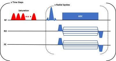 |
Golden-Angle Radial CEST MR Fingerprinting with Temporal Compressed Sensing Reconstruction
Ouri Cohen1 and Ricardo Otazo1
1Memorial Sloan Kettering Cancer Center, New York, NY, United States
Chemical Exchange Saturation Transfer (CEST) is a novel imaging technique that is sensitive to metabolite concentrations at imaging resolutions in clinically relevant scan times. These features have generated much interest and applications of CEST in assessing disease pathologies, progression and therapeutic response are currently being explored. A quantitative framework for CEST based on MR Fingerprinting (MRF) was recently proposed using an EPI readout. Here we describe preliminary work to leverage the inherent B0 and motion robustness of radial imaging to develop a clinical golden-angle radial CEST-MRF that doesn't suffer from the classic EPI drawbacks.
|
|||
1487.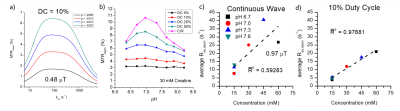 |
Improving Fidelity of Concentration Dependence in CEST- MRI using pH-insensitive Low Duty Cycle Saturation Pulse Trains
Julius Chung1 and Tao Jin1
1University of Pittsburgh, Pittsburgh, PA, United States
Low duty cycle π-pulsed CEST is an easy to implement method for exchange rate insensitive CE-MRI measurement that is dependent on labile proton concentration. In creatine phantoms with varying pH, decreasing duty cycle of π-pulse trains decreased pH dependence. In creatine phantoms of varying concentration and varied pH, exchange dependent relaxation asymmetry of π-pulsed CEST shows stronger linear dependence on concentration at lower duty cycles than continuous wave. Low duty cycle π-pulsed CEST may be useful in studies in which a change of chemical exchange rate (e.g., pH, catalyst concentration, and temperature changes) can interfere with accurate assessment of pathology.
|
|||
1488.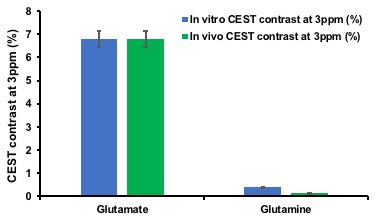 |
Glutamine contribution to GluCEST at 7.0T
Ravi Prakash Reddy Nanga1, Mohammad Haris2, Hari Hariharan1, and Ravinder Reddy1
1Radiology, University of Pennsylvania, Philadelphia, PA, United States, 2Research Branch, Sidra Medical and Research Center, Doha, Qatar
Glutamate chemical exchange saturation transfer (GluCEST) is an emerging imaging technique and in the recent years has shown promising applications in Alzheimer’s, Parkinson’s, Epilepsy, Brain tumors and Schizophrenia. Due to structural similarity of glutamine with glutamate, often there is a misconception of glutamine contribution to CEST effect of glutamate. In this study, we investigated the glutamine contribution to glutamate CEST at 3ppm offset.
|
|||
1489.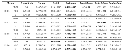 |
A comparison of recent motion-correction methods for CEST-MRI
Botao Zhao1, Ying Liu1, and Xiao-Yong Zhang1
1Institute of Science and Technology for Brain-Inspired Intelligence, Fudan University, Shanghai, China
The motion in chemical exchange saturation transfer (CEST) image was overlooked in many published studies. Fortunately, several works have been reported to do motion correction for CEST processing. However, the performance of these methods has not been evaluated. The main purpose of this study is to compare the performance of these methods on motion correction for CEST imaging. We used NMSE, MCC, and UI to evaluate the performance of these methods. Our simulation and preliminary in-vivo data showed that the method of Reg2mean works the best.
|
|||
1490.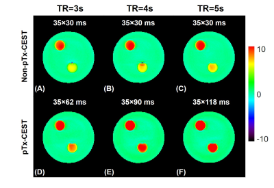 |
Sensitivity-enhanced and Shading-reduced Chemical Exchange Saturation Transfer Imaging of the Abdomen using Parallel Transmission
Ruibin Liu1, Zihua Qian2, Zhe Wu3, Yi-Cheng Hsu4, Caixia Fu5, Yi Sun4, Dan Wu1, and Yi Zhang1
1Key Laboratory for Biomedical Engineering of Ministry of Education, Department of Biomedical Engineering, College of Biomedical Engineering & Instrument Science, Zhejiang University, Hangzhou, China, 2Department of Radiology, The Children’s Hospital, Zhejiang University School of Medicine, National Clinical Research Center for Child Health, Hangzhou, China, 3Techna Institute, University Health Network, Toronto, ON, Canada, 4MR Collaboration, Siemens Healthcare Ltd., Shanghai, China, 5MR Application Development, Siemens Shenzhen Magnetic Resonance Ltd., Shenzhen, China
CEST imaging benefits from longer saturation duration and a higher saturation duty cycle. Dielectric shading effects occur when the RF wavelength approaches the object size. Here, we proposed a parallel-transmission-based CEST (pTx-CEST) sequence to extend the maximum saturation duration at 100% duty-cycle and mitigate shading effects. The maximum saturation duration in pTx-CEST was lengthened to 2170, 3150, and 4130ms compared to 1050ms in non-pTx-CEST at TR of 3s, 4s, and 5s, respectively, leading to a significant sensitivity enhancement. Besides, the optimal amplitude ratio and phase difference between RF channels, manifesting circular or elliptical polarization, help reduce the dielectric shading effects.
|
The International Society for Magnetic Resonance in Medicine is accredited by the Accreditation Council for Continuing Medical Education to provide continuing medical education for physicians.