Digital Posters
Contrast Mechanisms: Miscellaneous
ISMRM & SMRT Annual Meeting • 15-20 May 2021

Digital Posters
Contrast Mechanisms: Miscellaneous
| Concurrent 3 | 13:00 - 14:00 |
1824.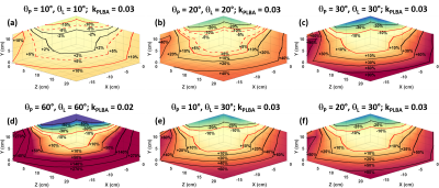 |
The Effect of Transmit B1 Inhomogeneity on Hyperpolarized [1-13C]-Pyruvate Metabolic MR Imaging Biomarkers
Collin J. Harlan1, Zhan Xu1, Christopher M. Walker1, Keith A. Michel1, Galen D. Reed 2, and James A. Bankson1,3
1Department of Imaging Physics, The University of Texas M.D. Anderson Cancer Center, Houston, TX, United States, 2GE Healthcare, Dallas, TX, United States, 3The University of Texas M.D. Anderson Cancer Center UT Health Graduate School of Biomedical Sciences, Houston, TX, United States
Accurate signal excitation is imperative when conducting hyperpolarized 13C MRI studies. It has been determined from previous work that greater than ±10% deviation in excitation angle can lead to significant excitation angle dependent errors in kinetic analysis of HP [1-13C] pyruvate to lactate conversion. This work was conducted to characterize the B1+ field homogeneity of a 13C volume transmit clamshell coil. Furthermore, an assessment of the impact of potential B1+ inhomogeneities on semi-quantitative and quantitative hyperpolarized metabolic MR imaging biomarkers was conducted.
|
|||
1825.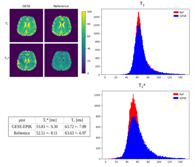 |
Simultaneous quantification of T2 and T2* by accelerated 10-echo GESE-EPIK sequence for carrageenan-phantoms and in vivo data
Fabian Küppers1,2,3, Seong Dae Yun1, and N. Jon Shah1,2,4,5
1Institute of Medicine and Neuroscience 4, Forschungszentrum Juelich GmbH, Jülich, Germany, 2Institute of Medicine and Neuroscience 11, Forschungszentrum Juelich GmbH, Jülich, Germany, 3RWTH Aachen University, Aachen, Germany, 4Department of Neurology, RWTH Aachen University, Aachen, Germany, 5JARA - BRAIN - Translational Medicine, Aachen, Germany
Combining GE and SE signals enables quantification of several pertinent parameters making it useful in various applications. An 10-echo GESE sequence based on EPIK, known as EPI with keyhole, has been presented in our earlier work. This work employs an accelerated sequence implementation and aims to perform quantification of T2/T2* for both phantom and in vivo data. The method was configured to provide improved resolution within feasible TEs for brain scans, i.e. 1 minute. The quantified T2/T2* values were in good agreement with values from reference methods. Moreover, an SNR/tSNR analysis proves the signal and time gain from multi-shot acceleration.
|
|||
1826.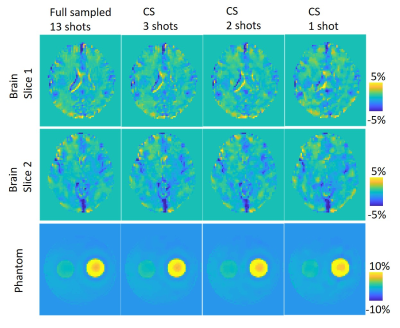 |
Highly accelerated compressed sensing chemical exchange saturation transfer
Ying-Hua Chu1, Patrick Alexander Liebig2, He Wang3, and Yi-Cheng Hsu1
1MR Collaboration, Siemens Healthcare Ltd., Shanghai, China, 2Siemens Healthcare GmbH, Erlangen, Germany, 3Institute of Science and Technology for Brain-Inspired Intelligence, Fudan University, Shanghai, China
We proposed a CEST imaging method using compressed sensing to accelerate image acquisition. Compared with the full sampled one with two averages, the compressed sensing method used only 7.6% scan time with around 0.5% error in amide proton transfer weighted (APTw) images. The whole-brain acquisition for one RF offset took only 6 seconds. This method can accelerate conventional APTw images to reduce motion artifact and make a high signal-to-noise ratio acquisition with large number offsets for Lorentzian fitting possible.
|
|||
1827.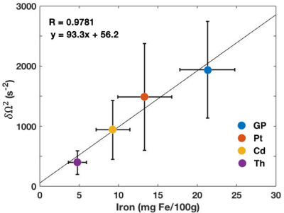 |
In Vivo Mapping of Non-heme Iron Using Time-dependent R2* Relaxation Measured with MGRE
Sohae Chung1,2, Dmitry S. Novikov1,2, Pippa Storey1,2, and Yvonne W. Lui1,2
1Center for Advanced Imaging Innovation and Research (CAI2R), Department of Radiology, New York University Grossman School of Medicine, New York, NY, United States, 2Bernard and Irene Schwartz Center for Biomedical Imaging, New York University Grossman School of Medicine, New York, NY, United States
Identification and quantification of imaging biomarkers sensitive to iron are important in understanding neurodegenerative disorders and aging. In this study, we introduce a microstructural parameter, $$$\delta\Omega^{2}$$$, the Larmor frequency variance, measured from the curvature in the logarithm of the signal from a 3D multiecho gradient echo sequence. Non-monoexponential relaxation originates from magnetic field perturbations due to the presence of mesoscopic susceptibility sources such as cellular-level iron. Our results in 26 healthy subjects show a strong linear correlation between $$$\delta\Omega^{2}$$$ and published values for iron concentration in deep gray matter regions, suggesting its potential as an imaging biomarker for iron.
|
|||
1828.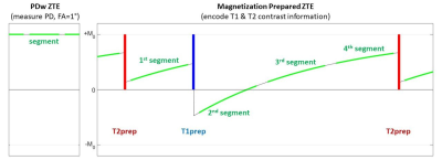 |
3D Silent Parameter Mapping: Further refinements & quantitative assessment
Florian Wiesinger1,2, Graeme McKinnon3, Sandeep Kaushik1, Ana Beatriz Solana1, Emil Ljungberg2, Mika Vogel1, Naoyuki Takei4, Rolf Schulte1, Carolin Pirkl1, Cristina Cozzini1, Laura Nuñez-Gonzalez5, Juan A. Hernandez Tamames5, and Mathias Engström6
1GE Healthcare, Munich, Germany, 2IoPPN, Department of Neuroimaging, King's College London, London, United Kingdom, 3GE Healthcare, Waukesha, WI, United States, 4GE Healthcare, Hino, Japan, 5Erasmus MC, Rotterdam, Netherlands, 6GE Healthcare, Stockholm, Sweden
Here we present further improvements of a 3D Silent Parameter Mapping method in terms of Deep Learning image reconstruction and synthetic CT image conversion. We evaluated its quantitative accuracy using the NIST/ISMRM phantom and illustrate healthy volunteer results at 1.5T and 3T.
|
|||
1829.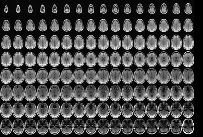 |
The “Prequence” Concept: Toward Significantly Faster Clinical MRI Exams
Vincent Schmithorst1, Ashok Panigrahy2, and Rafael Ceschin3
1Radiology, University of Pittsburgh, Pittsburgh, PA, United States, 2University of Pittsburgh, Pittsburgh, PA, United States, 3Bioinformatics, University of Pittsburgh, Pittsburgh, PA, United States
Availability of clinical fMRI would be much improved via reduction of clinical exam times. A major factor in length of exam times is that typical MRI pulse sequences are optimized for a single image contrast (T1, T2, FLAIR, etc.) necessitating multiple pulse sequences each with significant “dead time” to acquire all desired contrasts for a given protocol. We here propose instead the time-efficient “prequence” (protocol sequence) concept which aims at acquisition of all desired contrasts in one or only a few sequences with minimal dead time. Preliminary proof-of-concept is demonstrated via a simultaneous T1/T2 contrast whole-brain neuro acquisition.
|
|||
1830.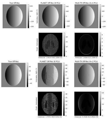 |
Balanced SSFP Parameter Estimation by Fitting Multi-TR Ellipses
Nicholas McKibben1, Michael Mendoza2, Neal K Bangerter2, and Michael N Hoff3
1University of Utah, Salt Lake City, UT, United States, 2Department of Bioenginnering, Imperial College London, London, United Kingdom, 3Deptartment of Radiology, University of Washington, Seattle, WA, United States
Multiple linearly phase-cycled, multiple-TR bSSFP can be used to effectively estimate off-resonance and condition input to the PLANET algorithm to make it robust to noise and lack of available phase-cycled images. We provide methods of estimating off-resonance given 6 or 8 phase-cycles distributed across multiple-TR acquisitions. Using the elliptical signal model, it is shown that similar ellipse shape at high flip angles allow rotated ellipses to be superimposed, leading to the construction of "virtual" ellipses. Virtual ellipses are shown to be robust to noise and perform well in simulation and phantom studies.
|
|||
1831.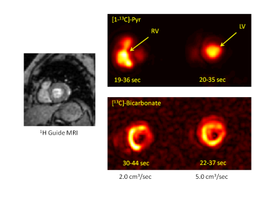 |
Pyruvate Infusion Rates for Hyperpolarized Metabolite Imaging of Human Heart
Jeffry R. Alger1,2,3,4, Jae Mo Park1, Junjie Ma1, Mahitha Roy1, Crystal Harrison1, James Ratnakar1, Albert Chen5, Galen Reed6, A. Dean Sherry1,7, Vlad Zaha1, and Craig R. Malloy1,8
1Advanced Imaging Research Center, University of Texas Southwestern Medical Center, Dallas, TX, United States, 2Neurology, University of California, Los Angeles, Los Angeles, CA, United States, 3NeuroSpectroScopics LLC, Sherman Oaks, CA, United States, 4Hura Imaging Inc, Los Angeles, CA, United States, 5GE Healthcare, Toronto, ON, Canada, 6GE Healthcare, Dallas, TX, United States, 7Chemistry, University of Texas at Dallas, Richardson, TX, United States, 8Cardiology, Veterans Affairs North Texas Healthcare System, Dallas, TX, United States
MRI/MRS metabolic tracing in human heart with hyperpolarized 13C-enriched pyruvate is feasible, but there remains a need to define the optimal infusion timing that accommodates both short polarization lifetime and subject comfort. Typical studies have used a 5.0 cm3/sec pyruvate infusion rate. We hypothesized that slower infusion is feasible because of the in vitro versus intravascular T1 difference and because vascular properties limit the rate of pyruvate delivery from the infusion site to the heart. Vascular dynamic simulations and preliminary human investigations suggest that a 2.0 cm3/sec pyruvate infusion rate be effectively used.
|
|||
1832.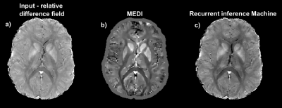 |
Dipole inversion by recurrent inference for quantitative susceptibility mapping
Samy Abo Seada1, Emanoel Ribeiro Sabidussi1, Sebastian Weingärtner2, Dirk H. J. Poot1, and Juan Antonio Hernandez-Tamames1
1Department of Radiology and Nuclear Medicine, Erasmus MC, Rotterdam, Netherlands, 2Department of Imaging Physics, TU Delft, Delft, Netherlands
QSM dipole inversion remains a challenge and recent machine learning approaches have not incorporated the known forward model directly. We propose using recurrent inference machines (RIM), a type of unrolled optimization technique, which are proposed for solving iterative inverse problems specifically. RIMs enable incorporating the forward dipole convolution directly in the learning process. Simulated data was used for training. The QSM reconstruction was tested on simulated data and healthy subject data acquired at 3T.
|
|||
1833.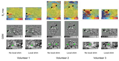 |
Improvements from local B0 shimming for QSM at 7 Tesla
Sina Straub1, Mark E. Ladd1,2, Paul Chang3, and Sahar Nassirpour3
1Division of Medical Physics in Radiology, German Cancer Research Center (DKFZ), Heidelberg, Germany, 2Faculty of Physics and Astronomy, Faculty of Medicine, University of Heidelberg, Heidelberg, Germany, 3MR Shim GmbH, Reutlingen, Germany
A 14-channel local array of shim coils was used to improve B0 homogeneity and reduce artifacts in quantitative susceptibility mapping at 7T. Using the local array of shim coils in addition to the standard 2nd order spherical harmonic shims, the B0 shim quality was improved by an average of 16% across all volunteers. Local inhomogeneities were significantly reduced and could be correlated to reduced artifacts in QSM.
|
|||
1834.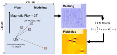 |
Development of a simulation method to evaluate T2* shortening due to susceptibility of fat in the liver using the finite element method
Daiki Tamada1, Noriaki Nagata1, Ryoichi Kose2, Katsumi Kose2, Utaroh Motosugi3, and Hiroshi Onishi1
1Department of Radiology, University of Yamanashi, Chuo, Japan, 2MRIsimulations Inc., Tokyo, Japan, 3Department of Radiology, Kofu-Kyoritsu Hospital, Kofu, Japan
A method to simulate T2* shortening by the susceptibility of fat in the liver using FEM and simple modeling was developed. The model consisting of water and lipid droplets (LD) placed randomly. Simulation and in vivo studies indicated the feasibility of our method. The results suggested the microscopic field inhomogeneity by LDs leads to non-neglectable T2* decay. Besides, the result of in vivo study was fairly agreed with the simulation.
|
|||
1835.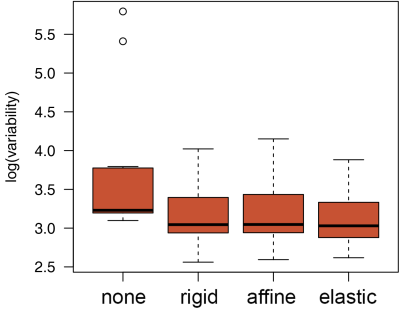 |
Software for T1ρ Mapping in the Knee: Addressing the Critical Role of Motion Correction
Artem Mikheev1, Louisa Bokacheva1, Azadeh Sharafi1, Ravinder Regatte1, and Henry Rusinek1
1Department of Radiology, New York University School of Medicine, New York, NY, United States
T1ρ mapping enables detecting early changes in the knee cartilage associated with osteoarthritis. Reproducible T1ρ measurements are challenging because of the motion artifacts arising from a long acquisition time of images at multiple spin-lock times. We implemented a new elastic motion correction method and applied it to T1ρ-weighted acquisitions in 20 subjects with normal knees and early osteoarthritis. The T1ρ voxel maps in articular cartilage generated from motion-corrected images showed significantly lower variability compared to maps derived from uncorrected images. The finding underscores the importance of motion correction in T1ρ mapping of the knee.
|
|||
1836.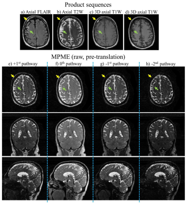 |
Early results from a patient study aimed at testing a quantitative and synthetic brain MRI method, for abbreviated neuro protocols
Cheng-Chieh Cheng1, Jeffrey P. Guenette2, and Bruno Madore2
1Computer Science and Engineering, National Sun Yat-sen University, Kaohsiung, Taiwan, 2Radiology, Brigham and Women's Hospital, Harvard Medical School, Boston, MA, United States
We developed a quantitative and synthetic MRI method capable of generating the most-commonly encountered contrasts in neuro protocols, along with quantitative parameter maps, with full brain coverage and 1-mm isotropic resolution, from only about 5 min of scanning. The eventual goal is for this sequence and associated processing to play a role in abbreviated neuro protocols. Promising results were obtained in healthy volunteers and we recently embarked on a larger patient study. After a few months of delay, largely associated with the pandemic, patient recruitment has started, and preliminary results are shown here.
|
|||
1837.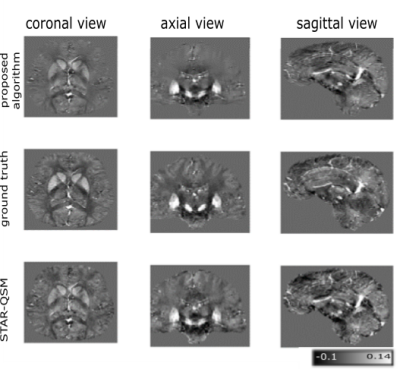 |
Shearlet-based susceptibility map reconstruction with additional TGV-regularization
Janis Stiegeler1,2 and Sina Straub1
1Medical Physics in Radiology, German Cancer Research Center (DKFZ), Heidelberg, Germany, 2Faculty of Physics and Astronomy, University of Heidelberg, Heidelberg, Germany
The 2016 Reconstruction Challenge urged the need of QSM algorithms which minimize several classical image quality measures relatively to a ground truth, but also achieve an acceptable human image perception by avoiding oversmoothing effects. A multicale shearlet system was used together with a total generalized variation (TGV) term to regularize the susceptibility-phase convolution problem. The results show that these regularizers are useful to obtain quantitative susceptibility maps which are rich in detail and simultaneously can achieve top five results in the ranking of the 2016 Reconstruction Challenge regarding all classical image quality measures used in this challenge.
|
|||
1838.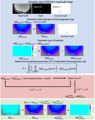 |
New method of estimating static field inhomogeneity for MR susceptometry-based oximetry
Alexander M Barclay1, Michael C Langham1, and Felix W Wehrli1
1Department of Radiology, University of Pennsylvania, Philadelphia, PA, United States
Large vein blood oxygenation level can be estimated by magnetic susceptibility-based oximetry. Calculation of the local field shift between intravascular blood and surrounding tissue using measurement of signal phase is done with field mapping. Measurement is confounded by background static field inhomogeneity. Correction by either high-pass filtering to remove spatially slow-varying inhomogeneity, or polynomial fitting of inhomogeneity to be subtracted-out require measuring reference tissue phase, imparting bias and constraining signal-to-noise ratio considerations to tissue, not lumen. An objective approach based on inhomogeneity estimation using a smoothness criterion is proposed. Tissue phase is eliminated, allowing measurement of intravascular phase alone.
|
|||
1839.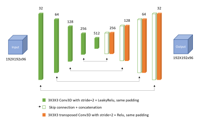 |
A Deep Learning Approach to QSM Background Field Removal: Simulating Realistic Training Data Using a Reference Scan Ground Truth and Deformations
Oriana Vanesa Arsenov1, Karin Shmueli1, and Anita Karsa1
1Medical Physics and Biomedical Engineering, University College London, London, United Kingdom
Current techniques for background field removal (BGFR), essential for quantitative susceptibility mapping, leave residual background fields and inaccuracies near air-tissue interfaces. We propose a new deep learning method aiming for robust brain BGFR: we trained a 3D U-net with realistic simulated and in-vivo data augmented with spatial deformations. The network trained on synthetic data predicts accurate local fields when tested on synthetic data, (median RMSE = 49.5%), but is less accurate when tested on in-vivo data. The network trained and tested on in-vivo data performs better, suggesting our synthetic set did not fully capture the complexity found in vivo.
|
|||
1840.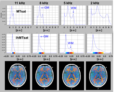 |
Measuring inhomogeneous MT (ihMT) in human brain by multi-parameter mapping (MPM)
Gunther Helms1,2, Lenka Vaculčiaková2, Kerrin J. Pine2, Harald E. Moeller3, and Nikolaus Weiskopf2,4
1Medical Radiation Physics, Clinical Sciences Lund, Lund University, Lund, Sweden, 2Neurophysics, Max Planck Institute for Human Cognitive and Brain Sciences, Leipzig, Germany, 3NMR Methods & Development Group, Max Planck Institute for Human Cognitive and Brain Sciences, Leipzig, Germany, 4Felix Bloch Institute for Solid State Physics, Leipzig University, Leipzig, Germany
Inhomogenous Magnetization Transfer (ihMT), the differential response to irradiation at single and dual frequency offsets, is sensitive to myelination. Quantification of ihMT in terms of MT saturation (MTsat), the percent reduction of Mz, corrects for underlying T1 and B1+ and allows comparing different implementations to optimize multi-parameter mapping (MPM) protocols. ihMTsat in white matter is roughly one twentieth of MTsat, merely 0.2 p.u. at 5kHz and 100% SAR at 3T. Noise propagation demands acquisition at 1.5mm resolution, large-array coils, and smaller offsets than suggested previously. Inverse MT ratios are highly correlated to ihMTs but obscure the true effect size.
|
|||
1841.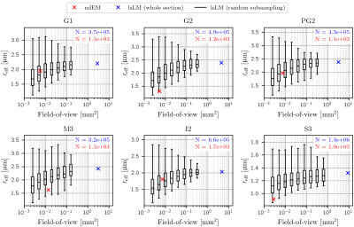 |
Reliable estimation of the MRI-visible effective axon radius using light microscopy: the need for large field-of-views
Laurin Mordhorst1, Maria Morozova2,3, Sebastian Papazoglou1, Björn Fricke1, Jan Malte Oeschger1, Henriette Rusch3, Carsten Jäger2, Markus Morawski2,3, Nikolaus Weiskopf2,4, and Siawoosh Mohammadi1,2
1Institute of Systems Neuroscience, University Medical Center Hamburg-Eppendorf, Hamburg, Germany, 2Department of Neurophysics, Max Planck Institute for Human Cognitive and Brain Sciences, Leipzig, Germany, 3Paul Flechsig Institute of Brain Research, University of Leipzig, Leipzig, Germany, 4Felix Bloch Institute for Solid State Physics, Faculty of Physics and Earth Sciences, Leipzig, Germany
MRI-based models enable non-invasive characterization of brain microstructure, e.g. the effective axon radius ($$$r_{\text{eff}}$$$). Often, these models were validated by small-field-of-view microscopy (sFoVM) images (~10³ axons). As $$$r_{\text{eff}}$$$ is dominated by large, sparsely occurring axons, sFoVM-based estimations may miss large axons and underestimate $$$r_{\text{eff}}$$$. We employed an in-house developed pipeline to estimate $$$r_{\text{eff}}$$$ on large-scale light microscopy (lsLM) sections with similar spatial extent as the voxel sizes in human MRI systems. Taking lsLM as a baseline, we showed that sFoVM is an unsuitable reference for $$$r_{\text{eff}}$$$ and verified the potential of $$$r_{\text{eff}}$$$ to capture relevant spatial, anatomical variation.
|
|||
1842.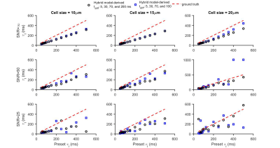 |
Simultaneous Quantification of Mean Intracellular Water Lifetime and Cell Size Using Temporal Diffusion Spectroscopy
xiaoyu jiang1, sean p devan1, john c. gore1, and junzhong xu1
1Vanderbilt University Institute of Imaging Science, nashville, TN, United States
There is an increasing interest in characterizing tissue microstructure using multi-compartment diffusion MRI models involving multiple b values and diffusion times ($$$t_{diff}$$$s). However, previous models usually ignore water exchange between intra- and extracellular compartments, which limits the accuracy of the fitted model parameters and potentially their interpretation as indicators of disease progression or treatment response. Here, we propose a simplified biophysical model of solid tissues that allows derivation of the mean intracellular water lifetime ($$$\tau_{i}$$$) and other microstructural parameters (e.g., cell size d) simultaneously from the $$$t_{diff}$$$ dependence of diffusion MRI signals.
|
|||
1843.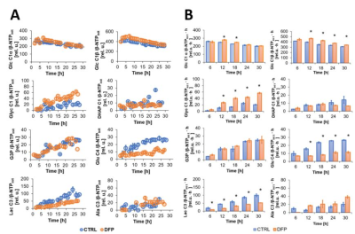 |
Metabolic Effects of Deferiprone on Triple Negative Breast Cancer
Paola Porcari1, Ellen Ackerstaff1, H. Carl Lekaye1, and Jason A. Koutcher1,2,3,4
1Medical Physics, Memorial Sloan Kettering Cancer Centre, New York, NY, United States, 2Department of Medicine, Memorial Sloan Kettering Cancer Center, New York, NY, United States, 3Molecular Pharmacology Program, Memorial Sloan Kettering Cancer Center, New York, NY, United States, 4Weill Cornell Medical College, Cornell University, New York, NY, United States
Triple-negative breast cancer (TNBC) metastasizes and is currently associated with poor prognosis and lack of effective targeted therapies. We are studying the potential of Deferiprone, an intracellular iron chelator clinically used for non-cancer-related diseases, to improve the chemotherapeutic treatment response in TNBC by altering cellular iron-dependent proliferation and metabolism. Here, cellular and energy metabolism changes in response to Deferiprone were assayed in live TNBC cells by multi-nuclear MRS in our MR-compatible bioreactor. Metabolic changes in a human TNBC cell line are compared to previous findings in murine TNBC cells.
|
The International Society for Magnetic Resonance in Medicine is accredited by the Accreditation Council for Continuing Medical Education to provide continuing medical education for physicians.