Digital Posters
Electrical Tissue Properties Mapping
ISMRM & SMRT Annual Meeting • 15-20 May 2021

Digital Posters
Electrical Tissue Properties Mapping
| Concurrent 2 | 15:00 - 16:00 |
3773.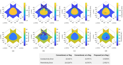 |
Global and Direct Electrical Properties Tomography Method Based on the Linear Integral Equations for Impedivity
Motofumi Fushimi1, Naohiro Eda1, and Takaaki Nara1
1The University of Tokyo, Tokyo, Japan
The present paper introduces novel reconstruction methods for electrical properties tomography (EPT), in which conductivity and permittivity are reconstructed from the complex B1 field, and phase-based EPT, in which only conductivity is reconstructed from the B1 phase. The proposed methods reconstruct electrical properties (EPs) by solving a linear integral equation derived from Helmholtz's identity, and does not require calculation of the second-order derivatives of the measured data nor iterative updating of the estimates. Numerical simulations and phantom experiments showed that the proposed method could retrieve EPs more stably than the conventional method that solves a linear partial differential equation.
|
|||
3774. |
New Approaches for Simultaneous Noise Suppression and Edge Preservation to Achieve Accurate Quantitative Conductivity Mapping in Noisy Images
Anita Karsa1 and Karin Shmueli1
1Department of Medical Physics and Biomedical Engineering, University College London, London, United Kingdom
Due to their extreme noise amplification, current Quantitative Conductivity Mapping (QCM) techniques require high SNR images. Simultaneous Quantitative Susceptibility Mapping and QCM uses low-SNR gradient-echo sequences, creating a need for QCM methods appropriate for noisy images. Here we proposed, optimised, and compared several new QCM methods (all shared on https://xip.uclb.com/i/software/MRI_conductivity.html) built on the widely-used phase-based formula and its equivalent, less popular integral form. We found that solving the integral equation provided lower errors and better edge preservation in both simulated and in-vivo images, and that edge preservation combining magnitude-based and image-segmentation-based techniques resulted in the best in-vivo conductivity map.
|
|||
3775.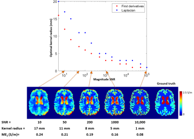 |
Optimal Kernel Radii for Calculating the Derivatives of Noisy B1 Phase for Accurate Phase-Based Quantitative Conductivity Mapping
Anita Karsa1, Patrick Fuchs1, and Karin Shmueli1
1Department of Medical Physics and Biomedical Engineering, University College London, London, United Kingdom
Quantitative conductivity mapping (QCM) techniques use the first spatial derivatives and/or the Laplacian of the B1 phase. These are commonly estimated by fitting a 3D quadratic function within a kernel around each voxel. However, small kernels lead to severe noise amplification and large kernels induce inaccuracies. Here we determined the optimal kernel radii across a range of magnitude SNR using an anthropomorphic, numerical brain phantom. The optimal kernel size decreased with increasing SNR. Calculating the first derivatives required smaller kernels than calculating the Laplacian making QCM methods using first derivatives likely more accurate than Laplacian-based techniques.
|
|||
3776.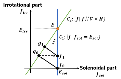 |
An Iterative Method for Electrical Properties Tomography Based on the Helmholtz Decomposition for the Electric Field
Naohiro Eda1, Motofumi Fushimi1, and Takaaki Nara1
1The University of Tokyo, Tokyo, Japan
This paper proposes a novel, integral-equation-based (IE-based) reconstruction method for magnetic resonance electrical properties tomography (MREPT). The proposed method can reconstruct the electric field and EPs simultaneously based on the Helmholtz decomposition of the electric field. In the proposed algorithm, we iteratively apply the projections onto the spaces in which the true electric field is contained. An advantage of the proposed method is that convergence is theoretically guaranteed in this iterative process. The efficacy of the proposed method is validated through numerical simulations.
|
|||
3777.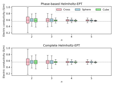 |
Uncertainty assessment under repeatability conditions in Helmholtz-based electric properties tomography
Alessandro Arduino1, Francesca Pennecchi1, Ulrich Katscher2, Luca Zilberti1, and Maurice Cox3
1Istituto Nazionale di Ricerca Metrologica (INRIM), Torino, Italy, 2Philips Research Laboratories, Hamburg, Germany, 3National Physical Laboratory (NPL), Teddington, United Kingdom
This work shows the uncertainty evaluation under repeatability conditions of the phase-based Helmholtz-electric properties tomography (EPT) technique. Repeated MRI scans of a homogeneous cylindrical phantom are analyzed with appropriate statistical techniques to evaluate the covariance matrix of the EPT input. This matrix is propagated through the EPT technique according to the law of propagation of uncertainty. The estimated electric conductivity is highly repeatable and exhibits a spatial dispersion, whose average value, within a central region, gives an accurate estimate of the phantom conductivity. The described approach will be applied in future to extend the characterization under reproducibility conditions.
|
|||
 |
3778.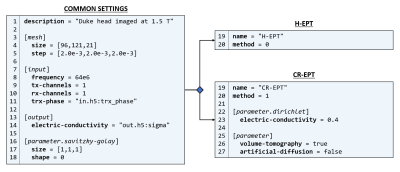 |
EPTlib: an open-source C++ library of electric properties tomography methods
Alessandro Arduino1, Umberto Zanovello1, Luca Zilberti1, and Oriano Bottauscio1
1Istituto Nazionale di Ricerca Metrologica (INRIM), Torino, Italy
This abstract introduces EPTlib, an open-source, extensible C++ library collecting electric properties tomography (EPT) methods. Currently, the library implements three methods (Helmholtz-EPT, convection reaction–EPT and gradient-EPT), but the list will be updated in the future. EPTlib comes along with a console application that allows to run all the implemented methods. After a brief description of the software architecture and of the EPT methods, an example of usage of each implemented method with simulated input data is provided.
|
||
3779.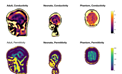 |
Evidence for tissue dielectric property differences between neonates and adults: a retrospective study using MR-EPT
Roy Kurtzbard1, Anthony Price2, Joseph V Hajnal1,2, Jeffrey W Hand2, and Shaihan J Malik1,2
1Biomedical Engineering Department, School of Biomedical Engineering and Imaging Sciences, King's College London, London, United Kingdom, 2Centre for the Developing Brain, School of Biomedical Engineering and Imaging Sciences, King's College London, London, United Kingdom
Electromagnetic simulations for RF safety calculation require knowledge of tissue dielectric properties, but these are not well understood for neonates whose tissues have much higher water content than adults. This work retrospectively used MR-EPT on archived raw data obtained from a study on 800+ neonates, to measure tissue conductivity and permittivity at 128MHz. Presented are initial results from 50 subjects. The measured properties are rather noisy at the individual level, but across the whole cohort the median brain conductivity in neonates is approximately 1.8 times greater than in adults, in line with expectations from scaling arguments.
|
|||
3780.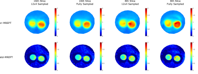 |
Fast 3D Undersampled Bloch-Siegert based B1+ Mapping for use in MREPT
Safa Ozdemir1, Efe Ilicak1, Carmen Stutz1, Mara Berger1, Jascha Zapp1, Lothar R. Schad1, Yusuf Z. Ider2, and Frank G. Zöllner1,3
1Computer Assisted Clinical Medicine, Medical Faculty Mannheim, Heidelberg University, Mannheim, Germany, 2Electrical and Electronics Engineering, Bilkent University, Ankara, Turkey, 3Mannheim Institute for Intelligent Systems in Medicine, Medical Faculty Mannheim, Heidelberg University, Mannheim, Germany
Magnetic Resonance Electrical Properties Tomography (MREPT) technique utilizes complex B1+ information to obtain conductivity (σ) and permittivity (ϵ). Since obtaining B1+ magnitude is rather time consuming, several phase-based MREPT methods were previously suggested. However, these methods contain inaccuracies especially towards the periphery of the imaged object. In this paper, we utilize one of the fastest B1+ magnitude mapping techniques in order to obtain fast and accurate B1+ magnitude maps to be used in conductivity imaging. Phantom measurements show that accurate 3D B1+ magnitude maps can be
acquired in less than 30 seconds, therefore improving the practicality of conductivity imaging.
|
|||
3781.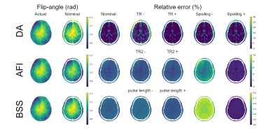 |
Fast virtual B1-mapping for the in silico characterization of EPT
Alessandro Arduino1 and Luca Zilberti1
1Istituto Nazionale di Ricerca Metrologica (INRIM), Torino, Italy
A fast modelling of B1-mapping techniques is proposed for the in silico characterization of electric properties tomography method. The performances of three B1-mapping techniques are analyzed on a realistic model problem, obtaining information on the systematic errors and on the random errors through the application of a Monte Carlo method. An Helmholtz-based electric properties tomography technique is tested on input provided by the B1-mapping techniques.
|
|||
3782.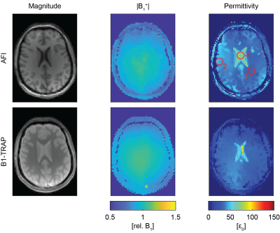 |
Permittivity Mapping with B1-TRAP
Santhosh Iyyakkunnel1,2, Carl Ganter3, Francesco Santini1,2, and Oliver Bieri1,2
1Department of Radiology, University Hospital Basel, Basel, Switzerland, 2Department of Biomedical Engineering, University of Basel, Basel, Switzerland, 3Department of Radiology, Technical University of Munich, Munich, Germany
Permittivity mapping strongly depends on the accuracy and the signal-to-noise ratio (SNR) of the underlying B1+ magnitude estimation method. In this work, we assess the suitability of a transient phase SSFP method, termed B1-TRAP, for permittivity mapping and compare this B1+ mapping method with the commonly-used Actual Flip Imaging (AFI) method.
|
|||
3783.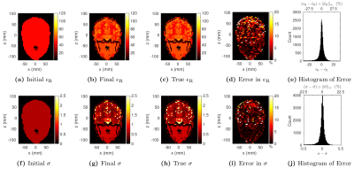 |
An Application of a Projected Newton Method to Electrical Properties Estimation via Global Maxwell Tomography
Jose EC Serralles1, Ilias I Giannakopoulos2, Jacob K White1, Luca Daniel1, and Riccardo Lattanzi2,3,4
1Electrical Engineering and Computer Science, Computational Prototyping Group (CPG), Research Laboratory of Electronics (RLE), Department of Electrical Engineering and Computer Science (EECS), Massachusetts Institute of Technology (MIT), Cambridge, MA, United States, 2Center for Advanced Imaging Innovation and Research (CAI2R), Department of Radiology, New York University Grossman School of Medicine, New York, NY, United States, 3The Bernard and Irene Schwartz Center for Biomedical Imaging (CBI), Department of Radiology, New York University Grossman School of Medicine, New York, NY, United States, 4Vilcek Institute of Graduate Biomedical Sciences, New York University Grossman School of Medicine, New York, NY, United States
Global Maxwell Tomography (GMT) is a recently introduced technique that estimates tissue electrical properties from magnetic resonance measurements by solving an inverse scattering problem. In this work, we propose a new implementation of GMT that uses a Projected Newton method to minimize the cost function, instead of the quasi-Newton method employed by the original GMT. We demonstrated the new approach with two numerical experiments, using a four--compartment phantom and a realistic head model. Compared to the results obtained with the original GMT, the number of iterations required for convergence was drastically reduced and the estimated EP were more accurate.
|
|||
3784.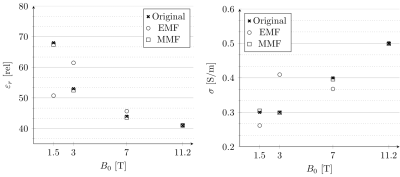 |
Electrical Properties Reconstruction from Free Induction Decay Measurements
Patrick Fuchs1 and Rob Remis1
1Microelectronics, Delft University of Technology, Delft, Netherlands
By incorporating scattering of dielectric tissue into the measurement model of the MRI signal we show a direct relationship between conductivity and permittivity of the tissue and the measured signal. Simplifying this for a known geometry a reconstruction of the dielectric properties is demonstrated based on the most simple of MRI signals, the finite induction decay or FID. This relationship can of course be exploited for more complicated applications such as local SAR, antenna design and optimisation and SNR computations, especially for high field applications.
|
|||
3785.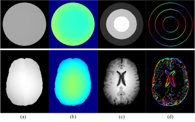 |
The electrical conductivity reconstruction using MR structural information
Xiangdong Sun1, Chunyi Liu2, Lijun Lu2, Xiaoyun Liu1, and Wufan Chen1,2
1School of Automation Engineering,University of Electronic Science and Technology of China, Chengdu, China, 2School of Biomedical Engineering, Southern Medical University, Guangzhou, China
The conductivity of biological tissues can be potentially used for a cancer diagnostic. Thus, imaging conductivity is useful for clinical applications. Here, we developed a conductivity imaging method based on the gradient conductivity imaging method by incorporating information from anatomical MR images. The characteristic of the defined prior was extracting the structure information and guided the conductivity reconstruction. To evaluate the performance of the proposed method, the electromagnetic field and magnitude images were simulated for a cylindrical phantom and brain model. The results demonstrated that the proposed method preserves more details of the conductivity images and mitigates the noise affection.
|
|||
3786.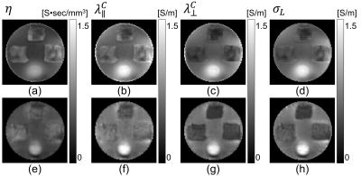 |
Comparison of MR-based low-frequency electrical conductivity tensor using DT-MREIT and CTI: a biological tissue phantom study
Saurav Zaman Khan Sajib1, Munish Chauhan1, Sulagna Sahu1, Enock Boakye1, and Rosalind J Sadleir1
1School of Biological and Health Systems Engineering, Arizona State University, Tempe, AZ, United States
Diffusion tensor magnetic resonance electrical impedance tomography (DT-MREIT) and electrodeless conductivity tensor imaging (CTI) are two emerging modalities that can quantify low-frequency tissue anisotropic conductivity properties by considering the relationship between ion mobility and water diffusion. While both methods have potential applications to estimating neuro-modulation fields or formulating forward models used for electrical source imaging, a direct comparison of these two modalities has not yet been performed. Therefore, the aim of this study to test the equivalence of these two modalities.
|
|||
3787.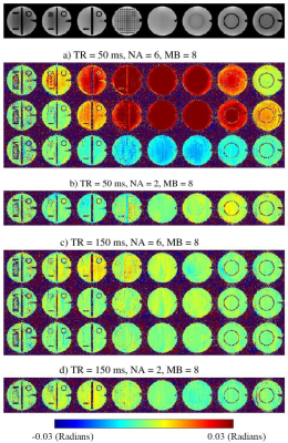 |
Phase Dispersion from Steady-State Signal Behavior in Phase-Sensitive Multiband Imaging with Application to MREIT
Guita Banan1, Munish Chauhan2, Manish Amin1, Sudhir Ramanna3, Zahra Hosseini4, Essa Yacoub3, Michael Schär5, Thomas H Mareci1, and Rosalind J Sadleir2
1Department of Biochemistry and Molecular Biology, University of Florida, Gainesville, FL, United States, 2School of Biological and Health System Engineering, Arizona State University, Tempe, AZ, United States, 3University of Minnesota, Minneapolis, MN, United States, 4MR R&D Collaboration, Siemens Medical Solutions USA, Atlanta, GA, United States, 5Department of Radiology, Johns Hopkins University, Baltimore, MD, United States
Incorporating multiband excitation into phase-sensitive imaging introduces distortions in the phase measurements. We used simulations and phantom images to show that a previously unreported phase dispersion problem arises within multiband-slice phase maps. We show that this problem can be understood in terms of the steady-state signal behavior and propose imaging protocols to resolve it. Human scans using these protocols show minimal phase dispersion. We are adopting this protocol for our studies of phase-sensitive magnetic resonance electric impedance tomography (MREIT).
|
|||
3788.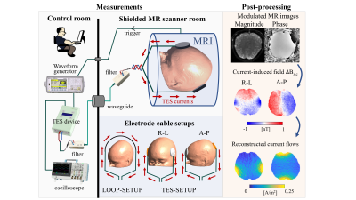 |
Sensitivity and resolution improvement for in-vivo magnetic resonance current density imaging (MRCDI) of the human brain
Cihan Göksu1,2, Klaus Scheffler2,3, Fróði Gregersen1,4,5, Hasan Hüseyin Eroğlu1,4, Rahel Heule2,3, Hartwig R. Siebner1,6,7, Lars G. Hanson1,4, and Axel Thielscher1,4
1Danish Research Centre for Magnetic Resonance, Centre for Functional and Diagnostic Imaging and Research, Copenhagen University Hospital, Amager and Hvidovre, Denmark, 2High-Field Magnetic Resonance Center, Max-Planck-Institute for Biological Cybernetics, Tübingen, Germany, 3Department of Biomedical Magnetic Resonance, University of Tübingen, Tübingen, Germany, 4Section for Magnetic Resonance, DTU Health Tech, Technical University of Denmark, Kgs Lyngby, Denmark, 5Sino-Danish Center for Education and Research, Aarhus, Denmark, 6Department of Neurology, Copenhagen University Hospital, Bispebjerg, Denmark, 7Institute for Clinical Medicine, Faculty of Medical and Health Sciences, University of Copenhagen, Copenhagen, Denmark
Effective use of transcranial electrical stimulation (TES) in clinical and neuroscience applications requires the exact knowledge of TES currents. MRCDI uses MRI to measure the TES-induced magnetic fields for estimating the underlying current flow distributions. Their accuracy highly depends on the sensitivity and spatial resolution of the MR measurements. Here, we propose an advanced gradient-echo-based MRCDI method utilizing an optimized spoiling, acquisition-weighting, and navigators to achieve a noise sensitivity of 84pT at 2×2×3mm3 resolution for a total scan time of less than five minutes. We test the method's performance by phantom and human in-vivo experiments for two TES injection profiles.
|
|||
3789.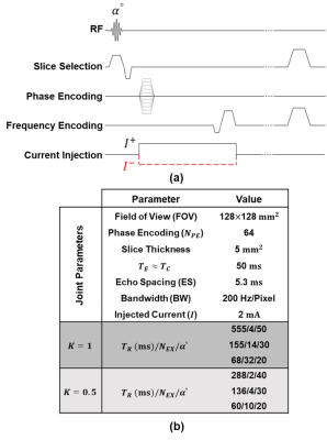 |
Optimization of SNR and the total acquisition time in MRCDI
Mehdi Sadighi1, Mert Şişman1, and B. Murat Eyüboğlu1
1Electrical and Electronics Eng., Middle East Technical University (METU), Ankara, Turkey
The clinical applicability of magnetic resonance current density imaging (MRCDI) is highly dependent on the sensitivity of the acquired current-induced magnetic flux density ($$$\widetilde{B}_z$$$) distribution. Here, the combined effect of relevant parameters of the ICNE-SPGE pulse sequence on the SNR level and the total acquisition time of the $$$\widetilde{B}_z$$$ images are analyzed. The optimized sequence parameters are estimated to acquire $$$\widetilde{B}_z$$$ images with the highest possible SNR for a given acquisition time or the desired SNR in the shortest scan time. Besides, alternative sequence parameters are estimated to acquire the same SNR level for a given acquisition time.
|
|||
3790.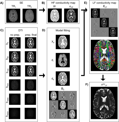 |
High-resolution whole-brain Conductivity Tensor Imaging of the human brain
Marco Marino1,2, Dante Mantini1,2, and Giulio Ferrazzi2
1Research Center for Motor Control and Neuroplasticity, KU Leuven, Leuven, Belgium, 2IRCCS San Camillo Hospital, Venice, Italy
We propose a framework to achieve high-resolution whole-brain conductivity tensor imaging (CTI) with MRI at low frequencies. Our methodology overcomes the limitations of previous approaches based on B1 mapping techniques, both in terms of spatial resolution and brain coverage. This was attained by combining high-frequency conductivity estimates derived from a water-mapping technique with multi-b-value diffusion tensor imaging data. We obtained conductivity values for grey matter and white matter that are in line with previous studies. Overall, our findings highlight noteworthy within- and between- subject variability of the conductivity values.
|
The International Society for Magnetic Resonance in Medicine is accredited by the Accreditation Council for Continuing Medical Education to provide continuing medical education for physicians.