Digital Posters
Quantitative MRI I
ISMRM & SMRT Annual Meeting • 15-20 May 2021

| Concurrent 2 | 19:00 - 20:00 |
4203.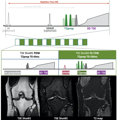 |
MIXTURE: A novel sequence for simultaneous morphological and quantitative imaging based on multi-interleaved 3D turbo-spin echo MRI
Masami Yoneyama1, Takayuki Sakai2, Shuo Zhang3, Daichi Murayama2, Hajime Yokota4, Yansong Zhao5, Shinji Saruya6, Masashi Suzuki6, Atsuya Watanabe7,8, Mamoru Niitsu6, and Marc Van Cauteren9
1Philips Japan, Tokyo, Japan, 2Department of Radiology, Eastern Chiba Medical Center, Chiba, Japan, 3Philips Healthcare, Hamburg, Germany, 4Department of Diagnostic Radiology and Radiation Oncology, Graduate School of Medicine, Chiba University, Chiba, Japan, 5Philips Healthcare, Cleveland, OH, United States, 6Department of Radiology, Saitama Medical University, Saitama, Japan, 7Department of Orthopaedic Surgery, Eastern Chiba Medical Center, Chiba, Japan, 8General Medical Services, Graduate School of Medicine, Chiba University, Chiba, Japan, 9Philips Healthcare, Best, Netherlands
3D high-resolution magnetic resonance imaging (MRI) is clinically essential for an accurate assessment of subtle pathophysiological changes in different anatomies. However, long exam time remains a practical challenge that requires additional scans for either complementary quantitative parametric mapping or morphological information for clinical diagnosis. In this study, we introduce a novel method termed Multi-Interleaved X-prepared TSE with inTUitive RElaxometry (MIXTURE) for simultaneous morphological and quantitative imaging with volumetric isotropic resolution within one single scan and demonstrate the approach in multi-contrast quantitative knee imaging with direct T2- or T1ρ-mapping.
|
|||
4204.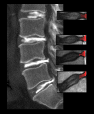 |
Annular tears in the intervertebral disc can be detected and highlighted using conventional MRI, texture analysis and neural networks
Christian Waldenberg1, Stefanie Eriksson1, Hanna Hebelka2, Helena Brisby3, and Kerstin Magdalena Lagerstrand1
1Department of Radiation Physics, University of Gothenburg, Gothenburg, Sweden, 2Department of Radiology, University of Gothenburg, Gothenburg, Sweden, 3Department of Orthopaedics, University of Gothenburg, Gothenburg, Sweden
Annular tears of intervertebral discs (IVDs) are associated with ingrowth of nerve endings with the potential to cause discogenic pain. We propose a novel non-invasive workflow for detecting annular tears and determining its location based on conventional magnetic resonance imaging (MRI). 124 IVDs in 44 patients with low back pain were included. Based on MRI, texture analysis and neural networks, a classification algorithm and workflow for determining the presence and position of annular tears were proposed. Excellent classification sensitivity of 100% and specificity of 93% were reached. The position of the tears was correctly determined in 67% of the IVDs.
|
|||
4205. |
Lumbar Fatty Acid Composition (FAC) Measurement in Osteoporosis Patients Using MRI
Lina Lin1, Lutian Bai1, Qun Cheng2, Manuel Schneider3, Marcel Dominik Nickel3, Caixia Fu4, Guangwu Lin1, and Shihong Li1
1Department of Radiology, Huadong Hospital, Fudan University, Shanghai, China, 2Department of Osteoporosis, Huadong Hospital, Fudan University, Shanghai, China, 3MR Application Predevelopment, Siemens Healthcare GmbH, Erlangen, Germany, 4Application Development, Siemens Shenzhen Magnetic Resonance Ltd, Shenzhen, China
This study is to explore the application value of fatty acid composition (FAC) measurements in the diagnosis of osteoporosis. 39 subjects who underwent dual-energy X-ray absorptiometry (DXA) and lumbar magnetic resonance imaging on a 3T scanner were included. We found that FF value was negatively correlated with T value (P < 0.05, r = -0.371), and SF value was positively correlated with T value (P < 0.05, r = 0.399). The PUF value of the osteopenia group was significantly higher than that of the osteoporosis group(P < 0.05)and normal bone mass group(P < 0.05).
|
|||
4206.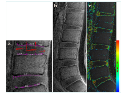 |
Association of cartilaginous endplate changes in degenerative intervertebral discs and vertebral marrow fat: a quantitative MRI study
Yayun Ji1, Jianqiang Fang1, Binyu Zhang1, Siyuan Mi1, Weiyin Vivian Liu2, Weian Zhao1, and Liheng Ma3
1Xianyang Central Hospital, Xianyang, China, 2MR research, GE Healthcare, Beijing, China, 3The First Affiliated Hospital of Guangdong Pharmaceutical University, Guangzhou, China
This study aims to investigate if advanced UTE T2* Mapping MRI quantitative sequence could sensitively assess the early changes in the biochemical components of the cartilage endplate. This might explain the pathological mechanism of cartilage endplate injury, that is, bone marrow fat content increased in vertebral body of degenerated intervertebral disc. To sum up, UTE T2* Mapping could a potential imaging tool to early diagnose cartilage endplate changes and assist clinical prevention of lumbar intervertebral disc degeneration at an earlier stage.
|
|||
4207.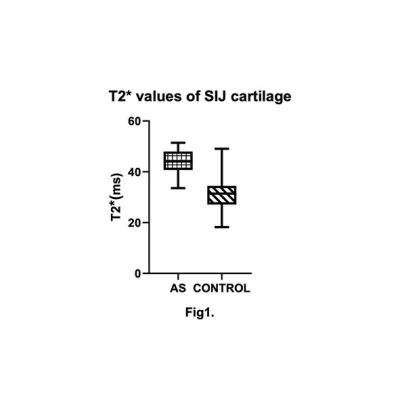 |
3D-ultrashort echo time imaging evaluation of the sacroiliac joint in patients with ankylosing spondylitis
Cui Ren1, Qing Li2, Stefan Sommer3,4, Qiao Zhu1, and Huishu Yuan1
1Radiology, Peking University Third Hospital, Beijing, China, 2MR Collaborations, Siemens Healthcare Ltd, Shanghai, China, 3Siemens Healthcare, Zurich, Switzerland, 4Swiss Center for Musculoskeletal Imaging (SCMI), Zurich, Switzerland
The aim of this study was to evaluate the performance of a 3D-UTE sequence in the detection of sacroiliac joint cartilage and bone cortex abnormalities in patients with clinically confirmed ankylosing spondylitis. T2* derived from 3D-UTE displayed potential as a quantitative measure to diagnose ankylosing spondylitis.
|
|||
4208. |
A Combined Solid-State 1H and 31P Magnetic Resonance Imaging to Assess Bone Mineral and Matrix Densities in Rat bones
Victor Babu Kassey1,2,3,4, Matthias Walle1, Jonathan Egan1, Diana Yeritsyan1, Yaotang Wu2,3,4, Brian D Snyder2,4, Edward Rodriguez1,2, Jerome Ackerman2,5, and Ara Nazarian1,2,4
1Carl J. Shapiro Department of Orthopaedic Surgery, Beth Israel Deaconess Medical Center, Boston, MA, United States, 2Harvard Medical School, Boston, MA, United States, 3Massachusetts General Hospital, Charlestown, MA, United States, 4Department of Orthopaedic Surgery, Boston Children's Hospital, Boston, MA, United States, 5Department of Radiology, Massachusetts General Hospital, Charlestown, MA, United States
Bone matrix and mineral densities (BMD) are important parameters to identify bone diseases such as osteoporosis and osteomalacia. Micro-CT, CT, and DXA scans provide bone mineral densities but not bone matrix. In this study, a non-invasive, radiation-free, and clinically proven combined 1H/31P MRI method was developed to measure bone matrix and mineral densities from rat bones from the same volume-of-interest sequentially in a single session. A custom-designed home-made double-tuned single volume coil was designed for 7T, and 1H/31P ZTE rat bone images were obtained, auto-registered, bone matrix and mineral densities were computed quantitatively, and osteoporosis and osteomalacia were successfully identified.
|
|||
4209. |
Monitoring changes of knee with Amateur Marathon athletes using Synthetic Magnetic Resonance Imaging :A preliminary study
Yijie Fang1, Wenjun Yu1, Long Qian2, and Shaolin Li1
1The Fifth Affiliated Hospital of Sun Yat-sen University, Zhuhai, China, 2MR Research, GE Healthcare, Beijing, China
To explore the value of Synthetic magnetic resonance imaging (Sy MRI) in the quantitative monitoring of knee joint structural changes and cartilage of amateur marathon runners before and after the whole marathon. Twenty-six knee joints of healthy volunteers who took part in the marathon were scanned by Sy MRI technology one week before the race and within 48 hours after the race. The values of T1, T2 and PD of knee cartilage in whole and different regions before competition were higher than those of 48 hours after the marathon, and the difference was statistically significant
|
|||
4210. |
Glycosaminoglycan detection in OA patients using AC-iTIP at 3.0T: A preliminary study
Baiyan Jiang1, Ki-wai, Kevin Ho2, Xiao Fan1, Jian Hou1, Chun-man Lawrence Lau2, James Griffith1, and Weitian Chen1
1Imaging & Interventional Radiology, The Chinese University of Hong Kong, Sha Tin, Hong Kong, 2Orthopaedics & Traumatology, The Chinese University of Hong Kong, Sha Tin, Hong Kong
The measurement of R1rho (1/T1rho) spectrum and its asymmetry have several advantages over conventional CEST MTR asymmetry in probing chemical exchange effect. Previous studies have reported that AC-iTIP is a robust approach to obtain R1rho-spectrum and R1rho asymmetry. However, an accurate B0 map is required to perform R1rho asymmetry. In this work, we fit the entire R1rho spectrum to a mathematical expression and directly acquire the pool population parameter pb. A preliminary in vivo scan on 4 osteoarthritis patients was performed and AC-iTIP shows promising results in probing GAG content in knee cartilage at 3.0T.
|
|||
4211.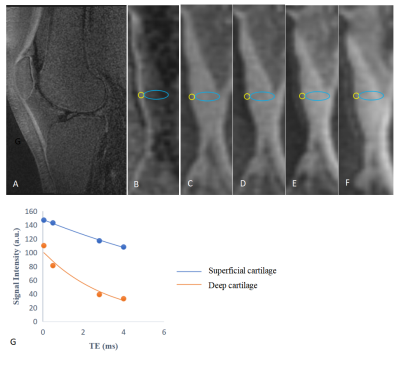 |
Measuring the effect of mechanical loading on deep and superficial cartilage using quantitative UTE MRI
Hanqi Wang1, Qing Li2, Stefan Sommer3,4, and Yong Lu1
1Ruijin Hospital, School of Medicine, Shanghai Jiao Tong University, Shanghai, China, 2MR Collaboration, Siemens Healthcare Ltd., Shanghai, China, Shanghai, China, 3Siemens Healthcare, Zurich, Switzerland, Zurich, Switzerland, 4Swiss Center for Musculoskeletal Imaging (SCMI), Balgrist Campus, Zurich, Switzerland, Zurich, Switzerland
UTE sequences were utilized to quantitatively assess deep and superficial cartilage. Deep cartilage T2* values demonstrated a significant difference in different load-bearing regions while there was no statistical difference for superficial cartilage. No significant change in T2* value of deep cartilage after activity was observed, while T2* values of the superficial layer decreased after activity in both the medial tibial and lateral femoral cartilage, indicating that different bio-mechanical properties exist within the deep and superficial cartilage.
|
|||
 |
4212.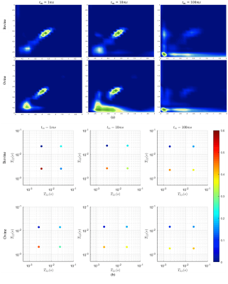 |
Intermediate exchange rate measured between bound and free water pools of tendon: implications on bi-component relaxation modeling
Muhammad Ali Raza Anjum1, Anshuman Swain 1, Johannes Leisen2, Felix Gonzalez1, and David Reiter 1
1Emory University, Atlanta, GA, United States, 2Georgia Tech, Atlanta, GA, United States
Bi-component T2* modelling is widely applied to tendon to elucidate structural and biochemical properties of tissue. This model does not incorporate magnetization exchange between collagen-bound water and interstitial water pools. This study reports intermediate exchange-rates compared with respective pool relaxation rate estimates, in ex vivo bovine and ovine Achilles tendon, measured using T2–T2 correlation NMR. Intermediate exchange causes the bi-component model to underestimate the population of the rapidly-relaxing pool and overestimate relaxation rates and the population of the slowly-relaxing pool. The two-pool exchange model could be more specific to structural and biochemical changes in tendon with pathology.
|
||
4213.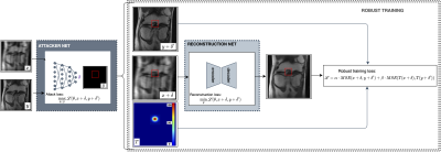 |
State of the ART (Adversarial Robust Training) to Reconstruct Clinically Relevant Features in Accelerated Knee MRI
Francesco Caliva1, Victor Kaiyang Cheng2, Rutwik Shah1, Misung Han1, Sharmila Majumdar1, and Valentina Pedoia1
1University of California San Francisco, San Francisco, CA, United States, 2University of California Berkeley, Berkeley, CA, United States
We propose an Adversarial Robust Training (ART) strategy to overcome the problem with accelerated MRI models, which are prone to missing small yet clinically relevant features. We introduced small, difficult to reconstruct synthetic features to undersampled MRIs and encouraged their reconstruction through robust training. To assess generalizability of our technique to real world applications, we annotated morphological features relevant to musculoskeletal disease diagnosis on images in the FastMRI dataset and tested ART. Overall, the approach has potential to reduce network instability and improve reliability and fidelity in image reconstruction.
|
|||
4214.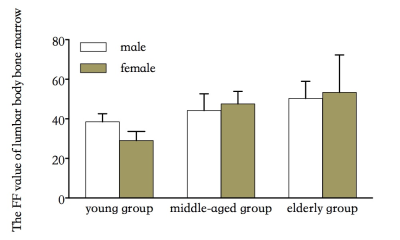 |
Quantitative Evaluation of Lumbar Bone Marrow Fat Content with Age,Gender and Body Mass Index by Using 3D mDixon Quant
Yu Song1, Qingwei qing Song2, Yingkun Guo1, Gang Ning1, Xuesheng Li1, and Yu Song3
1Department of Radiology, West China Second University Hospital, Sichuan University, Chengdu, China, 2Department of Radiology, the First Affiliated Hospital of Dalian Medical University, Dalian, China, 3Department of Radiology, West China Second University, Sichuan University, Chengdu, China
The changes of adipose tissue in bone marrow will directly affect the changes of bone marrow composition. 3D mDixon Quant technology is a new method for precise fat quantification developed by Philips. It automatically generates high-quality FF and T2 * mapping maps by multi-echo acquisition combined with 7 peak fat model and T2 * correction, so as to achieve accurate and rapid quantification of bone marrow fat content.
|
|||
4215.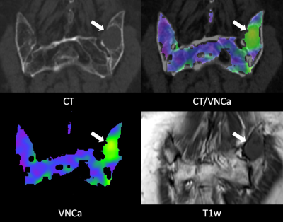 |
Dual-energy CT-based bone marrow imaging in multiple myeloma: Assessment of focal lesions in relation to disease status and MRI findings
Sebastian Werner1, Bernhard Krauss2, and Marius Horger1
1Radiology, University Hospital Tuebingen, Germany, Tuebingen, Germany, 2Siemens Healthineers, Forchheim, Germany
Retrospective quantitative evaluation of 103 focal osteolytic lesions of the axial skeleton in virtual-non-calcium bone marrow images of 32 multiple myeloma patients in correlation to hematologic disease status, T1w signal intensity and ADC. Virtual-non-calcium bone marrow imaging allows differentiation between overall active and inactive disease with higher attenuation signifying an increasing likelihood of active disease. This is supported by a significant positive correlation between the attenuation and the ADC, as well as a corresponding inverse correlation to T1w signal intensity.
|
|||
4216.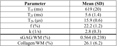 |
Quantitative Magnetization Transfer MRI of the Knee Meniscus: Correlations with Biochemistry and Histology
Kirstin D Olsen1, Lumeng Cui1, Brennan E Berryman2, Ives R Levesque3, and Emily J McWalter2
1Biomedical Engineering, University of Saskatchewan, Saskatoon, SK, Canada, 2Mechanical Engineering, University of Saskatchewan, Saskatoon, SK, Canada, 3Medical Physics, McGill University, Montreal, QC, Canada
The objective of this research was to determine if quantitative magnetization transfer (qMT) MRI can assess knee meniscus tissue health. Cadaver knee menisci underwent qMT assessment, biochemical content assessment, and histological scoring. Weak correlations were found between T1f and collagen content, T1f and histology score, and the histology scores and T2f and T2b. A moderate correlation was found between T2f and collagen content. qMT shows promise as a measure of meniscal tissue health but future studies with more severely damaged tissue are required to elucidate relationships between qMT and measures of meniscal content and health.
|
|||
4217.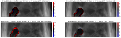 |
A multi-spectral imaging template of hip arthroplasty showing distribution of 3T vs. 1.5T artifacts in the acetabulum of varying implants
Andrew S. Nencka1, Peter S. Johnson1, and Kevin M. Koch1
1Radiology, Medical College of Wisconsin, Milwaukee, WI, United States
An average proton density weighted template of the hip with arthroplasty was developed, and multi-spectral images from patients acquired at both 1.5T and 3.0T were registered to it. As a proof-of-concept, maps of residual metal artifact were transformed into the template space and, for different implant compositions, probability maps of residual artifact were generated. Ceramic implants exhibit minimal cross-field artifact differences, while metal-on-metal implants show more profound differences and metal-on-poly implants exhibit more moderate differences. This template and registration software lays the foundation for future quantitative analysis of cross-session and cross-subject imaging of hip arthroplasty.
|
|||
4218.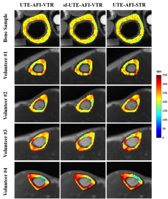 |
Fast T1 Measurement of Cortical Bone using 3D Ultrashort Echo Time Actual Flip Angle Imaging and Single Repetition Time Acquisition (UTE-AFI-STR)
Zhao Wei1,2,3, Hyungseok Jang1, Zubiad Ibrahim1, Mohammadamin Cheraghi1, Graeme M. Bydder1, Wenhui Yang2,3, and Ya-Jun Ma1
1Department of Radiology, UC San Diego, San Diego, CA, United States, 2Institute of Electrical Engineering, Chinese Academy of Sciences, Beijing, China, 3University of Chinese Academy of Sciences, Beijing, China
To improve the time efficiency for cortical bone T1 measurement on clinical scanners, we proposed a new method which fits the datasets of 3D ultrashort echo time actual flip angle imaging (UTE-AFI) and UTE with a single TR (UTE-STR) simultaneously (UTE-AFI-STR). The results show that the UTE-AFI-STR can measure the T1 of cortical bone accurately and more efficiently than the UTE-AFI-VTR method.
|
|||
4219.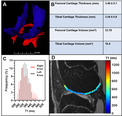 |
Quantitative 7T MRI for Post-traumatic Osteoarthritis Progression in a Rabbit Model
Rossana Terracciano1,2, Yareli Carcamo-Bahena1, Xiaowei Zou3, Joshua D. Harris4, Bradley Weiner4, John Scott Labis5, Nakul Gupta5, and Carly S. Filgueira1,6
1Nanomedicine, Houston Methodist Research Institute, Houston, TX, United States, 2Electronics and Telecommunications, Politecnico di Torino, Torino, Italy, 3Siemens Medical Solutions USA Inc, Malvern, PA, United States, 4Orthopedic Surgery, Houston Methodist Research Institute, Houston, TX, United States, 5Clinical Radiology, Houston Methodist Research Institute, Houston, TX, United States, 6Cardiovascular Surgery, Houston Methodist Research Institute, Houston, TX, United States
No medical therapies have been shown to halt or slow the progression of post-traumatic osteoarthritis (PTOA), which develops after acute/repetitive injury to a joint. Improvements in non-invasive methods to assess PTOA will help diagnose stages for early clinical interventions. Rabbits offer a translationally relevant animal model where anterior cruciate ligament (ACL) injury allows for monitoring PTOA progression, determining a treatment window for early invention. In this work, we leveraged the high signal-to-noise ratio (SNR) of magnetic resonance imaging at 7T to develop a quantitative, non-invasive, 3D-ultrahigh-resolution dGEMRIC MRI exam for femoral-tibial observations and T1 quantification of matrix changes in rabbits.
|
|||
4220.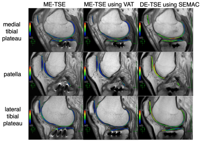 |
Quantitative T2 mapping MRI near metal: comparison of metal artifact reduction techniques
Takayuki Sakai1,2, Masami Yoneyama3, Atsuya Watanabe4,5, Daichi Murayama1, Shigehiro Ochi1, and Tosiaki Miyati6
1Radiology, Eastern Chiba Medical Center, Tonage, Japan, 2Division of Health Sciences, Graduate School of Medical Sciences, Kanazawa University, Kanazawa, Japan, 3Philips Japan, Tokyo, Japan, 4General Medical Services, Chiba University Graduate School of Medicine, Chiba, Japan, 5Orthopaedic Surgery, Eastern Chiba Medical Center, Chiba, Japan, 6Faculty of Health Sciences, Institute of Medical, Pharmaceutical and Health Sciences, Kanazawa University, Kanazawa, Japan
Cartilage degeneration can be evaluated non-invasively by qualitative T2 mapping MRI. Although cartilage evaluation using T2 mapping is desirable in cases which open reduction and internal fixation for fractures around the knee joint and osteotomy for knee osteoarthritis, evaluation is often difficult due to the existence of metal artifacts. Theoretically, metal artifacts are suppressed by metal artifact reduction techniques such as VAT and SEMAC. Therefore, we evaluated the usefulness of T2 mapping using VAT or SEMAC for cartilage around the knee joint that was metal-fixed after surgery.
|
|||
| 4221. | Monitoring the efficacy of TNF-α antagonists in the treatment of SpA preliminary study based on MRI biochemical Imaging technique
Yu Shun1, Lin Min-gui1, Zhongshuai Zhang2, Chen Xian-yuan1, and Ma Mingping1
1Radiology department, Fujian Provincial Hospital, fuzhou, China, 2Diagnostic Imaging, SIEMENS Healthcare, Shanghai, China
This study aimed to evaluate the feasibility of using MRI to monitor the efficacy of tumor necrosis factor-α antagonists in the treatment of SpA (axial spondyloarthritis).
|
The International Society for Magnetic Resonance in Medicine is accredited by the Accreditation Council for Continuing Medical Education to provide continuing medical education for physicians.