Digital Posters
Muscle MRI
ISMRM & SMRT Annual Meeting • 15-20 May 2021

| Concurrent 4 | 17:00 - 18:00 |
3165.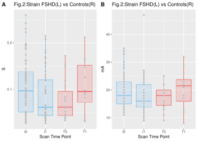 |
Dynamic MR imaging of muscle contraction during electrical stimulation in facioscapulohumeral muscular dystrophy: a longitudinal study
Xeni Deligianni1,2, Francesco Santini1,2, Matteo Paoletti3, Francesca Solazzo3, Niels Bergsland4,5, Giovanni Savini3, Arianna Faggioli3, Giancarlo Germani3, Mauro Monforte6, Giorgio Tasca6, Enzo Ricci6, and Anna Pichiecchio3,7
1Department of Radiology/ Division of Radiological Physics, University Hospital of Basel, Basel, Switzerland, 2Biomedical Engineering, University of Basel, Allschwil, Switzerland, 3Advanced Imaging and Radiomics Center, Neuroradiology Department, IRCCS Mondino Foundation, Pavia, Italy, 4Buffalo Neuroimaging Analysis Center, Department of Neurology, Buffalo Neuroimaging Analysis Center, Department of Neurology, Jacobs School of Medicine and Biomedical Sciences, Buffalo, NY, United States, 5IRCCS, Fondazione Don Carlo Gnocchi ONLUS, Milan, Italy, 6Unità Operativa Complessa di Neurologia, Fondazione Policlinico Universitario A. Gemelli IRCCS, Rome, Italy, 7Department of Brain and Behavioral Sciences, University of Pavia, Pavia, Italy
Quantitative muscle MRI (T2&fat mapping) is progressively more used to assess disease involvement in muscle disorders, but in a static way, with dynamic assessment usually performed by clinical and instrumental examinations. To characterize muscle deformation we applied dynamic MRI synchronized with neuromuscular electrical stimulation evaluating the quadriceps muscles in 34 ambulatory subjects affected by facioscapulohumeral muscular dystrophy (FSHD) and ten healthy controls, longitudinally. Maximum values and rates of the strain differed between the two groups. Our results suggest that the evaluation of muscle ability to contract could be potentially used to monitor the evolution of muscle involvement in FSHD.
|
|||
3166.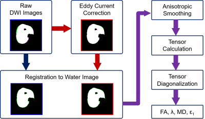 |
Effect of Eddy Current Correction on Muscle Diffusion Measurements
Xingyu Zhou1,2, Melissa T. Hooijmans1,3, Crystal L. Coolbaugh1, Mark K. George1, and Bruce M. Damon1,2,4,5
1Vanderbilt University Institute of Imaging Science, Nashville, TN, United States, 2Department of Biomedical Engineering, Vanderbilt University, Nashville, TN, United States, 3Department of Radiology & Nuclear Medicine, Amsterdam Movement Sciences, Amsterdam University Medical Center, Amsterdam, Netherlands, 4Department of Radiology & Radiological Sciences, Vanderbilt University Medical Center, Nashville, TN, United States, 5Department of Molecular Physiology & Biophysics, Vanderbilt University Medical Center, Nashville, TN, United States
Diffusion-tensor MRI (DTMRI) fiber tractography is a useful tool for reconstructing the structure of white matter tracts, nerves, and muscles. Eddy current correction is recognized as an important step in analysis of neuronal DTMRI data, while its effect on muscle DTMRI analysis has not been studied systematically. In this work we describe a preliminary comparison of muscle diffusion tensor imaging results with and without including eddy current correction in the pre-processing steps, including the morphological mismatch with anatomical image, the estimation of eigenvalues and fractional anisotropy, and the distribution of the first eigenvector within the selected muscle.
|
|||
3167.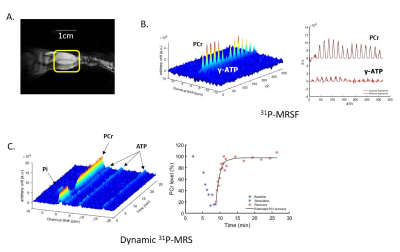 |
Altered creatine kinase activity and mitochondrial oxidative capacity in muscular dystrophic mdx mice after repeated muscle contraction
Kihwan Kim1, Yuning Gu1, Yuran Zhu1, Yudu Li2,3, Sherry Huang1, Zhi-Pei Liang 2,3, and Xin Yu1,4
1Biomedical Engineering, Case Western Reserve University, cleveland, OH, United States, 2Electrical and Computer Engineering, University of Illinois at Urbana-Champaign, Urbana, IL, United States, 3Beckman Institute for advanced Science and Technology, University of Illinois at Urbana-Champaign, Urbana, IL, United States, 4Case Center for Imaging Research, Case Western Reserve University, cleveland, OH, United States
This study investigated the creatine kinase activity and the effects of repeated muscle contraction on muscle metabolism in mdx mice, a mouse model for Duchenne Muscular Dystrophy (DMD). Using phosphorous-31 (31P) magnetic resonance fingerprinting and dynamic 31P magnetic resonance spectroscopy, we observed a significant decrease in creatine kinase (CK) reaction rate in mdx mice compared to control mice. Decrease in phosphocreatine (PCr) recovery rate after its depletion by stimulation-induced muscle contraction was also observed in mdx mice. These findings suggest altered mitochondrial function in mdx mice.
|
|||
3168.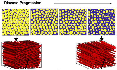 |
Establishing a computational approach to investigate the biophysical basis of relaxivity contrast imaging in the context of muscle degeneration
Natenael B Semmineh1, Sudarshan Ragunathan2, Laura C Bell2, and C Chad Quarles 2
1Imaging Research, Barrow Neurological Institute rrow Neurological Institute, Phoenix, AZ, United States, 2Imaging Research, Barrow Neurological Institute, Phoenix, AZ, United States
The relaxivity contrast imaging parameter, TRATE (tissue transverse relaxivity at tracer equilibrium), has been shown to decrease during ALS induced myofiber degradation, due to T1 and T2* effects that occur when the contrast agent distributes around myofibers. The goal of this study is to expand a validated computational method to investigate the biophysical basis of TRATE in muscles of ALS patients and to optimize RCI acquisition parameters.
|
|||
3169. |
Effect of moderate exercise on diffusion indices in skeletal muscle
Amy R McDowell1, Matthew T Lee2, Kiran K Seunarine2, and Chris A Clark2
1Centre for Neuromuscular Diseases, UCL Queen Square Institute of Neurology, London, United Kingdom, 2GOS UCL Institute of Child Health, London, United Kingdom
An examination of the effect of moderate exercise on diffusion indices in skeletal muscle in volunteers in order to quantify the effect of the exercise, and define when diffusion indices returned to baseline rest values. We found that there was no significant difference between pre- and post-exercise MD or FA. We believe that moderate exercise will have no evident effect on diffusion indices, and no period of rest pre-scan is required.
|
|||
3170. |
Quantification of gluteal and multifidus muscle asymmetry and fat infiltration in patients with unilateral lumbosacral nerve root compression
Mengyue Wang1, Weiqiang Dou2, Yu Zheng1, Yin Shi1, and Yuefen Zou1
1The First Affiliated Hospital of Nanjing Medical University, Nanjing, China, 2GE Healthcare, MR Research China, Beijing, China
Muscle asymmetry and fatty infiltration resulting from lumbosarcal nerve root compression are associated with movement ability directly. However, the specific rule remains unclear. In this study, we used three dimensional fast imaging empolying steady-state acquisition with phase cycling(3D-FIESTA-C) and iterative decomposition of water and fat with echo asymmetry and least-squares estimation intelligent quantification (IDEAL-IQ) technique to evaluate the changes quantitatively. We found muscle changes were side- and level-specific, affected by several other factors. Muscle content is suggested as a good parameter for clinical evaluation.
|
|||
3171.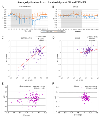 |
Interleaved multi-voxel 1H and 31P MRS for dynamic pH monitoring in the exercising calf muscle at 3 T with dynamic water reference.
Alfredo Liubomir Lopez Kolkovsky1,2, Beatrice Matot1,2, Eric Giacomini1, Martin Meyerspeer3, Benjamin Marty1,2, and Harmen Reyngoudt1,2
1NMR Laboratory, Institute of Myology, Paris, France, 2NMR Laboratory, CEA\DRF\IBFJ\MIRCen, Paris, France, 3High Field MR Center, CMPBME, Medical University of Vienna, Vienna, Austria
We extended an interleaved 1H/31P multi-voxel sequence for simultaneous pH monitoring based on carnosine (pH1H, PRESS) and Pi (pH31P, semi-LASER) with a co-localized dynamic water reference (PRESS). Simultaneous pH evaluations were done at 3 T in the soleus and gastrocnemius muscles during a straight-leg plantar flexion paradigm. A pH1H bias of ~0.05 units was removed by using the dynamic water reference. pH1H and pH31P time courses (1-minute temporal resolution) were comparable. Pi and Carnosine (C2-H) resonances broadened during exercise in the gastrocnemius. This method opens the way for new applications in functional studies targeting individual muscles at 3 T.
|
|||
3172. |
A Marker Controlled Active Contour Model for Thigh Muscle Segmentation in MR Images
Weihong Guo1, Michael Judkovich1, Richard Lartey2, Dongxing Xie2, Mingrui Yang2, and Xiaojuan Li2
1Mathematics, Applied Mathematics and Statistics, Case Western Reserve University, Cleveland, OH, United States, 2Biomedical Engineering, Program of Advanced Musculoskeletal Imaging (PAMI), Lerner Research Institute, Cleveland Clinic, Cleveland, OH, United States
Thigh muscle morphology and composition quantified from MR images are potential imaging biomarkers for diseases such as osteoarthritis and sarcopenia. MR thigh muscle segmentation is an important step in quantifying both muscle morphology and composition. Unfortunately, the thigh muscle groups are tightly bundled together, making them very hard to segment due to a lack of clear boundaries between different muscles. We proposed a novel geometric flow based semi-automatic scheme to effectively segment them. We combined reproducible kernel Hilbert space edge descriptor and geodesic distance maps from a set of markers and anti-markers to define the force for the geometric flow.
|
|||
3173. |
DT-MRI with the random permeable barrier model shows larger, more heterogeneous muscle fibre diameters in Becker muscular dystrophy patients
Donnie Cameron1,2, Jedrzej Burakiewicz1, Nienke M. van de Velde3, Celine Baligand1, Thom T.J. Veeger1, Melissa T. Hooijmans4, Jan J.G.M. Verschuuren3, Erik H. Niks3, and Hermien Kan1
1C.J. Gorter Centre for High Field MRI, Department of Radiology, Leiden University Medical Centre, Leiden, Netherlands, 2Norwich Medical School, University of East Anglia, Norwich, United Kingdom, 3Department of Neurology, Leiden University Medical Centre, Leiden, Netherlands, 4Biomedical Engineering and Physics, Amsterdam University Medical Centre, Amsterdam, Netherlands
Becker muscular dystrophy (BMD) is characterised by progressive muscle damage and weakness. Diffusion tensor imaging (DTI) represents a promising candidate for studying BMD; the random permeable barrier model (RPBM), in particular, gives estimates of muscle fibre diameters and membrane permeabilities. Here we study DTI and RPBM metrics in BMD patients and controls. Spin-echo and stimulated-echo DTI data were acquired in the lower leg and RPBM metrics calculated. DTI metrics showed time-dependence and intramuscular differences, but no inter-group differences. RPBM analysis, however, successfully showed differences between BMD patients and controls, with fibre diameters being larger and more variable in patients.
|
|||
3174.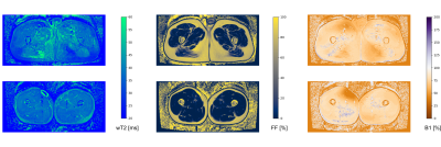 |
Optimized water T2 mapping from multi-echo spin-echo acquisitions with an external fat fraction constraint
Francesco Santini1,2, Xeni Deligianni1,2, Matteo Paoletti3, Francesca Solazzo3, Matthias Weigel1,4,5, Paulo Loureiro De Sousa6, Oliver Bieri1,2, Mauro Monforte7, Enzo Ricci7,8, Giorgio Tasca7, Anna Pichiecchio3,9, and Niels Bergsland10,11
1Radiological Physics, Department of Radiology, University Hospital of Basel, Basel, Switzerland, 2Department of Biomedical Engineering, University of Basel, Allschwil, Switzerland, 3Neurological Institute Foundation Casimiro Mondino (IRCCS), Pavia, Italy, 4Translational Imaging in Neurology, Department of Biomedical Engineering, University of Basel, Allschwil, Switzerland, 5Department of Neurology, University Hospital of Basel, Basel, Switzerland, 6UMR7357 Laboratoire des sciences de l'Ingénieur, de l'Informatique et de l'Imagerie (ICube), Strasbourg, France, 7Institute of Neurology, A. Gemelli University Hospital Foundation, Catholic University of the Sacred Heart, Rome, Italy, 8Institute of Neurology, Catholic University of the Sacred Heart, Rome, Italy, 9Department of Brain and Behavioral Sciences, University of Pavia, Pavia, Italy, 10Western New York Stem Cell Culture and Analysis Center, Jacobs School of Medicine and Biomedical Sciences, University of Buffalo, Buffalo, NY, United States, 11Fondazione Don Carlo Gnocchi Onlus (IRCCS), Milan, Italy
The measurement of the T2 of the water component is an important marker of neuromuscular diseases. Strategies exist to estimate it from conventional multi-echo spin-echo acquisitions, taking into account fat infiltration and B1 inhomogeneity. In this work, we present an open software that is able to efficiently perform this kind of fitting, and additionally to incorporate the information from an external fat fraction acquisition, for increased accuracy and reduced scan time.
|
|||
3175.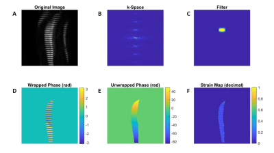 |
Elliptical filter optimization for HARP based strain quantification in skeletal muscle
Melissa T. Hooijmans1,2, Crystal L. Coolbaugh3, Xingyu Zhou2,4, Mark K. George2, and Bruce M. Damon4,5,6
1Department of Radiology & Nuclear Medicine, Amsterdam Movement Sciences, Amsterdam UMC, Location AMC, Amsterdam, Netherlands, 2Vanderbilt University Institute of Imaging Science, Nashville, TN, United States, 3Vanderbilt University Institute of Imaging Sciences, Nashville, TN, United States, 4Department of Biomedical Engineering, Vanderbilt University Medical Center, Nashville, TN, United States, 5Department of Radiology & Radiological Sciences, Vanderbilt University Medical Center, Nashville, TN, United States, 6Department of Molecular Physiology & Biophysics, Vanderbilt University Medical Center, Nashville, TN, United States
SPAMM, combined with HARP, is primarily used for strain quantification in the myocardium; but applications in other soft tissues, including skeletal muscle, are increasing. HARP suffers from artifacts due to usage of inappropriate filters and should therefore be optimized especially for skeletal muscle. We simulated strains ranging from -0.156 to +0.156 (decimal strain) in dynamically acquired line- and grid-tagged SPAMM images, and optimized elliptical filter parameters were determined for skeletal muscle. With this filter, differences between the measured strain and absolute strain were small for the low strain values and increased with the actual strain values and number of dynamics.
|
|||
3176.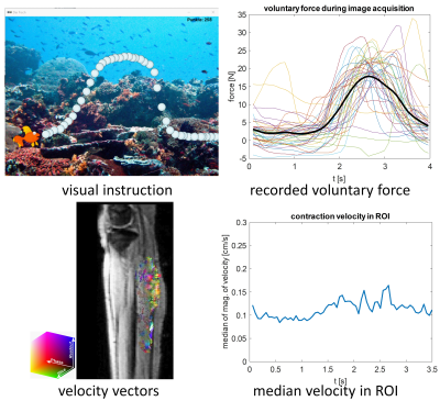 |
Imaging of calf muscle contraction in pediatric patients with cerebral palsy: comparison of voluntary motion and electrically evoked motion
Claudia Weidensteiner1,2, Xeni Deligianni1,2, Tanja Haas1, Philipp Madoerin1, Oliver Bieri1,2, Meritxell Garcia3, Jacqueline Romkes4, Erich Rutz5, Francesco Santini1,2, and Reinald Brunner6
1Department of Radiology, Division of Radiological Physics, University Hospital Basel, Basel, Switzerland, 2Department of Biomedical Engineering, University of Basel, Basel, Switzerland, 3Department of Radiology, Division of Neuroradiology, University Hospital Basel, Basel, Switzerland, 4Laboratory for Movement Analysis, University Children's Hospital Basel, Basel, Switzerland, 5Hugh Williamson Gait Laboratory, The Royal Children's Hospital Melbourne, Parkville, Australia, 6Department of Orthopedic Surgery, University Children's Hospital Basel, Basel, Switzerland
Aim of this study is to investigate the feasibility of phase contrast imaging at 3T for assessment of muscle function in children with cerebral palsy. Time-resolved cine phase contrast MRI was synchronized with (a) electrical muscle stimulation (EMS) of the calf muscle (b) voluntary plantarflexion following visual instructions. The achieved force was higher for the voluntary task, but its periodicity was lower compared to the stimulated contraction. Therefore, it was not possible to resolve two distinct velocity peaks for voluntary contraction and release, but it was during EMS, in case the tolerated current was high enough to evoke sufficient contraction.
|
|||
3177.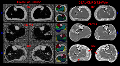 |
IDEAL-CPMG muscle T2-water and Dixon Fat-Fraction Maps in People with Inclusion Body Myositis and Charcot-Marie-Tooth disease Type 1A
Amy R. McDowell1, Stephen J. Wastling1,2, Lara Cristiano3, Jasper M. Morrow1, Matthew R.B. Evans4, Christopher D.J. Sinclair1, Pedro M. Machado1, Michael Hanna1, Mary M. Reilly1, Tarek Yousry1, and John S. Thornton1
1MRC Centre for Neuromuscular Diseases, UCL Queen Square Institute of Neurology, London, United Kingdom, 2Lysholm Department of Neuroradiology, National Hospital for Neurology and Neurosurgery, London, United Kingdom, 3Department of Pediatric Neurology and Radiology, Fondazione Policlinico Universitario, Rome, Italy, 4St Thomas Hospital, London, United Kingdom
Muscle T2 and fat-fraction are two promising metrics for sensitive measures of early disease-related changes in muscle for use in clinical trials. However, fat has a longer T2 than water in muscle and masks underlying increases in water T2 due to muscle oedema. The IDEAL-CPMG sequence with appropriate image-data processing produces a T2-water map uncontaminated by the fat signal. This sequence was acquired in healthy volunteers and two different muscular dystrophy disease types. It was found that elevated T2-water may be a predictor of later progression to fatty-atrophy in muscle, supporting the value of future longitudinal studies to test this.
|
|||
3178.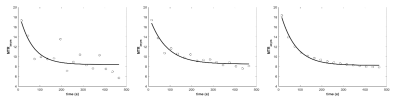 |
Denoising dynamic CrCEST in skeletal muscle following exercise using low rank tensor approximations
Neil Wilson1, Mark A Elliott2, Dushyant Kumar2, and Ravinder Reddy2
1Siemens Medical Solutions USA Inc, Malvern, PA, United States, 2CMROI, University of Pennsylvania, Philadelphia, PA, United States
Dynamic CrCEST allows for high resolution mapping of the creatine kinase reaction recovery kinetics following exercise. However, voxel averaging over large ROIs is usually done to get reliable fits because of high variation due to limited SNR. Here, we show that improved denoising utilizing low rank tensor approximations that exploit the full dimensionality of the scans allows for reliable fits over smaller volumes or even single voxels, making muscle response heterogeneity measurable.
|
|||
3179.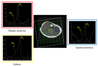 |
Feasibility Study of 31P Multivoxel Spectroscopy for the detection of Alkaline Inorganic Phosphate in multiple compartments of the lower leg at 3T
Rajakumar Nagarajan1 and Gwenael Layec2
1Human Magnetic Resonance Center, Institute for Applied Life Sciences, University of Massachusetts Amherst, Amherst, MA, United States, 2Kinesiology, University of Massachusetts Amherst, Amherst, MA, United States
In this study, we demonstrate the feasibility and sensitivity of 31P two-dimensional CSI sequence for the detection of alkaline Pi in the soleus, tibialis anterior, and gastrocnemius muscles in six healthy volunteers in rest. The major finding of the current study was the consistent detection of an alkaline Pi from the cytosolic Pi signal in the volume localized 31P MRS from the resting muscle in healthy human subjects at 3T using decoupling technique. This study provides the proof of concept for a non-invasive and localized method to determine the alkaline Pi, a potential index of mitochondrial density at rest.
|
|||
3180.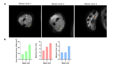 |
Measuring motor unit morphology in upper extremities using motor unit magnetic resonance imaging (MUMRI)
Matthew Birkbeck1,2,3, Linda Heskamp1, Ian Schofield1, Roger Whittaker1, and Andrew Blamire1
1Translational and Clinical Research Unit, Newcastle University, Newcastle upon Tyne, United Kingdom, 2Newcastle Biomedical Research Centre, Newcastle Biomedical Research Centre, Newcastle upon Tyne, United Kingdom, 3Northern Medical Physics and Clinical Engineering, Newcastle upon Tyne NHS Foundation Trust, Newcastle upon Tyne, United Kingdom
Motor unit (MU) magnetic resonance imaging (MUMRI) is a non-invasive technique which detects muscle fibre micro-contraction and is based on diffusion weighted MRI. To date MUMRI has been applied in conjunction with in scanner electrical stimulation to study MU activity in the lower leg. Here we present the first use of MUMRI in the upper limbs to study single human MUs. The acquired images show low levels of distortion and good fat suppression, allowing single human MU sizes and shapes to be determined. This is of interest in neuromuscular diseases as it is a non-invasive way to study MU morphology.
|
|||
3181.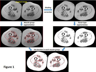 |
MR analysis of thigh muscle myopathy using texture features and supervised machine learning
Hon J Yu1, Saya Horiuchi1,2, Toshimi Tando1, Vincent J Caiozzo3, Virginia E Kimonis4, and Hiroshi Yoshioka1
1Radiological Sciences, University of California, Irvine, Orange, CA, United States, 2Radiology, St. Luke's International Hospital, Tokyo, Japan, 3Department of Orthopaedics, Physiology & Biophysics, University of California, Irvine, Irvine, CA, United States, 4Division of Genetic and Genomic Medicine, Department of Pediatrics, University of California, Irvine, Irvine, CA, United States
This study evaluates texture features to demonstrate their relationship with muscle classification based on a 5-grade scale and their value as classifier when trained in supervised machine-learning framework. The results suggest the texture features capture various image characteristics that are likely utilized during manual muscle classification by human being and can correctly predict with up to 81% accuracy when properly trained in supervised machine-learning setting. A further study with bigger data size would be necessary to fully examine such classification model and also to look into the possibility of selecting subset of features to make such an approach more practical.
|
|||
3182.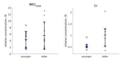 |
1H MRS: a tool to study age-related changes in intracellular metabolites in the deep spinal muscles?
Sarah Catherine Wayte1, Alexander Dallaway2, Andrew David Weedall1, John Hattersley3, and Adrian John Wilson3,4
1Radiology Physics, Department of Clinical Physics and Bioengineering, University Hospital, Coventry, United Kingdom, 2Centre for Sport, Exercise and Life Sciences, Coventry University, Coventry, United Kingdom, 3Coventry NIHR CRF, Human Metabolic Research Unit, University Hospital, Coventry, United Kingdom, 4Department of Physics, University of Warwick, Coventry, United Kingdom
1H MRS was used to investigate relative concentrations of methylene intracellular lipids [IMCLCH2]R and creatine [Cr]R in the psoas of groups of younger (19-30yrs) and older (61-81yrs) fit male volunteers. No difference was found between groups for either metabolite (p>0.05). However, the median value for [Cr]R in the older group (median;Q1-Q3=1.02;0.55-1.29%) was much higher than in the younger group (0.57;0.50-0.60%) suggesting [Cr]R could partially explain reduction in muscle strength with age being greater than predicted by loss of muscle mass. There was no correlation between metabolite concentrations (r2=0.21) so [Cr]R may possibly be a marker for age-related changes in muscle.
|
|||
3183.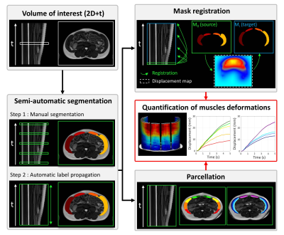 |
SEMI-AUTOMATIC QUANTIFICATION OF ABDOMINAL WALL MUSCLES DEFORMATIONS BASED ON DYNAMIC MRI IMAGE REGISTRATION
Arthur Jourdan1, Arnaud Le Troter2, Pierre Daude2, Stanislas Rapacchi2, Catherine Masson1, Thierry Bege1,3, and David Bendahan2
1Aix-Marseille Univ, Univ Gustave Eiffel, IFSTTAR, LBA, Marseille, France, 2Aix Marseille Univ, CNRS, CRMBM, Marseille, France, 3Department of General Surgery, Aix Marseille Univ, North Hospital, APHM, Marseille, France
A novel semi-automatic post-processing method dedicated to real-time dynamic MRI aiming at a fast and reliable quantification of abdominal wall muscles deformations. The described method combines a highly accurate supervised 2D+t segmentation procedure of the abdominal wall muscles (mean Dice similarity coefficient of 0.95 ± 0.03), the quantification of muscles deformations based on masks registration and the mapping of deformations within muscle compartments leveraging a dedicated parcellation. The present genuine method provides a quantitative analytical frame that could be used in further studies for a better understanding of abdominal wall deformations in physiological and pathological situations.
|
|||
3184. |
Feasibility and Reproducibility Study of Diffusion-Tensor Imaging in Rotator Cuff Muscles of Asymptomatic Volunteers.
Cyril Tous, PhD1, Alexandre Jodoin, MD2, Detlev Grabs, MD, PhD3, Elijah Van Houten, PhD4, and Nathalie J Bureau, MD MSc FRCP(C)1,2
1Radiology, Centre de recherche du Centre hospitalier de l’Université de Montréal, Montreal, QC, Canada, 2Radiology, Centre hospitalier de l’Université de Montréal, Montréal, QC, Canada, 3Anatomy, Université du Québec à Trois-Rivières, Trois-Rivières, QC, Canada, 4Mechanical Engineering, Université de Sherbrooke, Sherbrooke, QC, Canada
Surgical planning of rotator cuff tears does not benefit from quantitative measurement of muscles stiffness and microstructure leading to retears in the following years. Since stiffness is dependent on anisotropy, Diffusion Tensor Imaging can simplify the inversion algorithm of Magnetic resonance elastography. Repeatability of DTI metrics from three scans in six shoulders of six asymptomatic volunteers was achieved (coefficient of variance <10%) for the fractional anisotropy, mean diffusivity, mode and eigenvalues. Clear convergence of myocytes to the tendons was observed with tractography in the supraspinatus as known in dissection. This MRI protocol is promising for developing biomarkers for surgical planning.
|
The International Society for Magnetic Resonance in Medicine is accredited by the Accreditation Council for Continuing Medical Education to provide continuing medical education for physicians.