Digital Posters
Cartilage I
ISMRM & SMRT Annual Meeting • 15-20 May 2021

| Concurrent 4 | 15:00 - 16:00 |
 |
2946.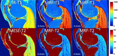 |
Relaxation Time Mapping of Knee Articular Cartilage Using Magnetic Resonance Fingerprinting
Sanam Assili1, Victor Casula1,2, Jaakko Ikäheimo1, Egor Panfilov1, Ari Väärälä1, Martijn A. Cloos3, Riccardo Lattanzi4, and Miika T. Nieminen1,2,5
1Research Unit of Medical Imaging, Physics and Technology, University of Oulu, Oulu, Finland, 2Medical Research Center, University of Oulu and Oulu University Hospital, Oulu, Finland, 3Centre for Advanced Imaging, University of Queensland, Brisbane, Queensland, Australia, 4Center for Advanced Imaging Innovation and Research, New York University Grossman School of Medicine, New York, NY, United States, 5Department of Diagnostic Radiology, Oulu University Hospital, Oulu, Finland
Knee osteoarthritis is a degenerative musculoskeletal disorder which is characterized by progressive damage of articular cartilage (AC). Quantitative MRI (qMRI), such as T1 and T2 maps, can be used to assess the AC compositional changes. However, long acquisition time and equipment dependency are the limitations of the current clinical qMRI techniques. In the present study, we validate magnetic resonance fingerprinting (MRF) against conventional T1 and T2 relaxation time mapping methods in cartilage-mimicking phantoms, and apply MRF in vivo for sub-regional analysis of femoral and tibial cartilage in human subjects.
|
||
2947.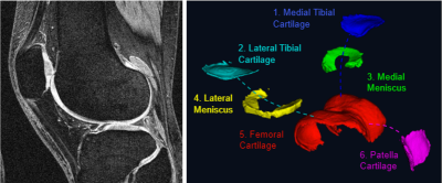 |
Deriving a Cartilage Signature to Predict Joint Replacement in Osteoarthritis
Edward Peake1,2,3, Stefan Pszczolkowski1,2,3, Christoph Arthofer2,3,4, and Dorothee P Auer1,2,3
1NIHR Nottingham Biomedical Research Centre, University of Nottingham, Nottingham, United Kingdom, 2Radiological Sciences, Division of Clinical Neuroscience, University of Nottingham, Nottingham, United Kingdom, 3Sir Peter Mansfield Imaging Centre, University of Nottingham, Nottingham, United Kingdom, 4Nuffield Department of Clinical Neuroscience, University of Oxford, Oxford, United Kingdom
Prediction of future knee replacement in osteoarthritis may support tailored decision making, and provide a surrogate outcome marker for clinical trials of diseases modifying osteoarthritis drugs. Fully automated cartilage segmentation is generated from knee MRI, and feature are extraction to create a radiomic signature for the prediction of knee replacement within 5 -years. The radiomic signature using univariate cox regression predicted surgery with a HR of 7.5 (p = 2e-28, 95% CI 7.1 – 7.9) which was higher than established clinico-demographic multivariate prediction model with HR 5.9 (p = 2e-28, 95%CI 5.6 – 6.2).
|
|||
 |
2948.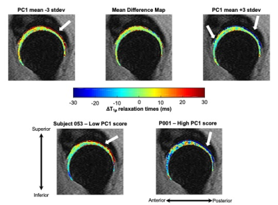 |
T1ρ relaxation times for voxel-wise characterization of longitudinal changes in hip cartilage biochemistry
Koren Roach1, Richard Souza1, Sharmila Majumdar1, and Valentina Pedoia1
1UCSF, San Francisco, CA, United States
Hip osteoarthritis coincides with or is preceded by biochemical changes in articular cartilage,1, 2 which can be measured on a voxel-wise basis with magnetic resonance T1ρ and T2 relaxation times. In this study, we employed principal component analysis to characterize variation in longitudinal changes of hip cartilage T1ρ relaxation times across healthy subjects and those with early-to-moderate hip osteoarthritis. Our results indicated that over two years, a majority of the variation in T1ρ relaxation time changes was located in the deep layers of the acetabular cartilage.
|
||
2949.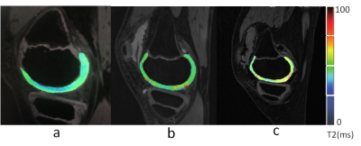 |
The value of T2 mapping to characterize early knee joint cartilage damage in hemophilia arthropathy
Shufang Wei1, Jiajia Li1, Xianchang Zhang2, Jing An3, and Yinghui Ge1
1Fuwai Central China Cardiovascular Hospital, Zhengzhou, China, 2MR Collaboration, Siemens Healthcare Ltd, Beijing, China, 3Siemens Shenzhen Magnetic Resonance Ltd, Shenzhen, China
Detecting early articular cartilage damage can help formulate preventive treatment plans for hemophilia arthropathy (HA). Magnetic responance imaging (MRI) T2 mapping has been used extensively to investigate early chondral changes in osteoarthritis; however, it has seldomly been applied to investigate HA. This study acquired T2-mapping results on 30 patients (54 knees) with HA and 15 volunteers (24 knees). Patients with HA had significantly higher knee cartilage T2 values than the healthy controls. The T2 values also increased with the disease severity, suggesting that MRI T2 mapping is a promising method for detecting early stage cartilage damage in patients with hemophilia.
|
|||
2950.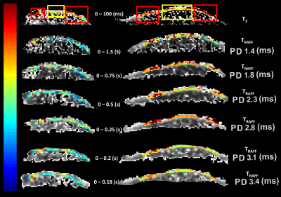 |
Optimization of Relaxation along Fictitious Field (RAFF) Contrast to Detect Osteoarthritis
Seyed Amir Mirmojarabian1, Victor Casula1, Olli-Pekka Aro1, Henning Henschel2, Miika T Nieminen1,3,4, and Timo Liimatainen1,3
1Research Unit of Medical Imaging, Physics and Technology, University of Oulu, Oulu, Finland, 2Department of Medicinal Chemistry, Uppsala University, uppsala, Sweden, 3Medical Research Center, Oulu University Hospital, Oulu, Finland, 4Department of Diagnostic Radiology,, Oulu University Hospital, Oulu, Finland
Relaxation along Fictitious Field (RAFF) at various pulse durations was exploited to study the contrast in collagen phantoms and cartilage-mimicking phantoms. RAFF Relaxation times from collagen phantoms agreed with Bloch McConnell simulations. The maximum contrast between collagen phantoms with concentration difference was maximized when pulse duration was ~2.8 ms. Given the optimized pulse duration, our simulation results combined with ex vivo measurements suggest that hydroxyl groups concentration through proton exchange between collagen and free water may have significant role in TRAFF contrast between intact and degraded cartilage.
|
|||
2951. |
The value of magnetic resonance T2*mapping for early detection of knee cartilage damage in hemophilia arthropathy
Jiajia Li1, Shufang Wei1, Xianchang Zhang2, Jing An3, and Yinghui Ge1
1Fuwai Central China Cardiovascular Hospital, Zhengzhou, China, 2MR Collaboration, Siemens Healthcare Ltd., Beijing, China, Beijing, China, 3Siemens Shenzhen Magnetic Resonance Ltd, Shenzhen, China
Early detection and therapy of articular cartilage damage are key to the management of hemophilia arthropathy (HA). Magnetic resonance (MR) T2* mapping has been used to detect iron deposits in the brain and liver but rarely in HA. We compared T2*mapping results of 15 HA patients (18 knees) and 10 volunteers (18 knees). We found that the average T2* values of the medial femoral condyle and medial tibial plateau cartilage in HA patients were significantly lower than in the normal group, suggesting the importance of T2* mapping in the quantitative assessment of iron deposition for early cartilage damage diagnosis in HA patients.
|
|||
2952.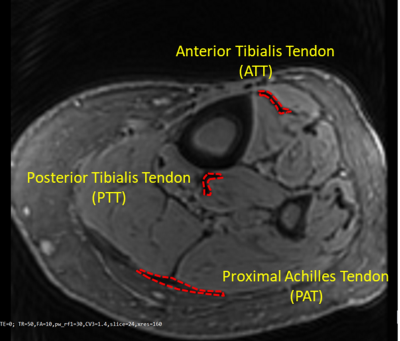 |
Decreased collagen content in tendons of patients with osteoporosis and osteopenia detected with ultrashort echo time cones MRI
Saeed Jerban1, Yajun Ma1, Amir Masoud Afsahi1, Douglas G Chang2, Zhao Wei1, Meghan Shen1, Mei Wu1, Alecio Lombardi1,3, Nicole Le4, Jiang Du1, and Eric Y Chang1,3
1Radiology, University of California, San Digeo, La Jolla, CA, United States, 2Orthopaedic Surgery, University of California, San Digeo, La Jolla, CA, United States, 3Radiology Service, VA San Diego Healthcare System, San Diego, CA, United States, 4Radiology, VA San Diego Healthcare System, La Jolla, CA, United States
Bone and tendon comprise a highly interactive mechanical unit. Investigating tendons quality during osteoporosis and osteopenia progress is of great interests. Clinical MRI sequences are not often capable of directly visualizing tendon because of the tissue’s short T2. Ultrashort echo time (UTE) MRI combined with magnetization transfer (MT) modeling (UTE-MT) has demonstrated promise as a quantitative technique that is resistant to the magic angle effect. Lower leg tendons in osteopenia and osteoporosis patients were compared with healthy subjects via UTE-MT. Macromolecular fraction (MMF) obtained from UTE-MT modeling showed a significant reduction in osteopenia and osteoporosis patients compared with healthy subjects.
|
|||
|
2953.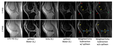 |
Highly Efficient Single-Point Dixon-Based Fat Suppression for Ultrashort Echo Time Double Echo Steady State (UTE-DESS) Imaging of the Knee Joint
Hyungseok Jang1, Yajun Ma1, Michael Carl2, Saeed Jerban1, Eric Y Chang1,3, and Jiang Du1
1University of California, San Diego, San Diego, CA, United States, 2GE Healthcare, San Diego, CA, United States, 3Veterans Affairs San Diego Healthcare System, San Diego, CA, United States
In this study, we explored the feasibility of applying single-point Dixon (spDixon)-based fat suppression to a novel ultrashort echo time-based double echo steady state (UTE-DESS) sequence for highly efficient morphological musculoskeletal (MSK) imaging. Compared to the two-point Dixon-based approach used with conventional UTE-DESS MSK imaging which requires two echoes for fat-water separation, we demonstrated a new technique which requires only a single image in, removing the need for additional data acquisitions. The feasibility and efficacy of our approach were demonstrated in human knee joints.
|
||
2954.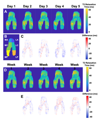 |
Examining short-term longitudinal and activity-based variability of femoral cartilage T2 relaxation times in healthy subjects
Lauren Watkins1,2, Andrew Schmidt1, Elka Rubin1, Marco Barbieri1, Arjun Desai1,3, Valentina Mazzoli1, Marianne Black1, Garry Gold1,2, Brian Hargreaves1,2,3, Akshay Chaudhari1,4, and Feliks Kogan1
1Radiology, Stanford University, Stanford, CA, United States, 2Bioengineering, Stanford University, Stanford, CA, United States, 3Electrical Engineering, Stanford University, Stanford, CA, United States, 4Biomedical Data Science, Stanford University, Stanford, CA, United States
Subtle variations in T2 relaxation times may be related to the risk of early osteoarthritis and progression. It is important to estimate T2 variability in healthy individuals as a basis for cross-sectional and longitudinal comparisons. We examine short-term T2 variability in femoral articular cartilage of healthy volunteers over 5 days and 5 weeks and consider the impacts of physical activity. Over both periods, mean T2 change, variance, and CVRMS were small and comparable between knees and cartilage subregions. There was a moderate positive correlation between T2 changes and weekly rated perceived exertion over 5 weeks
|
|||
2955.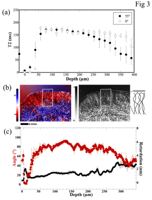 |
Morphologic Structure of Rabbit Suprapatella Cartilage by µMRI and PLM
Hannah Mantebea1, Syeda Batool1, Mouhamad Hammami1, and Yang Xia1
1Physics, Oakland University, Rochester, MI, United States
This study aims to quantity the structural morphology of sesamoid fibrocartilage in rabbits, using µMRI at 19.5-µm resolution and polarized light microscopy at 1-µm resolution. We show that the sesamoid fibrocartilage has one thin surface layer (~ 10 µm) of parallel-arranged fibers followed immediately by the majority of random fibers (~ 390 µm). T2 anisotropy was not observed in fibrocartilage. The collagen fibers in the surface layer are dense and in parallel with the surface, which allows it to withstand stress from the quadriceps tendon during flexion, while at the same time to reduce pressure load from the femur.
|
|||
2956.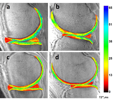 |
Deep Cartilage UTE-T2* Shows Compositional Heterogeneity in Patients with Degenerative Meniscus Tears
Ashley A. Williams1,2, Karyn E. Chappell1,2, and Constance R. Chu1,2
1Orthopaedic Surgery, Stanford University, Stanford, CA, United States, 2Veterans Affairs Palo Alto Health Care System, Palo Alto, CA, United States
This study evaluated deep cartilage and meniscus UTE-T2* against spatially targeted arthroscopic assessments in patients with degenerative meniscus tears to further inform interpretation and utility of in vivo UTE-T2* for different degrees of pathology. While arthroscopic evaluation confirmed that UTE-T2* was elevated in menisci with degenerative fraying and tears, increasing intra-operative cartilage grade was not associated with strictly increasing cartilage UTE-T2*. Instead, in osteoarthritic disease states where the articular surface of cartilage is disrupted, this report suggests that mono-exponential deep cartilage UTE-T2* may not be a reliable indicator of tissue pathology.
|
|||
2957.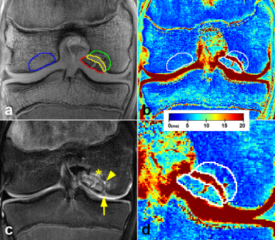 |
Evaluation of Lesion Tissue Composition in Patients with Juvenile Osteochondritis Dissecans (JOCD) of the Knee Using T2* Mapping at 3T
Stefan Zbyn1,2, Cassiano Santiago1, Casey P. Johnson1,3, Kai D. Ludwig1,2, Lin Zhang4, Shelly Marette2, Marc A. Tompkins5, Bradley J. Nelson5, Takashi Takahashi2, Gregory J. Metzger1, Cathy S. Carlson3, and Jutta M. Ellermann1,2
1Center for Magnetic Resonance Research, University of Minnesota, Minneapolis, MN, United States, 2Department of Radiology, University of Minnesota, Minneapolis, MN, United States, 3Department of Veterinary Clinical Sciences, University of Minnesota, St. Paul, MN, United States, 4Division of Biostatistics, School of Public Health, University of Minnesota, Minneapolis, MN, United States, 5Department of Orthopaedic Surgery, University of Minnesota, Minneapolis, MN, United States
Clinical MRI of patients with juvenile osteochondritis dissecans (JOCD) relies on morphological, spin echo imaging with long echo times that cannot unambiguously distinguish between the cartilaginous, fibrous and osseous tissues within lesions. This 3T study evaluates 34 JOCD lesions from 25 patients using morphological MRI and T2* mapping. JOCD progression was associated with a significant T2* decrease in progeny and interface. Additionally, significantly lower T2* in stage IV progeny and parent bone of stable lesions suggests increased bone density compared to the healthy, control bone. T2* mapping provides quantitative evidence of disease progression which may help inform clinical JOCD management.
|
|||
2958.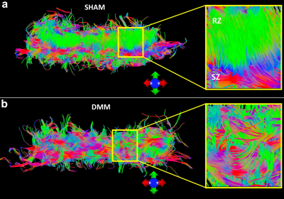 |
Disruption of collagen fiber architecture after DMM surgery using high-resolution DTI
Nian Wang1, Anthony J. Mirando2, Yi Qi3, Matthew J. Hilton2, and Charles E. Spritzer3
1Radiology and Imaging Sciences, Indiana University, Indianapolis, IN, United States, 2Department of Orthopaedic Surgery, Duke University, Durham, NC, United States, 3Department of Radiology, Duke University, Durham, NC, United States
Nondestructively probing the 3D collagen fiber architecture of articular cartilage is still challenging for both clinical and preclinical studies. Diffusion tensor imaging (DTI) is able to resolve the collagen fiber directions by quantifying the unique water diffusion properties in soft tissues. Applying DTI and tractography in cartilage is limited but may provide noninvasive imaging biomarkers for Osteoarthritis (OA). In this study we imaged rat knees after 8 weeks destabilization of the medial meniscus (DMM) surgery using a modified 3D diffusion-weighted spin-echo pulse sequence at both high spatial resolution and high angular resolution.
|
|||
|
2959.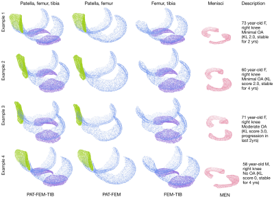 |
Learned knee cartilage and meniscus shape features are associated with osteoarthritis incidence
Claudia Iriondo1, Jinhee Lee1, Sharmila Majumdar1, and Valentina Pedoia1
1University of California, San Francisco, San Francisco, CA, United States
We derive cartilage and meniscus point clouds from 40,796 high resolution knee MR images and train point cloud networks to extract osteoarthritis shape features. We demonstrate the utility of these learned features by assessing their relative contributions in a Cox Proportional Hazard Regression model with existing clinical risk factors predicting incident radiographic osteoarthritis. Shape biomarkers for tibiofemoral joint cartilage and menisci had significantly increased hazard ratios. The best performing shape biomarker– tibial and femoral cartilage shape– combined with clinical risk factors achieved a concordance index of 0.759. Our findings suggest point cloud learned shape features are promising OA biomarkers.
|
||
2960.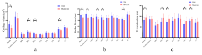 |
Assessment of knee cartilage volume, thickness and T2 relaxation times in patients with osteoarthritis
Hui Tan1, Bin Wang2, Wulin Kang1, Qiuju Fan1, Nan Yu1, Shaoyu Wang3, Esther Raithel4, Yue Li2, and Tuona Di2
1The affiliated hospital of Shaanxi University of Chinese Medicine, Xianyang, China, 2Shaanxi University of Chinese Medicine, Xianyang, China, 3Siemens Healthineers, Shanghai, China, 4Siemens Healthcare, Erlangen, Germany
This study aimed to quantitative measurements of the cartilage volume , thickness and T2 relaxation times in patients with symptomatic mild to moderate KOA by using T2 mapping in 3 T MR. The prototypic software were used to evaluate 17 mild KOA and 14 moderate KOA patients. Both biochemical imaging and morphologic examinations were sensitive to study the mild to moderate KOA. Compared with the mild, the moderate OA patients exhibited decreased CV and CT with the growth of K-L grade, and the T2 value increased with the growth of K-L grade.
|
|||
2961.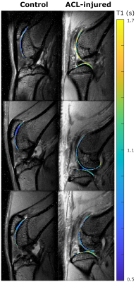 |
Imaging hyaluronan-mediated inflammation in articular cartilage
Jose M Raya1, Alejandra M Duarte1, Dalibel M Bravo1, Elisa M Ramos1, Chongda M Zahng1, Mary M Cowman2, Thorsten M Kirsch1, Mark Wilne3, Len Lyut3, and Amparo M Ruiz1
1New York University Langone Health, New York, NY, United States, 2New York University Tandon School of Engineering, New York, NY, United States, 3University of Western Ontario, Ontario, ON, Canada
The objective of this work is to develop and validate a molecular imaging approach to image inflammation in articular cartilage. We targeted hyaluronan (HA)-mediated inflammation, which is a common inflammatory pathway activated in cartilage, with contrast agents designed using a 15-mer peptide that modulates HA-cell interactions. We identify optimal imaging times and demonstrate that contrast agent accumulates in areas of macroscopic damage. In vivo, MRI detected accumulation of the contrast agent in cartilage of PTOA joints but not in controls. Cartilage in all but one ACL-injured joint showed T1-changes indicative of inflammation, demonstrating that HA-mediated inflammation is a common finding.
|
|||
2962.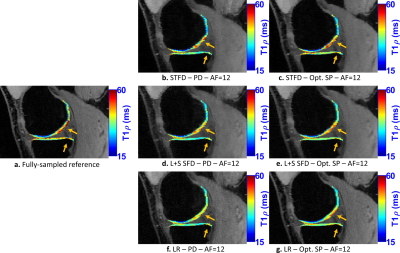 |
Improving Fast 3D-T1rho Mapping of Human Knee Cartilage with Data-Driven Learned Sampling Pattern
Marcelo Victor Wust Zibetti1, Azadeh Sharafi1, and Ravinder Regatte1
1Radiology, NYU Langone Health, New York, NY, United States
in evaluating the performance of optimized SP for accelerating T1rho mapping of the knee cartilage. In this study, we investigate the improvements in accelerating the T1rho mapping of knee joint by learning the SP in a data-driven manner. It was observed that the optimal learned SP depends on the selected spatial-temporal (k-t) data and the chosen reconstruction. Our preliminary results show that the learned SP improved the quality of the accelerated T1rho mapping of knee cartilage over Poisson disk for several different kinds of CS reconstructions.
|
|||
2963.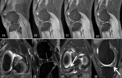 |
Application value of 3D-MRI based on compressed SENSE technology in meniscal injuries
Peiqi Ma1, Yushan Yuan1, Zongxi Zhang1, Bin Peng1, Jian Xu1, and Xiuzheng Yue2
1Fuyang People's Hospital,Anhui Province,China, Fuyang, China, 2Philips Healthcare, Beijing, China, Beijing, China
The compressed SENSE(CS-SENSE) technique, which is the combination of the parallel imaging technique SENSE with compressed sensing, has now been widely used in acceleration of MR examinations of various anatomies and anatomical contrasts. The purpose of this study was to explore the possibility of CS-SENSE in 3D knee joint imaging and its diagnostic value for meniscus injury types.
|
|||
2964.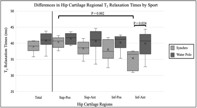 |
Regional Variations in T2 Relaxations Times in the Hip Cartilage of Female Water Polo Players and Synchronized Swimmers
Elka B Rubin1, Joanna L Langner2, Marianne S Black2, Arjun D Desai2, James W MacKay3, Carly Jones4, Kimberly E Hall2, Marc R Safran2, Feliks Kogan2, and Garry E Gold2
1Radiology, Stanford University, Stanford, CA, United States, 2Stanford University, Stanford, CA, United States, 3Radiology, Cambridge University, Cambridge, United Kingdom, 4Centre for Hip Health and Mobility, University of British Columbia, Vancouver, BC, Canada
Water polo players and synchronized swimmers have been found to have an increased prevalence of femoroacetabular impingement morphology compared to the general population. In this study, we used quantitative MRI to identify regional patterns in the composition of hip cartilage of Division 1 female water polo players and synchronized swimmers. Variations in T2 relaxation times showed regional hip cartilage differences between the water polo players and synchronized swimmers and within the sub-regions of the synchronized swimmer’s hip cartilage. This study suggests that microstructural differences are present in the various regions of these athletes’ hip cartilage.
|
|||
2965.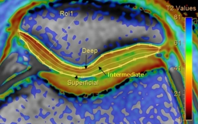 |
Deep, intermediate and superficial layers of patella cartilage assessment using T2 mapping in patients with chondromalacia.
Elena Voronkova1,2, Petr Menshchikov3,4, Ilya Melnikov1, Andrei Manzhurtsev1,4, Maxim Ublinskii1,4, Denis Vorobyev1, Dmitriy Kupriyanov3, and Tolib Akhadov1
1Clinical and Research Institute of Emergency Pediatric Surgery and Trauma, Moscow, Russian Federation, 2National Research Nuclear University MEPhI, Moscow, Russian Federation, 3Philips Healthcare, Moscow, Russian Federation, 4Emanuel Institute of Biochemical Physics of RAS, Moscow, Russian Federation
In this work, the values of the transverse relaxation times (T2) were measured for the first time in the deep, intermediate, and superficial layers of the patellar cartilage at different stages of chondromalacia as compared to control. The T2 values in the intermediate layer of cartilage show the best sensitivity to the grade of injury and have statistically significant differences for the groups of normal, mild, and severe cartilage damage. The T2-mapping technique is a potential tool for studying structural changes in cartilage and T2 values can be considered as a non-invasive biomarker for the assessment of the cartilage condition.
|
The International Society for Magnetic Resonance in Medicine is accredited by the Accreditation Council for Continuing Medical Education to provide continuing medical education for physicians.