Digital Posters
Cartilage II
ISMRM & SMRT Annual Meeting • 15-20 May 2021

| Concurrent 4 | 15:00 - 16:00 |
2966.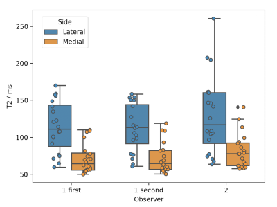 |
T2 mapping of normal and abnormal cartilage in the equine distal interphalangeal joint using low field MRI
Melissa E Baker1, Lucy E Kershaw2, Steve Roberts3, Richard Reardon1, Sionagh Smith1, and Sarah E Taylor1
1Royal (Dick) School of Veterinary Studies and The Roslin Institute, The University of Edinburgh, Edinburgh, United Kingdom, 2Centre for Cardiovascular sciences and Edinburgh Imaging, The University of Edinburgh, Edinburgh, United Kingdom, 3Hallmark Veterinary Imaging Ltd, Guildford, United Kingdom
Osteoarthritis is a common source of pain and lameness in horses of all ages. There are currently no diagnostic tests available to detect early cartilage lesions. A T2 mapping sequence was used on a low field 0.27 T open MR system to image the cartilage of 20 ex vivo equine distal interphalangeal joints. Results showed significantly increased mean T2 relaxation time 114±95 ms in lateral cartilage (higher OARSI grades) compared to 71±21 ms in medial cartilage (lower OARSI grades). This quantitative MRI technique could be used to diagnose early pathology in equine joints in the future.
|
|||
2967.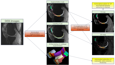 |
Automated Pipeline for Quantitative MRI Evaluation of Knee Articular Cartilage in Longitudinal Osteoarthritis Trials
Vladimir Juras1, Veronika Janáčová1, Pavol Szomolanyi1, Markus Schreiner2, Didier Laurent3, Celeste Scotti3, and Siegfried Trattnig1
1Department of Biomedical Imaging and Image-Guided Therapy, Medical University of Vienna, Vienna, Austria, 2Department of Orthopedics and Trauma Surgery, Medical University of Vienna, Vienna, Austria, 3Novartis Institutes for Biomedical Research, Basel, Switzerland
The purpose of this study was to develop a fully automated reproducible tool for analysis of morphological and compositional quantitative MR parameters in well-defined cartilage sub-fields across the human knee. This tool was deployed to proton and sodium MR images and the reproducibility was assessed by test re-test scan of ten patients at baseline and after eight days. Due to high reproducibility found in this study this tool may be considered as a good alternative to manual evaluation which, as being extremely expertise-dependent and time-consuming, could represents a considerable burden for large clinical osteoarthritis trials.
|
|||
2968.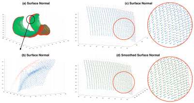 |
Automated Knee Cartilage Thickness Measurement from Magnetic Resonance Images
Yongcheng YAO1, Sheheryar KHAN1, Junru ZHONG1, Siyue LI1, and Weitian CHEN1
1Department of Imaging and Interventional Radiology, The Chinese University of Hong Kong, Hong Kong, China
We propose an automated method to measure knee cartilage thickness from MR images. The proposed method comprises the processes including cartilage and bone segmentation, cartilage boundary separation, surface normal estimation, and uncertainty estimation in thickness measurement. Compared with the conventional nearest neighbor approach, the proposed method provides more reliable measurement at the edges and the regions with thin cartilage.
|
|||
2969.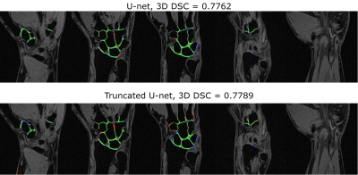 |
Wrist cartilage segmentation using U-Net convolutional neural networks
Nikita Vladimirov1 and Ekaterina A. Brui1
1Department of Physics and Engineering, ITMO University, Saint Petersburg, Russian Federation
Segmentation of wrist cartilage may be of interest for the detection of cartilage loss during osteo- and rheumatoid arthritis. In this work, U-Net convolutional neural networks were used for automatic wrist cartilage segmentation. The networks were trained on a limited amount of labeled data (10 3D VIBE images). The results were compared with the previously published for a planar patch-based archutecture (3D DSC = 0.71). Utilisation of U-Net archutecture and data augmentation allowed to significantly increase the segmentation accuracy in lateral slices. Truncation of the deepest level in the classical U-Net architecture provided the 3D DSC=0.77.
|
|||
2970.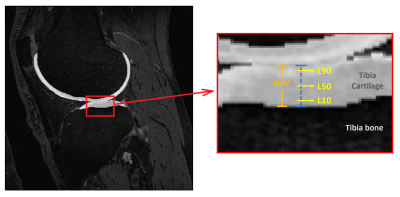 |
3D Texture Analysis of 3D DESS Cartilage Images for Prediction of Knee Osteoarthritis
Daniel Uher1, Ari Väärälä1, Antti Isosalo1, Victor Casula1,2, and Miika T. Nieminen1,2,3
1Research Unit of Medical Imaging, Physics and Technology, University of Oulu, Oulu, Finland, 2Medical Research Center, University of Oulu and Oulu University Hospital, Oulu, Finland, 3Department of Diagnostic Radiology, Oulu University Hospital, Oulu, Finland
In this study, a gray level co-occurrence matrix (GLCM) based 3D Texture Analysis method was utilized for early prediction of knee osteoarthritis using 3D DESS images. Twenty subjects were extracted from the Osteoarthritis Initiative (baseline) with Kellgren-Lawrence (KL) score = 0 at baseline. Ten of the selected subjects developed the disease and showed KL ≥ 2 at the 36-month visit. Knee DESS images were analyzed using various quantization schemes and three machine learning models were trained based on the output GLCM features. Naïve Bayes model trained on tibial features showed the highest accuracy (86.8%) for OA onset after 36 months.
|
|||
2971.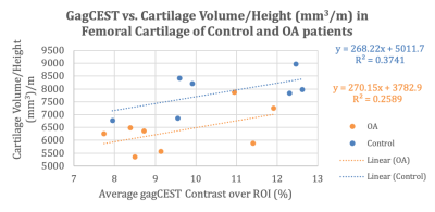 |
GagCEST Imaging of Healthy and OA Patients at 7 Tesla
Blake Alexander Benyard1, Ryan Armbruster1, Abigail Cember1, Neil Wilson2, Ravinder Reddy1, and Joshua Baker3
1Radiology, University of Pennsylvania, Philadelphia, PA, United States, 2Siemens Medical Solutions USA Inc, Malvern, PA, United States, 3Penn Medicine, Rheumatology, Philadelphia Veterans’ Affairs Medical Center, Philadelphia, PA, United States
Imaging of knee cartilage is of enormous significance, particularly for patients with osteoarthritis (OA). In this work, we applied Glycosaminoglycans-weighted Chemical Exchange Saturation Transfer (GagCEST) magnetic resonance imaging (MRI) at 7 Telsa (7T) to OA and healthy patients. GagCEST MRI revealed an increase in GagCEST (%) values for control patients in comparison to OA patients and can serve as a sensitive molecular biomarker for quantifying early metabolic changes in cartilage.
|
|||
2972.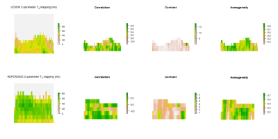 |
OPTIMIZATION OF KNEE CARTILAGE TEXTURE ANALYSIS FROM QUANTITATIVE MRI T2 MAPS
Veronika Janáčová1, Pavol Szomolanyi1, Siegfried Trattnig1, and Vladimir Juras1
1High Field MR Centre, Department of Biomedical Imaging and Image-Guided Therapy, Medical University of Vienna, Vienna, Austria
Texture analysis is currently being explored as an tool for evaluation of collagen specific T2 mapping in magnetic resonance imaging in osteoarthritis (OA) research. We assessed the effect of GLCM calculation offset and number of parameters used for T2 mapping of OA lesion and healthy cartilage on GLCM features. Significant differences in autocorrelation, contrast and homogeneity between lesion and reference when using combination of different offsets (0° and 90°) and 2- and 3-parameter T2 mapping were found. Considering architecture of healthy and OA cartilage and resultant T2 distribution, autocorrelation, contrast and homogeneity might be reliable indicators of cartilage damage.
|
|||
2973.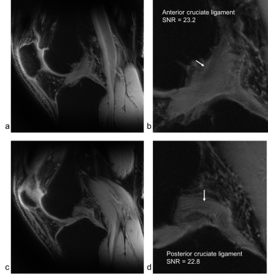 |
Ultra high resolution UTE imaging of the knee at 7T: Simultaneous view of cartilage, meniscus, ligament, tendon, and the chondro-osseous junction
Yongxian Qian1, Gregory Chang1, Eric J. Strauss2, and Fernando E. Boada1
1Radiology, New York University, New York, NY, United States, 2Orthopaedic Surgery, New York University, New York, NY, United States
This human subject study presents preliminary results demonstrating the great potential of ultra-high resolution (0.14mm) UTE imaging to simultaneously visualize daily movement active tissues in the knee joint, including cartilages, menisci, ligaments, tendons, and even the chondro-osseous junctions. Non-invasive observation of these functional tissues are critical to understanding mechanisms of the onset and progression of osteoarthritis (OA).
|
|||
2974.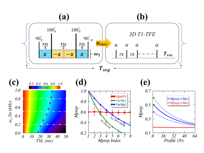 |
An efficient $$$R_{1\rho}$$$ dispersion imaging method for the human knee cartilage using constant magnetization prepared turbo-FLASH
Yuxi Pang1, Riann Palmieri-Smith2,3, and Tristan Maerz3
1Dept. of Radiology, University of Michigan, Ann Arbor, MI, United States, 2School of Kinesiology, University of Michigan, Ann Arbor, MI, United States, 3Dept. of Orthopaedic Surgery, University of Michigan, Ann Arbor, MI, United States
An efficient $$$R_{1\rho}$$$ dispersion imaging method is presented for clinical studies of the human knee cartilage at 3T. Eight constant magnetizations were prepared by tailoring both duration and amplitude of a fully-refocused spin-lock preparation pulse. The limited initial magnetization dynamic range was expanded by the measure from the magic angle location in the deep femoral cartilage. The proposed method was applied to four asymptomatic knees from three subjects. The results from repeated scans and from comparisons with the literature indicate that the proposed method is a promising tool to further explore $$$R_{1\rho}$$$ dispersion of human knee cartilage in clinical settings.
|
|||
2975.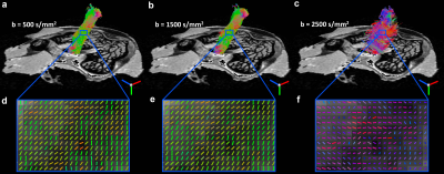 |
Effects of Angular Resolution and b Value on Diffusion Tensor Imaging in Knee Joint
Qi Zhao1, Rees P. Ridout1, Jikai Shen1, and Nian Wang2
1Duke University, Durham, NC, United States, 2Radiology and Imaging Sciences, Indiana University, Indianapolis, IN, United States
Diffusion MRI and tractography in the musculoskeletal system has recently attracted more and more attention to investigate the connective tissue microstructure, local collagen fiber alignment, and the 3D collagen fiber network. Although diffusion tractography was successfully performed in some individual regions of knee, investigating the effects of the acquisition parameters for the whole joint are still lack.
|
|||
2976.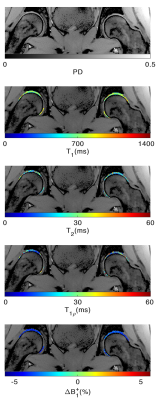 |
Simultaneous Bilateral T1, T2, and T1ρ Relaxation Mapping of the Hip Joint with Magnetic Resonance Fingerprinting
Azadeh Sharafi1, Marcelo V. W. Zibetti1, Gregory Chang1, Martijn Cloos2, and Ravinder Regatte1
1Radiology, NYU Langone Health, New York, NY, United States, 2University of Queensland, Brisbane, Australia
MR imaging has an essential role in the diagnosis of hip disorders such as femoroacetabular impingement (FAI) and osteoarthritis (OA). An increase in pre-contrast T1 is reported in association with cartilage edema and fibrillation and both T2 and T1ρ are shown to be elevated in OA and FAI patients . This work investigates the feasibility of using the MRF sequence introduced in for simultaneous bilateral T1, T2, and T1ρ mapping of the hip joint and assessing the difference between the left and right side cartilage subregions.
|
|||
2977.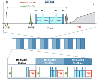 |
3D sub-millimeter isotropic knee cartilage T1ρ mapping using multi-interleaved fluid-attenuated TSE acquisition (MIXTURE)
Shinji Saruya1, Masashi Suzuki1, Masami Yoneyama2, Kaiji Inoue1, Eito Kozawa1, and Mamoru Niitsu1
1Department of Radiology, Saitama Medical University, Saitama, Japan, 2Philips Japan, Tokyo, Japan
For T1ρ quantification, a three-dimensional (3D) acquisition is desired to obtain high-resolution images. In this study, we introduced a new sequence, termed MIXTURE (Multi-Interleaved X-prepared TSE with inTUitive RElaxometry), based on 3D magnetization-prepared multi-interleaved TSE, to achieve rapid and robust acquisition of 3D T1ρ mapping data. MIXTURE might be useful for improving the quality and efficiency of 3D T1ρ-mapping.
|
|||
2978.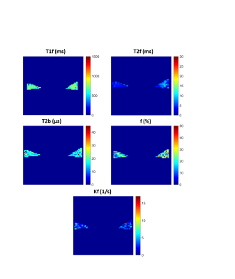 |
Quantitative Magnetization Transfer in the Knee Meniscus: Minimizing Data Collection Protocols
Fatemeh Mostafavi1, Lumeng Cui1, Brennan Berryman2, Ives R. Levesque3, and Emily J. McWalter1,2
1Division of Biomedical Engineering, University of Saskatchewan, Saskatoon, SK, Canada, 2Department of Mechanical Engineering, University of Saskatchewan, Saskatoon, SK, Canada, 3McGill University, Montreal, QC, Canada
A major challenge in the use of quantitative magnetization transfer (qMT) is the long scan time. One method to reduce scan time is acquiring fewer MT-weighted images. qMT mapping of the meniscus was performed in six cadaver knees. Fitting was repeated after systematically decreasing the number of input scans and comparing the results to a reference dataset (10 MT-weighted images). A set of eight MT-weighted images fit to a two-pool model with Gaussian lineshape was found to have an acceptable level of agreement (a priori of 10%) with the reference dataset, saving approximately 8 minutes in scan time.
|
|||
2979. |
A generalized magic angle effect model for better characterizing anisotropic T2W signals of human knee femoral cartilage
Yuxi Pang1
1Dept. of Radiology, University of Michigan, Ann Arbor, MI, United States
Collagen fibril microstructural distributions in articular cartilage are extremely complex. The simplistic two halves radially segmented cartilage in clinical T2W sagittal knee images may not truly represent histologically defined deep and superficial zones. Furthermore, the normal to femoral cartilage surface varies considerably within imaging slices from the lateral to medial side of knee. Consequently, the standard magic angle effect (MAE) functions become inadequate for characterizing the observed anisotropic T2W signals in knee cartilage. Thus, a generalized MAE model is presented for better quantifying anisotropic T2W signals in the clinical studies of human knee articular cartilage.
|
|||
2980.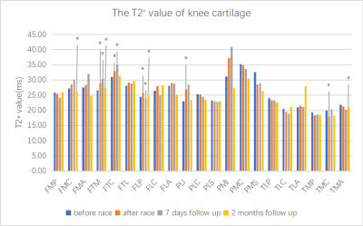 |
The morphology and T2* value changes of 21 knee joint cartilage sub-regions before and after marathon assessed by prototype software
Ranxu Zhang1, Xiaoshuai Chen1, Jian Zhao1, Ping Zhang1, Xiaoyue Zhou2, Baohai Yu1, and Jianping Ding3
1Department of Radiology, The third Hospital of Hebei Medical University, Hebei Province Biomechanical Key Laboratory of Orthopedics, Shijiazhuang, Hebei 050051, China, Shijiazhuang City, China, 2Siemens Healthineers Ltd., Shanghai, 201318, China., Shanghai, China, 3Department of Radiology, Affiliated Hospital of Hangzhou Normal University,Hangzhou 310015,China, Hangzhou, China
This study is designed to assess the changes of articular cartilage volume, thickness and T2* value before and after marathon in non-professional athletes by semi-automatic cartilage segmentation prototype software.All volunteers underwent MR scan before and after marathon and accepted follow-up observation.We confirmed that MR biochemical imaging combined with morphological changes can be used to assess the internal structure and general changes of cartilage. The anterior part of medial tibial plateau may be a high risk area for cartilage degeneration under the long-distance marathon.
|
|||
2981. |
T1 and T1rho Relaxation in Equine Groove Model of Cartilage Damage
Olli Nykänen1, Nina E Hänninen1,2, Swetha Pala1, Ali Mohammadi1, Mohammadhossein Ebrahimi1,2, Nikae CR te Moller3, Harold Brommer3, Rene van Weeren3, Janne TA Mäkelä1, Rami K Korhonen1, Juha Töyräs1,4,5, and Mikko J Nissi1,2
1Department of Applied Physics, University of Eastern Finland, Kuopio, Finland, 2Research Unit of Medical Imaging, Physics and Technology, University of Oulu, Oulu, Finland, 3Department of Clinical Sciences, Faculty of Veterinary Medicine, Utrecht University, Utrecht, Netherlands, 4Diagnostic Imaging Center, Kuopio University Hospital, Kuopio, Finland, 5School of Information Technology and Electrical Engineering, The University of Queensland, Brisbane, Australia
In this study, we examined the T1 and T1rho relaxation times of surgically damaged articular cartilage of equine carpal joints. Two different damage models (sharp and blunt grooves), inducing variation into the type of cartilage damage, were examined. The study revealed that adiabatic T1rho relaxation time is significantly increased in the bluntly damaged cartilage when compared to control cartilage. No significant differences between different damage models or between sharply damaged and control groups were seen for either T1 or T1rho relaxation. Moreover, both relaxation times correlated moderately (R≈0.65) with equilibrium modulus of articular cartilage.
|
|||
2982. |
Clinical MSK Imaging and Challenges at 7T
Garret M. Powell1, Robert J. Spinner2,3, Benjamin M. Howe1, Matthew A. Frick1, Andrew J. Fagan1, Venkata V. Chebrolu4, Peter D. Kollasch4, Eric G. Stinson4, Joel P. Felmlee1, and Kimberly K. Amrami1,2
1Radiology, Mayo Clinic, Rochester, MN, United States, 2Neurologic Surgery, Mayo Clinic, Rochester, MN, United States, 3Orthopedic Surgery, Mayo Clinic, Rochester, MN, United States, 4Siemens Medical Solutions USA, Inc., Rochester, MN, United States
Clinical MSK imaging at 7T is advantageous as a result of increased signal to noise ratio, contrast resolution, and spatial resolution compared to lower field strengths. Nevertheless, we have identified several challenges when performing MSK imaging at this field strength including inferior image uniformity, artifacts, coil limitations, and concealed pathology. The purpose of this exhibit is to introduce our early clinical experience with MSK imaging at 7T and illustrate the challenges we have identified.
|
|||
2983. |
Quantitative correlation of BMD and T2 using high-resolution CT and MRI imaging of rabbit knee joints.
Yang Xia1, Farid Badar1, and Sarah Salem1
1Oakland University, Rochester, MI, United States |
|||
2984. |
HR-MAS 1H-NMR investigations of ovine Achilles tendon and rat rotator cuff tendon
Anshuman Swain1, Johannes Leisen2, Muhammad A. R. Anjum1, Joe Pearson3, Johnna S. Temenoff3, Felix M. Gonzalez1, and David A. Reiter1
1Department of Radiology and Imaging Sciences, Emory University, Atlanta, GA, United States, 2Department of Chemistry, Georgia Institute of Technology, Atlanta, GA, United States, 3Department of Biomedical Engineering, Georgia Institute of Technology, Atlanta, GA, United States
Recent work quantifying tendon chemical shift information has reported resonances consistent with proteoglycan and lipid chemical groups. Currently, there is a lack of prior NMR studies of tendon available to interpret these signals and make assignments to matrix constituents. HR-MAS 1H-NMR is an ideal tool for resolving chemical composition and structure in semisolid tissues like tendon. This study reports 1H-NMR spectral assignments using 1D and 2D HR-MAS experiments in ex vivo tendon samples. Preliminary results show peaks attributed to residues comprising collagen fibrils, proteoglycans, lipids, and lactate. Ongoing work seeks to characterize 1H NMR signatures of healthy and injured tendon.
|
|||
2985. |
ATP Resynthesis after Exercise in Human Skeletal Muscle: Assessment by Phosphocreatine vs. Inorganic Phosphate Recovery in Elderly Subjects
Jimin Ren1,2, Craig R Malloy1,2, Wanpen Vongpatanasin3, and Jarett Berry3
1Advanced Imaging Research Center, UT Southwestern Medical Center, Dallas, TX, United States, 2Department of Radiology, UT Southwestern Medical Center, Dallas, TX, United States, 3Department of Internal Medicine, UT Southwestern Medical Center, Dallas, TX, United States
In this study, we investigated both phosphocreatine (PCr) and Pi as biomarkers of ATP resynthesis after exercise. We developed an efficient 1-min protocol of modest exercise, suitable for subjects in a wide range of age (from 20s to 80s), to generate sufficient large (~50%) PCr depletion and minimal pH drop (< 0.1) for quantitative measurement of ATP metabolism. We compared the rate constant for ATP production measured by Pi-based direct ATP synthase reaction (Pi + ADP → ATP) to PCr-based indirect CK-mediated reaction (ATP + Cr ↔ PCr + ADP), in the elderly population (aged 73±5 years).
|
The International Society for Magnetic Resonance in Medicine is accredited by the Accreditation Council for Continuing Medical Education to provide continuing medical education for physicians.