Digital Posters
Spine
ISMRM & SMRT Annual Meeting • 15-20 May 2021

| Concurrent 4 | 17:00 - 18:00 |
3185.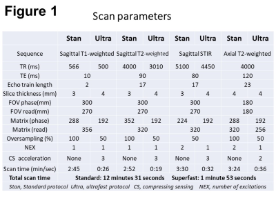 |
Clinical feasibility of ultrafast lumbar spine magnetic resonance imaging: a preliminary report
Nobuo Kashiwagi1, Yuichi Yamashita2, Hitoshi Watanabe3, Hisashi Tanaka1, Katsusuke Kyotani2, Hiroto Takahashi1, Chisato Matsuo1, Takehisa Sakisuka1, Azusa Miura1, Masahiro Fujiwara1, Atsuko Arisawa 1, and Noriyuki Tomiyama1
1Osaka University Graduate School of Mediine, Suita, Japan, 2Canon Medical Systems Corporation, Otawara, Japan, 3Department of Radiology, Yukoukai General Hospital, Ibaraki, Japan Using deep learning reconstruction, we developed an ultrafast lumbar MRI protocol with a total acquisition time of 1 minute 53 seconds. Compared with the standard MRI protocol, the interprotocol agreement for the classification of degenerative lumbar changes was almost perfect or substantial agreement with weighted kappa values of 0.72 to 0.84. The two protocols showed 100% concordance for the detection of clinically significant pathologies. Our preliminary study demonstrated that the ultrafast lumbar MRI protocol adequately preserved diagnostic equivalency with the standard protocol, suggesting that it could be an alternative protocol for restless patients and in an emergency setting. |
|||
3186.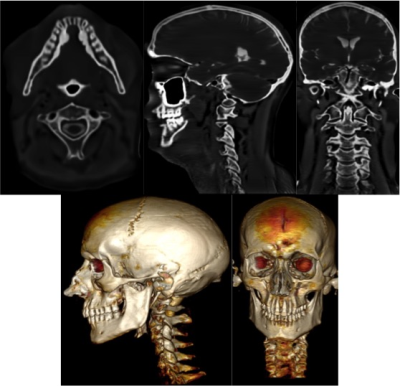 |
High-resolution head and C-spine MRI using a Zero-TE (ZTE) sequence at 3T
Aiming Lu1, Carrie M Carr1, Norbert G Campeau1, Steven A Messina1, and David F Kallmes1
1Radiology, Mayo Clinic, Rochester, MN, United States
Zero TE (ZTE) sequences have been demonstrated to be capable of achieving CT-like images of the skull and C-spine. To obtain high-resolution images of the skull and c-spine, two separate acquisitions are often performed. Although in general cortical bone, soft tissue and air could be differentiated based on the signal intensity, fatty tissues and connective tissues can be problematic due to the chemical shift artifacts and/or having similar signals to that of cortical bones. This work demonstrated that with optimized protocol and advanced post-processing, ZTE MRI allows for high quality skull and C-spine images in a single acquisition.
|
|||
3187.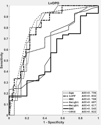 |
A Novel Hybrid Score Integrating Clinical and MR Features to Predict Lumbar Vertebral Osteoporosis in Female
Shao-Chieh Lin1, Chun-Jung Juan2, Yi-Jui Liu3, Chun-Wen Chen4, Chien-Yuan Wang5, Wu-Chung Shen6, Der-Yang Cho7, and Kai-Yuan Cheng8
1Ph.D. program in Electrical and Communication Engineering, Feng Chia University, Taichung, Taiwan, 2Department of Medical Imaging, China Medical University Hsinchu Hospital, Hsinchu, Taiwan, 3Department of Automatic Control Engineering, Feng Chia University, Taichung, Taiwan, 4Department of Radiology, Taichung Armed Forces General Hospital, Taichung, Taiwan, 5Department of Orthopedics, China Medical University Hsinchu Hospital, Hsinchu, Taiwan, 6Department of Medical Imaging, China Medical University Hospital, Taichung, Taiwan, 7Department of Neurosurgery, China Medical University Hospital, Taichung, Taiwan, 8Department of Medical Imaging and Radiological Sciences, Central Taiwan University of Science and Technology, Taichung, Taiwan
Lumbar vertebral osteoporosis (LvOPO) in women is clinically important. It is affected by mixed causes, including but not limited to the aging, lumbar vertebral fat fraction (LvFF), and estrogen. In the current study, the LvOPO in women was diagnosed by a newly proposed hybrid scoring system, i.e., the LvOPO score (LvOPOS). By integrating the independent predictors including age, LvFF, and menopause-MR interval (MMI), the LvOPOS achieved an AUC higher than the LvFF (0.802), age (0.794), and MMI (0.788). Our results provide a new insight in diagnosing the LvOPO specific to women with relevant to both clinical and MR features.
|
|||
3188.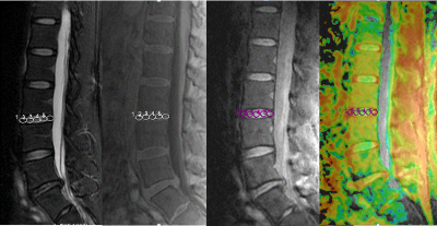 |
A comparative study of R2*, UTE-T2* and T1rho mapping for evaluation of degenerative alterations in human intervertebral discs
LiLan Wu1, JianJun Zhou2, and Pu-Yeh Wu3
1Department of Radiology, Fudan University Affiliated Zhongshan Hospital Xiamen Branch, Xiamen, China, 2Department of Radiology, Fudan University Affiliated Zhongshan Hospital, Shanghai, China, 3GE Healthcare, Beijing, China
This study aims to investigate whether human lumbar IDD can be detected using R2*, UTE-T2* and T1rho mapping. We systematically compared the efficacy of R2*, UTE-T2* and T1rho values in the diagnosis of early IDD. Specifically, we found that T1rho value is superior to UTE-T2* and R2* values for diagnosis of the early IVD, while UTE-T2* value is optimal for diagnosis of advanced IVD. Overall, we concluded that R2* and UTE-T2* mapping provides another promising method for quantitatively evaluated lumbar IDD, and T1rho mapping can be considered an effective tool for distinguishing IDD at earlier stage of the degenerative process
|
|||
3189.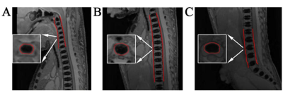 |
Normal fetal development of the cervical, thoracic and lumbar spine: a post-mortem study based on magnetic resonance imaging
Shuai Zhang1, Xiangtao Lin2, Ximing Wang2, Xiang Feng3, and Rui Diao2
1School of Medicine, Shandong First Medical University, School of Medicine, Shandong First Medical University, Jinan, China, 2Department of Radiology, Shandong Provincial Hospital affiliated to Shandong First Medical University, Jinan, China, 3MR Scientific Marketing, Diagnosis Imaging, Siemens Healthcare Ltd, Beijing, China
Before evaluating spinal pathology, it is essential to have knowledge of the normal spinal development at different gestational ages. Postmortem magnetic resonance imaging was performed on 55 fetuses (gestational ages, 17–42 weeks) by using three-dimensional T2-weighted sequences. The volumes of interest were manually outlined for the cervical, thoracic, lumbar, and L1–L5 centrum ossification centers (COCs), and the COC volumes (COCVs) were calculated. The cervical, thoracic, and lumbar COCVs showed a positive relationship with gestational age.The cervical, thoracic, lumbar, and L1–L5 COCVs show good correlation with gestational age in the second and third trimesters.
|
|||
3190.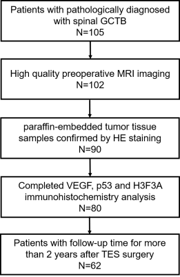 |
Spinal Giant Cell Tumor of Bone: Immunohistochemistry and Preoperative Magnetic Resonance Imaging Features for Prognostic Prediction
Qizheng Wang1, Siyuan Qin1, Yang Zhang2, Enlong Zhang3, Xiaoying Xing1, Min-Ying Su2, and Ning Lang1
1Radiology, Peking University Third Hospital, Beijing, China, 2Center for Functional Onco-Imaging, Irvine, CA, United States, 3Radiology, Peking University International Hospital, Beijing, China
A new evaluating system based on tumor immunohistochemistry and preoperative MRI features in spinal giant cell tumor of bone to predicting overall survival of total en bloc spondylectomy patients with over 2 years follow up. The largest lesion diameter (>4.2 cm) and the vertebral compression were independent predictors of postoperative recurrence. According to Kaplan-Meier survival analysis, the cystic change in the lesion and the degree of compression ≥50% suggest a worse clinical outcome. The expression levels of vascular endothelial growth factor and p53 gene have no obvious clinical significance on the survival outcome. H3F3A was positively expressed in our cohort.
|
|||
3191.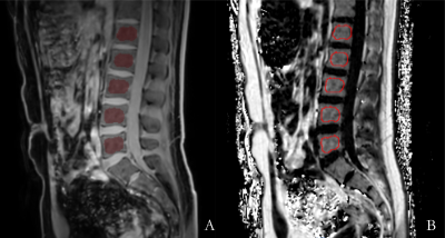 |
Effects of different acceleration factors on lumbar body fat quantification using 3D mDixon Quant technique
Yu Song1, Qingwei qing Song2, Yingkun Guo1, Gang Ning1, and Xuesheng Li1
1Department of Radiology, West China Second University Hospital, Sichuan University, Chengdu, China, 2Department of Radiology, the First Affiliated Hospital of Dalian Medical University, Dalian, China
Compressed SENSE (CS) is a cutting-edge signal acquisition and processing innovation technology based on applied mathematics. By directly collecting the compressed signal, the amount of sampled data is greatly reduced, and the optimal acceleration factor can be passed. The realization of the method can shorten the clinical magnetic resonance scanning time while ensuring the spatial resolution.
|
|||
3192.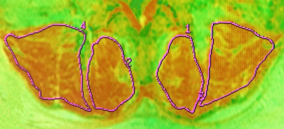 |
Quantitative T2 parametric value in assessment of abnormal lumbar paraspinal muscle in patients with low back pain
Yinqi Liu1, Huiting Deng1, Weiyin Liu2, and Kun Zhang1,3
1Department of Radiology, First Affiliated Hospital of Hunan University of Chinese Medicine, Changsha, China, 2MR Research, GE Healthcare, Beijing, China, 3College of Integrated Traditional Chinese and Western Medicine, Hunan University of Chinese Medicine, Changsha, China
Chronic low back pain (CLBP) has been reported to be associated with low back dysfunction with the characteristics of decreased cross-section area (CSA) of paraspinal muscles and increased adipose content. In our study, T2 value at L4/L5 multifidus muscle was significantly higher in LBP group than in HCs. The multifidus muscle is the most medial in the low back extensor group and has the maximum strain stress when spinal movement and balance maintenance, so it might easily cause the abnormality of the low back muscle group. T2 mapping could be a good predictor of detecting minimal variation of multifidus muscles.
|
|||
3193.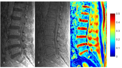 |
Evaluation of Lumbar Disc Degeneration Using Ultrashort Echo Time Magnetization Transfer (UTE-MT) Imaging in Cartilaginous Endplate
Jin Liu1, Yajun Ma2, Jianwei Liao1, Xiaojun Chen1, Wei Li1, Lin Yao1, Zhihai Su3, Long Qian4, Jiang Du2, and Shaolin Li1
1Department of Radiology, The Fifth Affiliated Hospital of Sun Yat-Sen University, Zhuhai, China, 2Department of Radiology, University of California, San Diego, CA, United States, 3Department of Orthopedics, The Fifth Affiliated Hospital of Sun Yat-Sen University, Zhuhai, China, 4MR Research, GE Healthcare, Guangzhou, China
Ultrashort echo time magnetization transfer (UTE-MT) sequence can quantify magnetization transfer ratio (MTR) values of short T2 tissues, which has potential to evaluate early disc degeneration. The current study aims to assess the feasibility of UTE-MT quantification of cartilaginous endplate for evaluation of early disc degeneration. In addition, the latent associations between the UTE-MTR and grade of disc degeneration, age, gender, the oswestry disability index (ODI) were evaluated, respectively. Our results demonstrated that the UTE-MT sequence is feasible to quantify cartilaginous endplate in lumbar spine. UTE-MTR may be a potentially useful biomarker in the assessment of early lumbar disc degeneration.
|
|||
3194.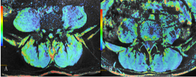 |
BOLD and IDEAL-IQ evaluation for lumbarparaspinal muscle changes in patients with chronic low back pain
Zeng Xiao min1, Huang Yi long2, Nie Li sha3, and He Bo1
1The First Affiliated Hospital of Kunming Medical University, Kunming, China, 2The First Affiliated Hospital of Kunming Medical University, kunming, China, 3GE healthcare, China, Beijing, China
This study analyzed the relationship between the cross-sectional area (CSA), effective lateral relaxation rate (R2* value), fat fraction (FF value) of lumbar paraspinal muscles of patients with chronic low back pain (CLBP group) and asymptomatic control group (Control group). The multifidus muscle and erector spinae muscle at the central level of the L4/L5 intervertebral disc were the target muscles.BOLD and IDEAL-IQ provided a new idea for imaging evaluation of lumbar paraspinal muscles. Our study concluded that BOLD and IDEAL-IQ are feasible to evaluate the changes of lumbar muscle morphology and function in patients with CLBP.
|
|||
3195.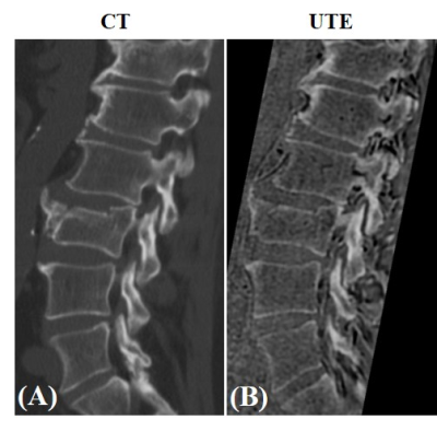 |
High-Contrast Lumbar Spinal Bone Imaging Using a 3D Slab-Selective UTE Sequence
Amir Masoud Afsahi1, Zhao Wei1, Michael Carl2, Saeed Jerban1, Hyungseok Jang1, Nicole Le1, Jiang Du1, Eric Y. Chang1,3, and Ya-Jun Ma1
1Department of Radiology, University of California, San Diego, San Diego, CA, United States, 2GE HealthCare, San Diego, CA, United States, 3Radiology Service, Veterans Affairs, San Diego Healthcare System, San Diego, CA, United States
To explore the application of a 3D slab-selective UTE sequence as a superior technique to 3D ZTE at 3T field strength and to compare with 3D CT as a gold standard in Lumbar spinal bone assessment.
|
|||
3196.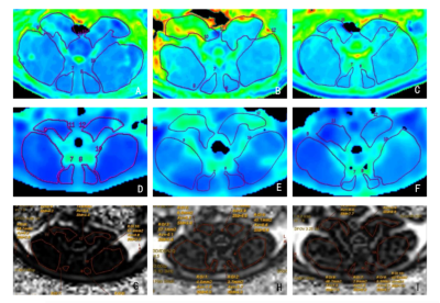 |
Multimodal MRI evaluation of paraspinal muscles and TNF- α in rats with discogenic low back pain after rehabilitation training
Kaiwen Yang1, Baofa Luo1, Yilong Huang1, Lisha Nie2, and Bo He1
1Medical imaging department, The First Affiliated Hospital of Kunming Medical University, Kunming, China, 2GE Healthcare,MR Research China, Beijing, China
Based on functional MRI, the current study aims to explore the pathological morphology, fat infiltration and the expression of TNF-α in paraspinal muscles of discogenic low back pain(DLBP). And to further explore the relation of rehabilitation exercise on paraspinal muscles and intervertebral discs in rats. It is concluded that rehabilitation exercise can reduce the content of TNF-α in serum and paraspinal muscles, relieve the symptoms of DLBP in rats, and improve paraspinal muscle dysfunction.
|
|||
3197.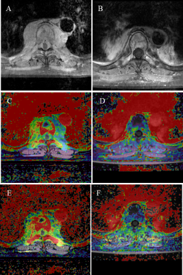 |
DCE-MRI for the Assessment of Outcome of CyberKnife Stereotactic Radiosurgery for Patients with Spinal Metastases
Yongye Chen1, Enlong Zhang 2, Qizheng Wang 1, Huishu Yuan1, Huishu Yuan1, Hongqing Zhuang1, and Ning Lang1
1Peking University Third Hospital, Beijing, China, 2Peking University International Hospital, Beijing, China
The imaging methods used to evaluate the efficacy of radiosurgery in spinal metastases have certain limitations. DCE-MRI can help determine the vascularity and hemodynamics of tumors in vivo, and can be used to evaluate local tumor response. Twenty-seven patients with 39 lesions were included. Post-treatment Kep, ΔKtrans and ΔKep in the non-progressive disease group were significantly lower than the corresponding values in progressive disease group. Post-treatment Ve and ΔVe in the non-progressive disease group were significantly higher than that of the progressive disease group. ΔKtrans had the highest diagnostic efficiency, with an AUC of 0.821.
|
|||
3198.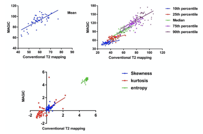 |
Comparison of T2 relaxation time from synthetic MRI and conventional T2 mapping in the diagnosis of intervertebral disc degeneration
Xiaoqing Liang1, Weiyin Vivian Liu2, Yitong Li1, and Xiaoming Li3
1Radiology, Tongji Hospital, Tongji Medical College, Huazhong University of Science and Technology, Wuhan, China, 2MR Research, GE Healthcare, Beijing, China, 31Radiology, Tongji Hospital, Tongji Medical College, Huazhong University of Science and Technology, Wuhan, China
Synthetic MRI has become a recent research hotspot due to its advantages of short-time acquiring multiple sets of weighted images and quantitative maps in one time. Synthetic MRI has been successfully used for the diagnosis of many cerebral diseases, but its feasibility and stability in other tissues and lesions need to be further studied. The current study demonstrates that the utility of synthetic MRI is slightly better than conventional T2 mapping in diagnosing intervertebral disc degeneration. In addition, one-time MAGiC scan on lumbar vertebrae should be concerned for clinical diagnosis.
|
|||
3199. |
Histogram analysis of IDEAL-IQ and UTE images better assists to detect abnormalities of the lumbar vertebral bone marrow in patients with different stages of chronic kidney disease
Yan Xiong1, Weiyin Vivian Liu 2, Fan He1, Yanan Wang1, Yao Zhang1, Shuang Hu1, and Xiaoming Li1
1Wuhan Tongji Hospital, Wuhan, China, 2MR Research, GE Healthcare, Beijing, China
Mineral bone disease (MBD) has an extremely high incidence in patients with chronic kidney disease (CKD) and significantly affects the prognosis and quality of life of patients. However, the current diagnostic methods are difficult to determine the type and severity of CKD-MBD. In our study, histogram analysis of IDEAL-IQ showed kurtosis and R2* could distinguish bone marrow changes in patients with CKD at different stages in indication of altered skeletal health and bi-component analysis of UTE images demonstrated short T2* value possibly in reflection of trabecular bone was highly correlated with blood biochemical indicators.
|
|||
3200.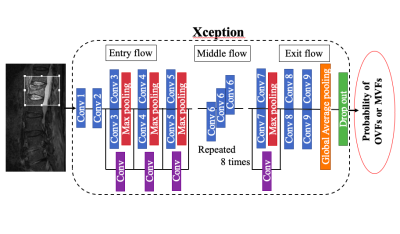 |
Automated differentiation between benign and malignant vertebral compression fracture using a deep convolutional neural network on MRI
Takafumi Yoda1, Satoshi Maki2, Koji Matsumoto1, Hajime Yokota3, Yoshitada Masuda1, and Takashi Uno3
1Department of Radiology, Chiba University Hospital, Chiba, Japan, 2Department of Orthopedic Surgery, Graduate School of Medicine, Chiba University, Chiba, Japan, 3Diagnostic Radiology and Radiation Oncology, Graduate School of Medicine, Chiba University, Chiba, Japan
The Differentiating between osteoporotic vertebral fractures (OVFs) and malignant vertebral compression fractures (MVFs) due to spinal metastasis is a challenging problem for the spine surgeons and the radiologists. We evaluated the performance of our CNN model in differentiating between OVFs and MVFs on short-TI inversion recovery (STIR) and T1-weighted (T1WI) images compared with the performance of three spine surgeons. The sensitivity, specificity, and accuracy of the CNN on both STIR and T1WI were equal to or better than those of the three spine surgeons.
|
|||
3201.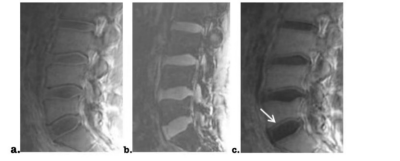 |
The assessment of cartilage endplate and its relationship with the corresponding disc degeneration using UTE imaging
Zhilin Ji1, Weiqiang Dou2, Yuefen Zou1, Yin Shi1, Yu Zheng1, and Hongyuan Ding1
1The First Affiliated Hospital of Nanjing Medical University, Nanjing, China, 2GE Healthcare, MR Research China, Beijing, P.R. China, Beijing, China
In this study, we aimed to investigate the feasibility of ultra-short echo time (UTE) imaging for assessing the cartilage endplate (CEP) damage and evaluate the relationship between the grading of CEP and lumbar disc degeneration. UTE images with short and long TEs were performed on patients with low back pain. The resultant subtracted UTE images between short and long TEs showed well structural CEP clearly. Strong correlation was observed between CEP degree based on UTE imaging and lumbar intervertebral disc degeneration. With these findings, UTE imaging might thus be considered an effective tool to assess the CEP damage in clinic.
|
|||
3202.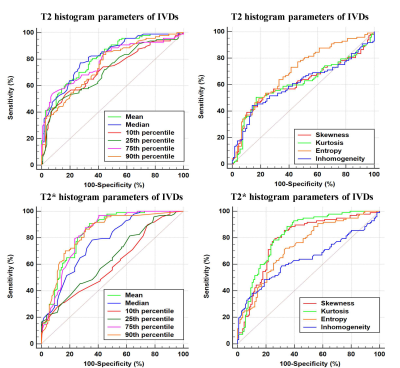 |
Evaluation of lumbar facet joint and intervertebral disc degeneration using histogram analysis of T2 and T2*values
Xiaoqing Liang1, Weiyin Vivian Liu2, and Xiaoming Li1
1Tongji Hospital, Tongji Medical College, Huazhong University of Science and Technology, Wuhan, China, 2MR Research,GE Healthcare, Beijing, China
Lumbar intervertebral disc (IVD) and lumbar facet joint (LFJ) are the primary and important load-bearing structures in spine. Any abnormality of the anterior IVD and the two posterior LFJs may lead to spinal instability and low back pain (LBP). The current study demonstrates that LFJ degeneration occurs earlier and progresses more slowly than IVD degeneration. Histogram analysis of T2 and T2* values is feasible for detecting and grading IVD but not LFJ degeneration.
|
|||
3203.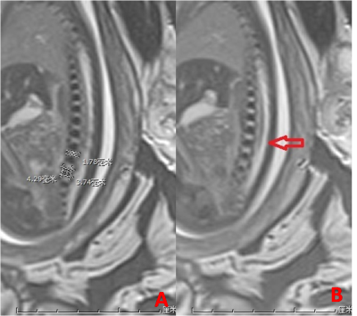 |
Fetal magnetic resonance imaging of normal lumbar spine development in vivo
Xing Yin1, Xin Zhao1, Liying Zhang1, Qingna Xing1, Rui Yuan2, and Zhijun Niu3
1Radiology, the Third Affiliated Hospital of Zhengzhou University, Zhengzhou, China, 2Ultrasound, the Third Affiliated Hospital of Zhengzhou University, Zhengzhou, China, 3Obstetrics and Gynecology, the Third Affiliated Hospital of Zhengzhou University, Zhengzhou, China
In the last few years, fetal magnetic resonance (MR) has been more and more used in the obstetrics and gynecology. We measure the height and length of each lumbar vertebra body ossification center and the height of each intervertebral gap, and our results show a good linear correlation between normal lumbar vertebra body ossification center development and gestational age in vivo
|
|||
3204.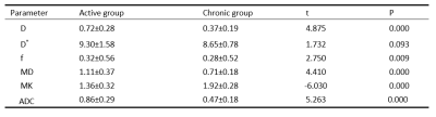 |
Diffusion Kurtosis Imaging and Intravoxel Incoherent Motion in diagnosis of active sacroiliitis with ankylosing spondylitis
Xiaolin Guo1,2, lixia Qian2, Yueluan Jiang3, and Zhongshuai Zhang4
1Medical Imaging, Shanxi Medical University, Taiyuan, China, 2Department of Radiology, Shanxi Bethune Hospital, Taiyuan, China, 3MR Scientific Marketing, Diagnosis Imaging, Siemens Healthineers China, Beijing, China, 4MR Scientific Marketing, Diagnosis Imaging, Siemens Healthineers China, Shanghai, China
Advanced DWI techniques, diffusion kurtosis imaging (DKI) and intravoxel incoherent motion (IVIM), can complement physiological information to morphological information that was obtained with conventional MRI This study investigated the application of DKI and IVIM in diagnosis of active sacroiliitis with ankylosing spondylitis.
|
The International Society for Magnetic Resonance in Medicine is accredited by the Accreditation Council for Continuing Medical Education to provide continuing medical education for physicians.