Digital Posters
Bone Tendon Inflammation
ISMRM & SMRT Annual Meeting • 15-20 May 2021

| Concurrent 4 | 17:00 - 18:00 |
4065.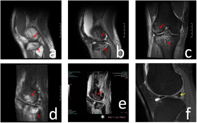 |
Discussion the Value of ADC in Bone Contusions and Bone Marrow Lesions in Osteoarthritis of Knee Joint
Weixin He1, Qi Zeng1, Ziwei Zhang1, Xia Zhu1, Zhaoshu Huang2, Lisha Nie3, and Lingling Song1
1Affiliated Hospital of Guizhou Medical University, Guiyang, China, 2592159673@qq.com, Guiyang, China, 3GE Healthcare, MR Research China, Beijing, China
The current study aims to investigate the application value of ADC in Bone Contusions and Bone Marrow Lesions in Osteoarthritis of Knee Joint. It was concluded that ADC value has the potential value to differentiate the two kinds of lesions.
|
|||
4066.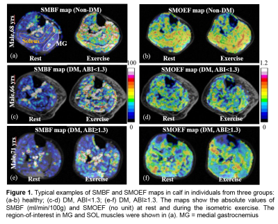 |
Pilot contrast-free MRI reveals significantly impaired calf skeletal muscle perfusion in diabetes with incompressible peripheral arteries
Jie Zheng1, Sara Gharabaghi2, Ran Li1, Yongsheng Chen3, Hongyu An1, E Mark Haacke2, Mohamed A Zayed1, and Mary K Hastings1
1Washington University in St. Louis, St. Louis, MO, United States, 2MR Innovations Inc, Bingham Farms, MI, United States, 3Wayne State University, Detroit, MI, United States
The purpose of this study is to investigate whether individuals with diabetes mellitus and incompressible arteries will have lower skeletal muscle microcirculation during a moderate isometric exercise. Healthy volunteers, diabetes with normal artery, and diabetes with incompressible arteries underwent MR angiography and calcification imaging, and skeletal muscle microcirculation imaging at rest and during an isometric contraction exercise. Significantly lower perfusion reserve and its association with higher ABI were observed in diabetes with incompressible arteries. Despite lack of apparent arterial stenosis, calcification can be readily visualized in 2 patients.
|
|||
4067.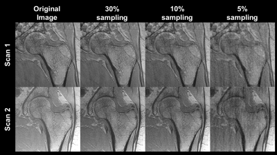 |
Acceleration of high-resolution proximal femur MRI using compressive sensing and sparsity in a retrospective study
Brian-Tinh Duc Vu1,2, Brandon Jones1,2, Winnie Xu2, Gregory Chang3, and Chamith Rajapakse2,4
1Bioengineering, University of Pennsylvania, Philadelphia, PA, United States, 2Radiology, University of Pennsylvania, Philadelphia, PA, United States, 3Radiology, Center for Biomedical Imaging, New York University, New York, NY, United States, 4Orthopaedic Surgery, Perelman School of Medicine, University of Pennsylvania, Philadelphia, PA, United States
Retrospective compressive sensing techniques demonstrate promise accelerating acquisition of high-resolution images of the femur while maintaining sufficient image quality to assess fracture risk. While the trabecular bone microstructure was preserved at an undersampling rate of 30%, lower sampling rates of 10% and 5% exhibited visually apparent artifacts and image degradation. Similarly, bone stiffness at 30% resembled fully sampled data but the error increased as sampling rate decreased. Nevertheless, the results show that compressive sensing is a promising candidate for accelerating the acquisition rate, and further prospective studies are needed to further validate this finding.
|
|||
4068. |
3T MRI Distribution of Textural Features in Bone Marrow for Osteoporosis.
Anmol Monga1, Dimitri Martel1, Stephen Honig2, and Gregory Chang1
1Department of Radiology, NYU Langone Health, New York, NY, United States, 2Osteoporosis Center, Hospital for Joint Disease, NYU Langone Health, New York, NY, United States
In this preliminary study we aim to analyze the relationship of first-order textural feature of Fat, water, Fat Fraction maps, clinical features (Age, height, and weight) and BMD (Hip, Spine and Femoral Neck). Radiomic features calculated on Fat parametric maps explains variability in Bone Mineral Density to higher extend than water and fat fraction parametric maps.
|
|||
4069.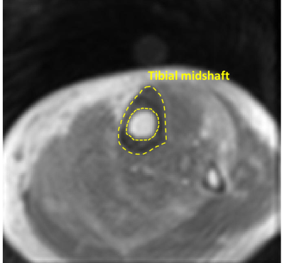 |
Significantly reduced collagen and increased water in tibia of patients with osteopenia and osteoporosis detected with ultrashort echo time MRI
Saeed Jerban1, Yajun Ma1, Zhao Wei1, Meghan Shen1, Amir Masoud Afsahi1, Zubiad Ibrahim1, Alecio Lombardi1,2, Douglas G Chang3, Eric Y Chang1,2, and Jiang Du1
1Radiology, University of California, San Digeo, La Jolla, CA, United States, 2Radiology Service, VA San Diego Healthcare System, San Diego, CA, United States, 3Orthopaedic Surgery, University of California, San Digeo, La Jolla, CA, United States
Assessment of water and collagen content in bone is missing from DEXA evaluation, in standard osteoporosis diagnosis. Bone signals in ultrashort echo time MRI (UTE-MRI) and inversion recovery UTE-MRI against a known external reference signal were used to measure total, bound, and pore water proton densities. Macromolecular proton density was estimated by multiplying total water proton density with macromolecular fraction derived from UTE magnetization transfer (UTE-MT) modeling. The UTE-evaluated MRI measures demonstrated significantly reduced collagen and increased water content in the tibia of patients with osteoporosis and osteopenia compared with healthy subjects. Hip T-scores showed significant correlation with UTE-MRI measures.
|
|||
4070.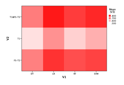 |
A comparative study on MRI-based radiomics model selection for a high-risk cytogenetics prediction in multiple myeloma
jianfang liu1 and huishu yuan1
1peking university third hospital, beijing, China
We aim to develop a radiomics model based on MRI to predict high-risk cytogenetics (HRC) status in patients with multiple myeloma (MM) and identify optimal machine learning methods. We retrospectively analyzed 89 patients’ (37 HRC, 52 non-HRC) radiomics features extracted from fat suppression T2W and TIW image. The following classification methods, including, support vector machine, random forest, logistic regression (LR) and decision tree were used to construct radiomics models. LR model showed the highest performance with an AUC of 0.82 ± 0.02. Radiomics model based on LR classifier can be used to predict HRC status in patients with MM effectively.
|
|||
4071.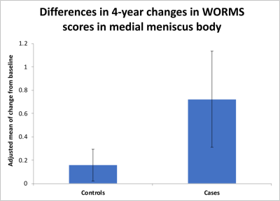 |
Impact of sustained Synovitis on Knee Joint Structural Degeneration: 4-Year MRI Data from the Osteoarthritis Initiative
Sara Ramezanpour1, Thanat Kanthawang 1,2, John Lynch 3, Thomas M Link 1, and Gabby B Joseph1
1Department of Radiology and Biomedical Imaging, University of California, San Francisco, San Francisco, CA, United States, 2Department of Radiology, Faculty of Medicine, Chiang Mai University, Thailand, Chiang Mai, Thailand, 3Department of Epidemiology and Biostatistics, University of California, San Francisco, San Francisco, CA, United States
Synovial inflammation is a well-known risk factor of osteoarthritis (OA). Several semi-quantitative scores have been developed to grade synovitis using non-contrast enhanced MRI studies of the knee. However, the role of inflammatory processes in the progression of knee OA is yet to be fully understood. One hundred and eighty individuals with right knee MRIs at baseline, 2 and 4 years were studied using MRI-based semi-quantitative synovitis scores measuring presence/absence as well as severity of synovitis Subjects with sustained synovitis had greater progression of meniscus, bone marrow and cartilage abnormalities compared to controls without sustained synovitis.
|
|||
4072.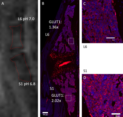 |
AcidoCEST MRI pH is strongly correlated with GLUT1 immunohistochemistry in multiple myeloma
Alecio Lombardi1,2, Jonathan Wong1,2, Rachel High1,2, Ya-Jun Ma2, Adam Searleman2, Saeed Jerban2, Qingbo Tang1,2, Jiang Du1,2, Patrick Frost3,4, and Eric Y. Chang1,2
1Radiology Service, Veterans Affairs San Diego Healthcare System, San Diego, CA, United States, 2Radiology, University of California, San Diego, CA, United States, 3Greater Los Angeles Veteran Administration Healthcare System, Los Angeles, CA, United States, 4University of California, Los Angeles, CA, United States
Multiple myeloma (MM) is a malignant plasma cell disease. Adaptive responses to hypoxia may be an essential element in its progression. The purpose of this study was to determine the feasibility of acidoCEST MRI for pHe measurement on a mouse model of MM with comparison with GLUT1 staining.
|
|||
4073.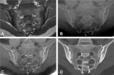 |
Diagnostic performance of zero echo time imaging and T1-weighted fast spin echo on sacroiliac joint bone erosions using CT as the gold standard
Yitong Li1, Shuang Hu1, Weiyin Vivian Liu2, and Xiaoming Li1
1Tongji Hospital, Tongji Medical College, Huazhong University of Science and Technology, Wuhan, China, 2MR Research, GE Healthcare, Beijing, China
Detection of sacroiliac joint (SIJ) bone erosions may improve diagnostic performance of axial spondyloarthritis (axSpA) as CT is the gold standard. Zero echo time (ZTE) MRI technique can acqire CT-like bone contrast and has been applied to bone imaging of many other body parts. Our research explored the ability of ZTE to detect SIJ bone erosions and conduct comparison of ZTE and conventional T1-weighted fast spin echo (T1 FSE). We found that ZTE had superior detection performance to T1 FSE and might be an effective supplement to routine MRI protocol in SIJs.
|
|||
 |
4074.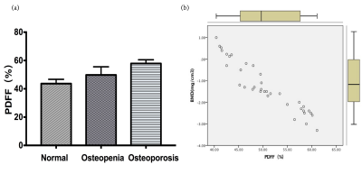 |
Evaluation of the risk of osteoporosis in diabetic patients by IDEAL-IQ
Yu Song1, Qingwei Song2, Yingkun Guo1, Gang Ning1, and Xuesheng Li1
1Department of Radiology, West China Second University Hospital, Sichuan University, Chengdu, China, 2the First Affiliated Hospital of Dalian Medical University, Dalian, China
Osteoporosis is a progressive metabolic bone lesion characterized by a decrease in bone mass per unit volume, which results in increased risk of bone fractures. Noninvasive and reliable assessment of osteoporosis is essential for clinical diagnosis and treatment. Studies have reported that precise measurement of fat content in the bone marrow is a biomarker for quantifying osteoporosis. IDEAL-IQ is a method used for quantitative measurement of fat fraction and iron content related parameters by multi-echo acquisition.
|
||
4075.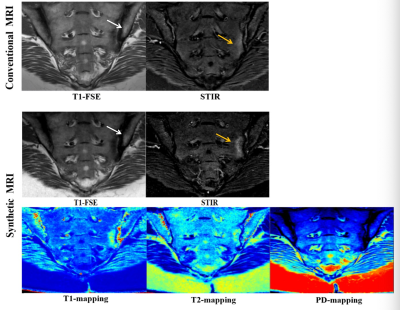 |
Detection of Sacroiliac Joint Lesions in Axial Spondyloarthritis:Utility of Synthetic MRI
Ke Zhang1 and Guobin Hong1
1Radiology, the Fifth Affiliated Hospital, Sun Yat-sen University, Zhuhai, China The current diagnosis of the sacroiliac joint lesions with axSpA is most concentrated on qualitative or semi-quantitative methods.There is an urgent need for a quantitative method that can objectively and accurately evaluate the severity of the disease.Synthetic MRI(MAGiC) can generate multiple contrast images and quantitative maps simultaneously based on the same scan.In this study the synthetic MRI can achieve similar qualitative diagnostic performance in detection of sacroiliac joint lesions compared with conventional MRI.And it could be used for distinguishing BME and fat metaplasia. |
|||
4076.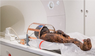 |
MRI and CT in an Ancient Child Mummy: Contrast Combination to Increase Tissue Differentiation
Agazi Samuel Tesfai1, Johannes Fischer1, Ali Caglar Özen1,2, Patrick Eppenberger3, Lena Öhrström3, Frank Rühli3, Ute Ludwig1, and Michael Bock1
1Dept. of Radiology, Medical Physics, Medical Center – University of Freiburg, Faculty of Medicine, University of Freiburg, Freiburg, Germany, 2German Consortium for Translational Cancer Research Partner Site Freiburg, German Cancer Research Center (DKFZ), Heidelberg, Germany, 3Institute of Evolutionary Medicine, Faculty of Medicine, University of Zurich, Zurich, Switzerland
MR images of a child mummy were acquired on a clinical 3T system using a dedicated RF coil with optimized RF switching hardware and 3D UTE sequence. In addition, dual-energy CT images were sampled and co-registered to compare MRI signal intensities and T2* relaxation times with CT Hounsfield Units and effective atomic numbers in bone, soft tissue and embalming material.
|
|||
4077.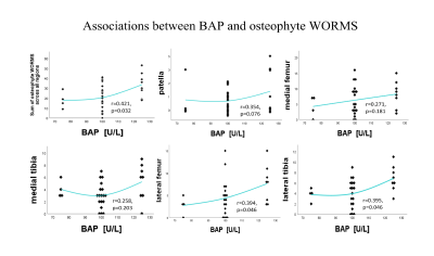 |
Associations between Bone Turnover Biomarkers and MR-based Knee composition and WORMS scores:cross-sectional
WANG BIN1, TAN HUI1, HE Taiping1, YU Nan1, and WANG Shaoyu2
1Shaanxi University of Chinese Medicine, SHAANXI, China, 2Siemens Healthineers, shanghai, China
We attempted to explore the relationship between bone markers (including bone formation and bone absorption) and cartilage, meniscus morphology, and WORMS socres in patients with osteoarthritis.
|
|||
4078.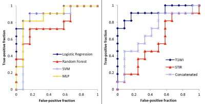 |
MRI-based radiomic features and machine learning for differentiating myelodysplastic syndrome and aplastic anemia
Miyuki Takasu1, Makoto Iida1, Yasutaka Baba2, Yuji Akiyama1, Yuji Takahashi1, Takashi Abe3, and Kazuo Awai1
1Department of Diagnostic Radiology, Hiroshima University Hospital, Hiroshima, Japan, 2Department of Radiology, International Medical Center, Saitama Medical University, Saitama, Japan, 3Department of Radiology, Nagoya University Hospital, Aichi, Japan, Nagoya, Japan
We assessed the feasibility of a method of radiomic analysis based on machine learning (ML) and lumbar MRI to differentiate between MDS and aplastic anemia (AA). Regions of interest were drawn in the L3 vertebral body on the mid-sagittal images of sagittal T1-weighted and STIR images of patients with MDS (n=62) or AA (n=78) from six institutions. The model of ML with logistic regression resulted in the best performance for differentiating MDS from AA when using T1-weighted images. The model was not predictive for STIR or concatenated images. The radiomics-based ML model enabled the differentiation of MDS and AA.
|
|||
4079.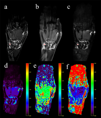 |
Evaluation of Synovitis of Hand in Patients with Rheumatoid Arthritis Using Diffusion Kurtosis Imaging: Initial Findings
Kaifang Liu1, Jie Meng1, and Zhengyang Zhou1
1Departments of Radiology, Nanjing Drum Tower Hospital Clinical College of Nanjing Medical University, Nanjing, China
Thirty patients with RA and 10 patients suspected of RA were enrolled to explore the application value of DKI in the noninvasive identification of synovitis in hand arthritis. The suspected synovitis or joint effusion was scored on a scale of 0 (joint effusion) to 3 (mild, moderate, severe synovitis), referring to RAMRIS (RA-MRI-Scoring) system. ADC, D, and K from DKI were recorded and compared. There were significant differences in ADC, D, and K values among different enhancing degree scores. The diagnostic performance provided by the D values is similar to the ADC value and higher than the K value.
|
|||
4080.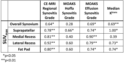 |
A Comparison of Synovitis Severity in the Knee Assessed Using Contrast-Enhanced MRI and FDG-PET
Jacob Thoenen1, James W. MacKay2,3, Kathryn J. Stevens1, Tom D. Turmezei4, Akshay Chaudhari1, Lauren E. Watkins1, Brian A. Hargreaves1, Garry E. Gold1, and Feliks Kogan1
1Department of Radiology, Stanford University, Stanford, CA, United States, 2Department of Radiology, University of Cambridge, Cambridge, United Kingdom, 3Norwich Medical School, University of East Anglia, Norwich, United Kingdom, 4Department of Radiology, Norfolk and Norwich University Hospital, Norwich, United Kingdom
T1-weighted contrast-enhanced MRI (CE-MRI) is used to evaluate synovitis, but fluctuations in severity of synovial inflammation may be slow to manifest as volumetric changes seen on CE-MRI. We compared metabolic activity using FDG-PET to CE-MRI synovitis gradings, MOAKS effusion- and Hoffa-synovitis gradings, and the transfer constant Ktrans in the overall synovial region and four subregions. Moderate to very strong correlation was seen between maximum FDG uptake (SUVmax) and corresponding synovitis grades, MOAKS effusion synovitis, and corresponding regional median Ktrans both within the entire synovium and in four synovial subregions, with the exception of median Ktrans at one subregion.
|
|||
4081.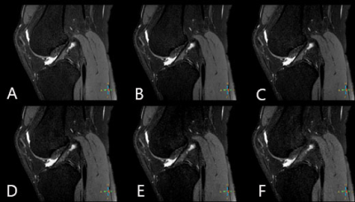 |
A preliminary study on the effect of compressed SENSE with multiple acceleration factors on knee examination
Nan Zhang1, Qingwei Song2, Ailian Liu2, Renwang Pu2, Haonan Zhang2, Jiazheng Wang3, and Liangjie Lin3
1The First Affilliated Hospital of Dalian Medical University, Dalian, China, 2The First Affiliated Hospital of Dalian Medical University, Dalian, China, 3Philips Healthcare, Beijing, China, Beijing, China
2D scans remain as standard for routine examination of joint lesions due to the short scan time. This study aims to explore feasibility of compressed SENSE with different acceleration factors in 3D high-resolution PD weighted imaging of knee joint.
|
|||
4082.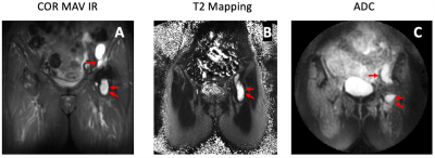 |
Diffusion MRI of THAs for the Classification of Synovial Reactions
Madeleine A. Gao1, Ek T. Tan1, John Neri1, Bin Lin1, Alissa J. Burge1, Hollis G. Potter1, Kevin M. Koch2, and Matthew F. Koff1
1Radiology and Imaging, Hospital of Special Surgery, New York, NY, United States, 2Medical College of Wisconsin, Milwaukee, WI, United States
The classification of synovial reactions caused by adverse local tissue reactions or prosthetic joint infections in total hip arthroplasty patients has remained a challenge due to the in-plane and through-plane distortions caused by metallic components. MAVRIC-based diffusion weighted imaging (DWI) is an innovative MRI technique sensitive to microstructural fluid changes and utilizes apparent diffusion coefficient values for the differentiation of diseased and non-diseased tissues. This study evaluated the effectiveness of MAVRIC-based DWI and T2 mapping in differentiating between types of synovial reactions. Our DWI ADC biomarker showed promise in differentiating between normal and abnormal synovial reaction subtypes within THA patients.
|
|||
4083.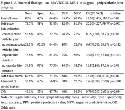 |
Clinical value of MAVRIC-SL MRI for the assessment of periprosthetic joint infection
Tsutomu Inaoka1, Masayuki Sugeta1, Noriko Kitamura1, Tomoya Nakatsuka1, Rumiko Ishikawa1, Takamitsu Uchi1, Rui Iwata1, Akinori Yamamoto1, Hisanori Tomobe1, Ryosuke Sakai1, Hidetoshi Yamana1, Shusuke Kasuya1, and Hitoshi Terada1
1Radiology, Toho University Sakura Medical Center, Sakura, Japan
MAVRIC-SL enabled to improve the image quality by decreasing susceptibility artifacts and the assessment of periprosthetic joint infection after joint arthroplasty. Joint effusion, soft-tissue fluid collection, and soft-tissue edema were suggestive of periprosthetic joint infection. Soft-tissue fluid collection and soft-tissue edema were indicative of therapeutic surgical intervention for periprosthetic joint infection. Using MAVRIC-SL, MRI can become a useful tool to suggest periprosthetic joint infection in arthroplasty patients and to determine the management of periprosthetic joint infection.
|
|||
4084.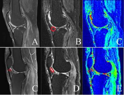 |
Diagnostic value of intro-voxel incoherent movement (IVIM) for infrapatellar fat pad high signal intensity in patient with osteoarthritis
Hui Tan1, Bin Wang1, Wulin Kang1, Nan Yu1, Yong Yu1, Shaoyu Wang2, Yue Li1, and Tuona Di1
1Shaanxi University of Chinese Medicine, Xianyang, China, 2Siemens Healthineers, Shanghai, China
This study aimed to investigate the role of IVIM in assessing infrapatellar fat pad high signal intensity in patients with osteoarthritis. Totally 43 patients with mild to moderate (K/L score = 1,2 and 3) KOA were included. The results show that D value, VAS, WOMAC, K/L score with T2FS-hyperintense regions were significantly higher than those without T2FS-hyperintense regions. Furthermore, D value was significantly positive associated with VAS and K/L score.
|
The International Society for Magnetic Resonance in Medicine is accredited by the Accreditation Council for Continuing Medical Education to provide continuing medical education for physicians.