Digital Posters
Neurofluids/Perivascular Spaces/White Matter Hyperintensities
ISMRM & SMRT Annual Meeting • 15-20 May 2021

Digital Posters
Neurofluids/Perivascular Spaces/White Matter Hyperintensities
| Concurrent 6 | 17:00 - 18:00 |
 |
2358.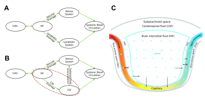 |
Brain Parenchymal Venous System Plays a Substantial Role in Cerebral Waste Clearance
Yongsheng Chen1, Jiani Hu2, Yimin Shen2, Lara M Fahmy3,4, Li Zhang3, E. Mark Haacke1,2, and Quan Jiang1,3
1Department of Neurology, Wayne State University School of Medicine, Detroit, MI, United States, 2Department of Radiology, Wayne State University School of Medicine, Detroit, MI, United States, 3Department of Neurology, Henry Ford Health System, Detroit, MI, United States, 4Department of Psychiatry and Behavioral Neurosciences, Wayne State University School of Medicine, Detroit, MI, United States
The current understanding of cerebral waste clearance (CWC) involves CSF participation but lacks the direct participation of the parenchymal vascular system. We used SPIO-enhanced SWI on rats to simultaneously study parenchymal veins, arteries and their corresponding para-vascular spaces, and observed not only CSF participation, but also parenchymal venous participation in CWC following intra-cisterna magna infusion of CSF tracers. These results lead to the speculation of a new CWC mechanism with directional brain parenchyma-to-blood permeability. The substantial role of the parenchymal venous system in CWC raises intriguing questions on the differential contributions of parenchymal venous versus CSF pathways in neurodegenerative disease.
|
||
2359.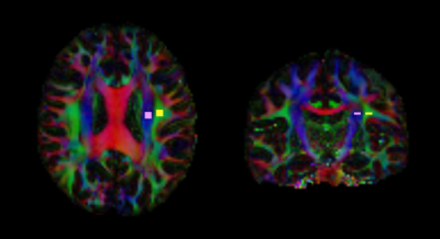 |
An investigation of the change in water diffusivity along the perivascular space in hypertensive patients
Junko Kikuta1, Koji Kamagata1, Kaito Takabayashi1, Toshiaki Taoka2, Hajime Yokota3, Yuki Someya4, Yoshifumi Tamura4,5, Hirotaka Watada4,5, Ryuzo Kawamori4,5, Shinji Naganawa6, and Shigeki Aoki1
1Department of Radiology, Juntendo University School of Medicine, Bunkyo-ku, Japan, 2Department of Innovative Biomedical Visualization, Graduate School of Medicine, Nagoya University, Nagoya, Japan, 3Department of Diagnostic Radiology and Radiation Oncology, Graduate School of Medicine, Chiba University, Chiba, Japan, 4Sportology Center, Juntendo University School of Medicine, Bunkyo-ku, Japan, 5Department of Metabolism & Endocrinology, Juntendo University School of Medicine, Bunkyo-ku, Japan, 6Department of Radiology, Nagoya University Graduate School of Medicine, Nagoya, Japan
We used the analysis along the perivascular space (ALPS) index, which has been suggested as a noninvasive method to measure water diffusivity along the perivascular space in vivo. We assessed the change in water diffusivity in living patients with hypertension for the first time.
|
|||
2360.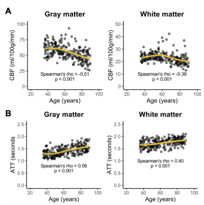 |
Assessment of cerebral white matter hemodynamics across the adult lifespan
Meher R. Juttukonda1,2, Randa Almaktoum1, Kimberly A. Stephens1, Kathryn Yochim1, Essa Yacoub3, Randy L. Buckner4, and David H. Salat1,2
1Athinoula A. Martinos Center for Biomedical Imaging, Massachusetts General Hospital, Charlestown, MA, United States, 2Radiology, Harvard Medical School, Boston, MA, United States, 3Center for Magnetic Resonance Research, University of Minnesota, Minneapolis, MN, United States, 4Psychology, Harvard University, Cambridge, MA, United States
Arterial spin labeling (ASL) approaches for measuring perfusion are challenging in white matter due in part to longer blood arrival times. We implemented a cross-correlation-based processing approach on multi-delay ASL data acquired in Human Connectome Project-Aging (HCP-A) to quantify white matter arterial transit time (ATT) and used these ATT values to analytically compute white matter CBF. Using this approach, we found that white matter CBF decreases (ρ=0.39) and white matter ATT elongates (ρ=0.42) with increasing age (p<0.001). We also found that CBF and ATT values are spatially heterogeneous, with periventricular white matter exhibiting the lowest CBF and longest ATT.
|
|||
2361.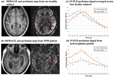 |
High resolution imaging of choroid plexus blood flow with multi-delay pseudo-continuous arterial spin labeling
Xingfeng Shao1, Chenyang Zhao1, Matthew Borzage2, Catherine Mark3, Elizabeth Joe4, Jonathan Russin3,5, Charles Liu3,4,5, Darrin Lee3,5,6, and Danny JJ Wang1,4
1Laboratory of FMRI Technology (LOFT), Mark & Mary Stevens Neuroimaging and Informatics Institute, Keck School of Medicine, University of Southern California, Los Angeles, CA, United States, 2Fetal and Neonatal Institute, Division of Neonatology, Children's Hospital Los Angeles, Department of Pediatrics, Keck School of Medicine, University of Southern California, Los Angeles, CA, United States, 3Department of Neurological Surgery, Keck School of Medicine, University of Southern California, Los Angeles, CA, United States, 4Department of Neurology, Keck School of Medicine, University of Southern California, Los Angeles, CA, United States, 5USC Neurorestoration Center, Keck School of Medicine, University of Southern California, Los Angeles, CA, United States, 6Department of Psychiatry, Keck School of Medicine, University of Southern California, Los Angeles, CA, United States
In this study we developed an arterial spin labeling (ASL) sequence with high spatial/temporal resolution and utilized a post-processing denoising method to measure dynamic choroid plexus (CP) perfusion signal. Quantified CP blood flow was evaluated in heathy subjects and one normal pressure hydrocephalus (NPH) patient and compared with water exchange rate across the blood-brain barrier (BBB). Our preliminary results show that CP blood flow may be altered in NPH and has a potential relation with water exchange rate across the BBB.
|
|||
2362.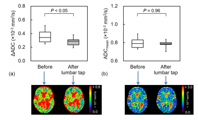 |
ADC change during cardiac cycle in idiopathic normal pressure hydrocephalus before and after tap test and shunt surgery
Ryo Yagawa1, Naoki Ohno1, Tosiaki Miyati1, Mitsuhito Mase2, Tomoshi Osawa2, Harumasa Kasai2, Yuta Shibamoto2, and Satoshi Kobayashi1
1Division of Health Sciences, Kanazawa University, Kanazawa, Japan, 2Nagoya City University Hospital, Nagoya, Japan
To assess the dynamic changes in the apparent diffusion coefficient (ADC) during the cardiac cycle (ΔADC) of the brain before and after the lumbar tap and shunt surgery for the purpose of determining changes in hydrodynamic and biomechanical properties in the brain after cerebrospinal fluid (CSF) drainage for iNPH. The frontal white matter ΔADC in iNPH decreased after the lumbar tap and shunt surgery. ΔADC analysis may provide detailed information regarding changes in the hydrodynamic and biomechanical properties through CSF drainage.
|
|||
2363. |
Long term evaluation of ventricular volume change associated with shunt-responsiveness in idiopathic normal pressure hydrocephalus
Romtheera Kamronritthisorn1, Kritdipha Ningunha2, Peeratat Suppapanya1, Sunee Bovonsunthonchai3, Doonyaporn Wongsawaeng 1, Yudthaphon Vichianin4, Theerapol Witthiwej5, Weerasak Muangpaisan6, Panida Charnchaowanish1, Siriwan Piyapittayanan1, Orasa Chawalparit1, and Chanon Ngamsombat1
1Department of Radiology, Faculty of Medicine Siriraj Hospital, Mahidol University, Bangkok, Thailand, 2Department of TELE-Radiology, Bangkok Hospital Headquarters, Bangkok, Thailand, 3Faculty of Physical Therapy, Mahidol University, Bangkok, Thailand, 4Department of Radiological Technology, Faculty of Medical Technology, Mahidol University, Bangkok, Thailand, 5Division of Neurosurgery, Department of Surgery, Faculty of Medicine Siriraj Hospital, Mahidol University, Bangkok, Thailand, 6Department of Preventive and Social Medicine, Faculty of Medicine Siriraj Hospital, Mahidol University, Bangkok, Thailand
Shunt surgery is an effective treatment for iNPH, however some patients showed unsustainable response. We investigated how reduction of ventricular size after shunt surgery correlated with clinical outcome, as potential predictor of long-term shunt responsiveness using automated segmentation of ventricular volume from T1W sequence. A reduction of ventricular volume is found to negatively correlate with improved cognition. These findings might be one of helpful imaging predictors for the shunt responders in iNPH.
|
|||
2364.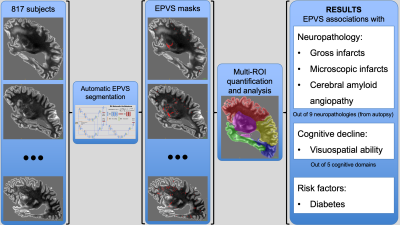 |
Neuropathologic and Cognitive Correlates of Automatically Segmented Enlarged Perivascular Spaces in Community-based Older Adults
Carles Javierre-Petit1, Ashish A. Tamhane2, Arnold M. Evia2, Marinos Kontzialis2, Nazanin Makkinejad1, Gady Agam1, David A. Bennett2, Julie A. Schneider2, and Konstantinos Arfanakis1,2
1Illinois Institute of Technology, Chicago, IL, United States, 2Rush University Medical Center, Chicago, IL, United States
Perivascular spaces form a network that enables clearance of waste products from the brain. Abnormal enlargement of perivascular spaces is common in older adults and has been linked to cognitive impairment and dementia. However, the neuropathologic and cognitive correlates of enlarged perivascular spaces (EPVS) are not well understood yet. In this work, we first developed an algorithm to automatically segment and quantify EPVS in brain MR images, and then investigated the neuropathologic correlates of total and regional EPVS, as well as the contributions of EPVS on cognitive decline in a large community-based cohort of 817 older adults.
|
|||
2365. |
ALL CENTRAL NERVOUS SYSTEM (CNS) NEURO- AND VASCULAR-COMMUNICATION CHANNELS ARE SURROUNDED WITH CEREBROSPINAL FLUID (CSF)
Lara M Fahmy1, Yongsheng Chen2, Stephanie Xuan3, E Mark Haacke3, Jiani M Hu3, and Quan Jiang4
1Psychiatry and Behavioral Neurosciences, Wayne State University School of Medicine, Detroit, MI, United States, 2Neurology, Wayne State University School of Medicine, Detroit, MI, United States, 3Radiology, Wayne State University School of Medicine, Detroit, MI, United States, 4Neurology, Henry Ford Health System, Detroit, MI, United States
It's well-established that the CNS is completely submerged in CSF; but to what extent is this true regarding the finer spaces within the CNS itself? We used MRI to simultaneously map the presence of CSF within all peri-neural and peri-vascular spaces in vivo in humans. Our findings indicated that all peri-neural spaces surrounding cranial and spinal nerves, as well as all peri-vascular spaces, were filled with CSF. These findings suggest that anatomically, substance exchange between the brain parenchyma and outside tissues (i.e. lymphatics) can only occur through CSF, warranting further investigation into its cerebral waste clearance and immunomodulation implications.
|
|||
2366.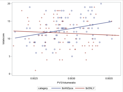 |
Association of Enlarged Perivascular Spaces and Sleep Disturbances in Military-related Mild Traumatic Brain Injury
Ping-Hong Yeh1, J. Kent Werner2,3, Rujirutana Srikanchana1, Kimbra Kenney1,2, Treven Pickett1,2, Grant Bonavia1,2, Gerard Riedy1,2, and John Ollinger1
1National Intrepid Center of Excellence, Bethesda, MD, United States, 2Uniformed Services University of the Health Sciences, Bethesda, MD, United States, 3Center for Neuroscience and Regenerative Medicine, Bethesda, MD, United States
Sleep disturbances are common following traumatic brain injury (TBI). We sought to quantify changes in perivascular space (PVS) in combat-related mild TBI (mTBI) patients and explore the association between normalized PVS volume and sleep measures. We found mTBI patients with previous potential concussive event (PCE) (TBIPCE) had larger PVS fraction than controls. A subjective sleep measure, The Pittsburgh Sleep Quality Index (PSQI), was positively associated with PVS in TBIPCE patients, which had mean higher PSQI than those of TBI patients who had only mTBI but no PCE. This result suggests enlarged PVS may be modulated by sleep and TBI.
|
|||
2367.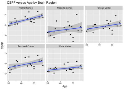 |
Cerebrospinal fluid water fraction increases with age in normal aging
Thanh D Nguyen1, Liangdong Zhou1, Elizabeth Sweeney1, Xiuyuan Wang1, Susan A Gauthier1, Yi Wang1, Amy Kuceyeski1, and Yi Li1
1Weill Cornell Medicine, New York, NY, United States Perivascular spaces (PVS) play a key role in the glial-lymphatic waste clearance pathway in the brain. We applied FAST-T2 multi-component T2 relaxometry to 20 healthy volunteers between 30 and 60 years old and found a significant relationship between CSF water fraction and age in the frontal and temporal cortex, while the brain tissue water content measured by T1 relaxation time remains stable over time. Our results suggest that CSF fraction may be a useful quantitative biomarker of PVS dilation in normal aging. |
|||
2368.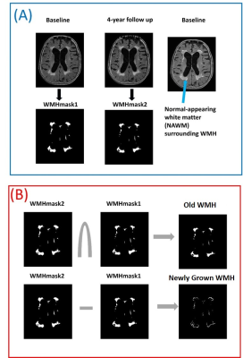 |
Diffusion and perfusion characteristics of brain white matter prone to hyperintensities: a four-year longitudinal study
Shruti Agarwal1, Jay J. Pillai1,2, and Hanzhang Lu3,4
1Division of Neuroradiology, Russell H. Morgan Department of Radiology and Radiological Science, Johns Hopkins University School of Medicine, Baltimore, MD, United States, 2Department of Neurosurgery, Johns Hopkins University School of Medicine, Baltimore, MD, United States, 3Division of MR Research, Russell H. Morgan Department of Radiology and Radiological Science, Johns Hopkins University School of Medicine, Baltimore, MD, United States, 4F. M. Kirby Research Center for Functional Brain Imaging, Kennedy Krieger Institute, Baltimore, MD, United States
White matter hyperintensities (WMH) are white matter brain lesions found as areas of increased signals on T2-weighted and FLAIR MRI scans. A large majority of elderly individuals have a certain degree of WMH which may be associated with cognitive decline, decline in physical function and a higher risk of stroke and death. To date, neurobiological mechanisms underlying and predictive of WMH is not fully characterized. In this study, we aim to use a longitudinal design to elucidate hallmarks of brain tissue at baseline that will predict a “conversion” from normal-appearing WM (NAWM) to WMH over a four-year period.
|
|||
2369.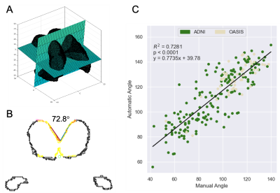 |
Callosal Angle Biomarker for Normal Pressure Hydrocephalus Calculated for 4,980 T1-Weighted MR Exams
Alexander Saunders1,2, Stefan Bluml1,2, Kevin S King3, and Matthew Borzage2,4
1Radiology, Children's Hospital Los Angeles, Los Angeles, CA, United States, 2Rudi Schulte Research Insitute, Santa Barbara, CA, United States, 3Barrow Neurological Institute, Phenoix, AZ, United States, 4Children's Hospital Los Angeles, Los Angeles, CA, United States
Normal pressure hydrocephalus is a treatable dementia often misdiagnosed as Alzheimer’s Disease. Callosal angle measurement distinguish normal pressure hydrocephalus from other dementias but measurements are time-consuming and subjective. Therefore, we developed an algorithm for automatic callosal angle measurement and calculated callosal angles for 4,980 T1-weighted MRIs from databases of patients with purportedly healthy aging brains or Alzheimer’s. Based on the published guidelines, 1.8-4.3% of exams in these databases indicate patients likely have normal pressure hydrocephalus and thus a treatable form of dementia. Our automatic measurement of the callosal angle biomarker can rapidly and objectively screen patients for normal pressure hydrocephalus.
|
|||
2370.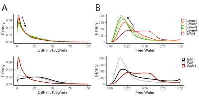 |
Perfusion and free water show opposite trends across tissue layers surrounding white matter hyperintensities in elderly participants
Corinne A. Donnay1, Pauline Maillard1, Charles DeCarli1, and Audrey P. Fan1,2
1Neurology, University of California Davis, Davis, CA, United States, 2Biomedical Engineering, University of California Davis, Davis, CA, United States
We investigated white matter (WM), cerebral blood flow (CBF) and free water content (FW) in the penumbra region surrounding white matter hyperintensities (WMH) in elderly participants. The tissue surrounding WMH clusters were defined as four 1mm concentric layers and compared to WM and WMHs. The findings of this study reveal that WMHs and adjacent layers have lower CBF than healthy WM and CBF increases from lower layers to higher layers. The opposite associations were found with FW.
|
|||
2371. |
Reduced cerebrovascular reactivity and cerebral blood flow in White Matter Hyperintensities (WMHs)
Chenyang Li1,2, Marco Muccio1, Dengrong Jiang3, Peiying Liu3, Jiangyang Zhang1, Arjun Masurkar4, Thomas Wisniewski4, Hanzhang Lu3, and Yulin Ge1
1Department of Radiology, Center for Biomedical Imaging, NYU Grossman School of Medicine, New York, NY, United States, 2Vilcek Institute of Graduate Biomedical Sciences, NYU Grossman School of Medicine, New York, NY, United States, 3Department of Radiology and Radiological Science, Johns Hopkins University, Baltimore, MD, United States, 4Department of Neurology, NYU Grossman School of Medicine, New York City, NY, United States
Understanding of in vivo vascular pathophysiology in white matter hyperintensities (WMHs) is still incomplete. Compared to grey matter, white matter is considerably less vascularized and tends to have lower blood flow. Therefore, sensitive detection and accurate estimation for white matter hemodynamics is crucial for early prevention and mechanistic understanding of WMHs pathogenesis and progression. In this study, we implemented advanced neurovascular MRI techniques to evaluate the white matter hemodynamics including cerebral blood flow and cerebrovascular reactivity in patients with varying degrees of WMHs.
|
|||
2372.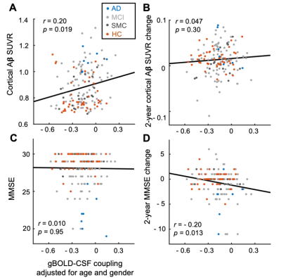 |
Decoupling between global brain activity and cerebrospinal fluid flow is associated with Alzheimer's disease pathologies
Feng Han1, Jing Chen1, Aaron Belkin-Rosen1, Yameng Gu1, Liying Luo2,3, Orfeu M Buxton4, and Xiao Liu1,5
1Department of Biomedical Engineering, The Pennsylvania State University, State College, PA, United States, 2Department of Sociology & Criminology, The Pennsylvania State University, State College, PA, United States, 3Population Research Institute, The Pennsylvania State University, State College, PA, United States, 4Department of Biobehavioral Health, The Pennsylvania State University, State College, PA, United States, 5Institute for Computational and Data Sciences, The Pennsylvania State University, State College, PA, United States
Glymphatic system responsible for brain waste clearance may play an important role in the development of Alzheimer's disease (AD). However, this possibility has not been sufficiently studied in AD patients due to a lack of non-invasive tools for gauging glymphatic function. Low-frequency (<0.1 Hz) fMRI signals have been recently linked to glymphatic function, and its global signal was found to be coupled with CSF flow known to be essential for glymphatic clearance. Here, we used the coupling of global BOLD signal and CSF to quantify glymphatic function and found this BOLD-CSF coupling metric is significantly correlated with various AD pathologies.
|
|||
2373.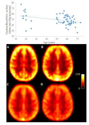 |
Cerebrovascular Reactivity and Cerebral Blood Flow across lifespan in females
Safa Sanami1, Brittany Intzandt2,3,4, Fatemeh Razavipour1, Julia Huck1, Richard D Hoge5, Louis Bherer3,4,6,7, and Claudine J Gauthier1,4,6
1Physics, Concordia University, Montreal, QC, Canada, 2INDI, Concordia University, Montreal, QC, Canada, 3Centre de Recherche de l'Institut Universitaire de Geriatrie, Montreal, QC, Canada, 4Centre de Recherche, l'Institut de Cardiologie de Montréal, Montreal, QC, Canada, 5Department of Neurology and Neurosurgery, McGill University, Montreal, QC, Canada, 6PERFORM Centre, Concordia University, Montreal, QC, Canada, 7Départment de Médicine, Université de Montréal, Montreal, QC, Canada
Aging is associated with cerebrovascular impairments in males and females, yet this impairment develops nearly one decade later in females. Although cerebral blood flow (CBF) is consistently reported as higher in females, results on cerebrovascular reactivity (CVR) have not been uniform in studies comparing females to males. Here, given that much less is known about cerebrovascular changes in females than males, we examined CBF and CVR during aging in healthy females only. Our results revealed that both CBF and CVR decline across the lifespan in females. Future work should include hormone levels, arterial stiffness, other vascular risk factors, and males.
|
|||
2374.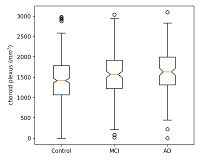 |
Changes of Choroid Plexus Volume in Alzheimer’s Patients
Li Zhao1, Xue Feng2, Yueqin Hu3, Dan Wu1, Craig H. Meyer2, and David C. Alsop4
1College of Biomedical Engineering & Instrument Science, Zhejiang University, Hangzhou, China, 2Biomedical Engineering, University of Virginia, Charlottesville, VA, United States, 3Psychology, Beijing Normal University, Beijing, China, 4Radiology, Beth Israel Deaconess Medical Center and Harvard Medical School, Boston, MA, United States
Choroid plexus volume was investigated using a deep learning segmentation method, which has demonstrated higher accuracy than the previous atlas-based method. Therefore, a higher sensitivity was expected with the new method. MR images of 733 subjects were selected retrospectively, including healthy volunteers, MCI and AD patients. The results show that 1) the choroid plexus volume increases with age; 2) is smaller in the female subjects; and 3) is larger in the MCI and AD patients compared to the healthy volunteers. Our preliminary results suggest that the choroid plexus volume changes with brain aging.
|
|||
2375.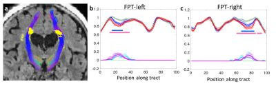 |
Effect of white matter hyperintensities on tractometry and its relationship with white matter connectivity
Tae Kim1, Howard J Aizenstein1, and James T Becker1
1University of Pittsburgh, Pittsburgh, PA, United States
The effect of white matter hyperintensities (WMHs) on 50 major WM tractometries was investigated. Elevated WMHs load on white matter tract was associated with lower apparent fiber density (AFD) on tractometery, showing the co-localization of WMH with AFD change. Reduced AFD is associated with lower connectivity of the WM tract. Differences in these metrics between lower WMH and higher WMH groups, and between lower WMH and MCI groups are significant. Moreover, AFD is lower in individuals with MCI, supporting a role of white matter microstructure in cognitive decline.
|
|||
2376.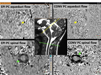 |
Cerebro spinal fluid dynamic in front of cardiac and breathing influence
Olivier Baledent1, Pan Liu1, Serge Metanbou1, Cyrille Capel1, Sidy Fall1, and Roger Bouzerar1
1university hospital Jules Verne, Amiens, France
it is still debated how breathing interact with the CSF. New Phase contrast MRI sequence based on Echo Planar imaging (EPI-PC) can now produce continuously during minutes a velocity map, more or less every 100 ms. We did not found in the literature quantitative evaluation of the CSF stroke volume change during breathing. The aim of this work is to quantify CSF dynamics change in the aqueduct and in the spinal canal during the breathing and cardiac period using EPI-PC.
|
|||
2377.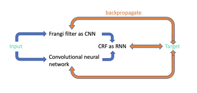 |
UPSS - Unsupervised perivascular spaces segmentation method with salient guidance of frangi filter
Haoyu Lan1 and Farshid Sepehrband1
1USC Stevens Neuroimaging and Informatics Institute, University of Southern California, Los Angeles, CA, United States
Automatic perivascular spaces (PVS) segmentation from magnetic resonance images enables whole brain morphometric assessment of glia-lymphatic network. While conventional deep learning segmentations are shown to produce reliable results, they heavily rely on the presence of ground truth for training. In this work we introduce an unsupervised learning method for PVS segmentation, by combining a rule-based image processing approach with a deep learning algorithm. The experiment results showed that the proposed method increased segmentation accuracy by effectively increasing the true positive rate compared to the rule-based method and decreasing false positive rate compared to the deep learning method.
|
The International Society for Magnetic Resonance in Medicine is accredited by the Accreditation Council for Continuing Medical Education to provide continuing medical education for physicians.