Digital Posters
fMRI: Applications with Clinical Relevance
ISMRM & SMRT Annual Meeting • 15-20 May 2021

Digital Posters
fMRI: Applications with Clinical Relevance
| Concurrent 5 | 17:00 - 18:00 |
1491.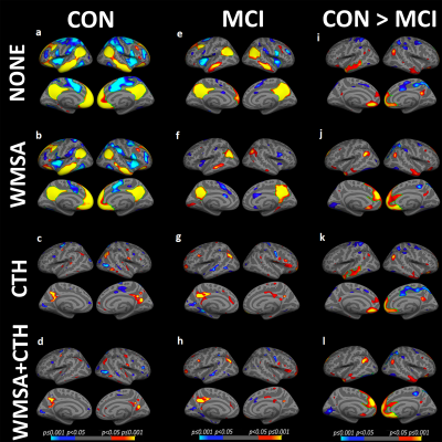 |
The effect of white matter signal abnormalities on default mode network connectivity in mild cognitive impairment
Zhuonan Wang1, Victoria J Williams2, Kimberly A Stephens3, Chan-Mi Kim3, Ming Zhang4, and David Salat3
1PET/CT Unit, Medical Imaging, The First Affiliated Hospital of Xi'an Jiaotong University, Xi'an, China, 2Alzheimer's Clinical and Translational Research Unit, Department of Neurology, Massachusetts General Hospital, Charlestown, MA, United States, 3Athinoula A. Martinos Center for Biomedical Imaging, Department of Radiology, Massachusetts General Hospital, Charlestown, MA, United States, 4Medical Imaging, The First Affiliated Hospital of Xi'an Jiaotong University, Xi'an, China
The striking spatial overlap between regions of default mode network (DMN) and cortical areas most susceptible to Alzheimer's disease (AD)-related pathology and neurodegeneration, with alterations in DMN functional connectivity routinely observed among individuals with mild cognitive impairment (MCI). We examine the relative associations between white matter lesions of presumed vascular origin and cortical thinning typical of AD pathology with DMN integrity to elucidate mechanisms of disease. The degree of white matter damage may have a specific influence on precuneus and mPFC coupling and the observed preferential associations with white matter lesions support a vascular etiology to subtle impairment in MCI.
|
|||
1492.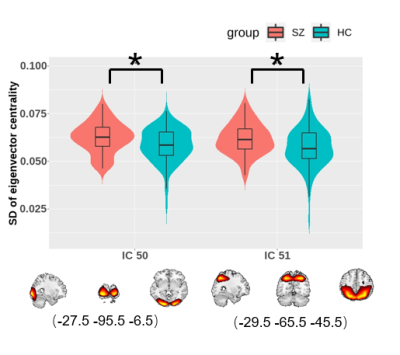 |
Altered brain networks dynamics in first-episode drug-free schizophrenia
Wanfang You1, Lekai Luo1, Qian Li1, Yuxia Wang1, Qiyong Gong1, and Fei Li1
1Huaxi MR Research Centre (HMRRC), Department of Radiology, West China Hospital of Sichuan University, chengdu, China
We investigated disturbances in dynamic functional connectivity (dFC) in whole-brain networks and altered dynamic functional topology of the brain regarding eigenvector centrality in first-episode drug-free patients with schizophrenia. By using resting-state functional magnetic resonance imaging (rf-MRI) data, schizophrenia was mainly manifested as prolonged dwell time in a state characterized by sparsely connected FCs and increased temporal variability of nodal centrality in the visual network, which may help us better interpret the mechanisms underlying visual and auditory hallucination in schizophrenia.
|
|||
1493.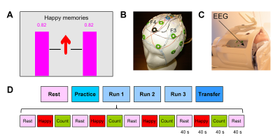 |
Investigation of Frontal Alpha Asymmetry EEG Neurofeedback in Major Depression Using Simultaneous fMRI
Vadim Zotev1, Aki Tsuchiyagaito1, and Jerzy Bodurka1,2
1Laureate Institute for Brain Research, Tulsa, OK, United States, 2Stephenson School of Biomedical Engineering, University of Oklahoma, Norman, OK, United States
We report a controlled study of emotion self-regulation training in patients with major depressive disorder (MDD) using frontal alpha asymmetry (FAA) EEG neurofeedback (EEG-nf) with simultaneous fMRI. MDD patients learned to significantly upregulate the FAA using EEG-nf while inducing happy emotion. Temporal correlations between the FAA and BOLD activity were significantly enhanced during the EEG-nf for many brain regions involved in emotion regulation, including the left DLPFC and the amygdala. Behavioral responses showed stronger approach bias after the training. Our study provides the first independent-modality evidence that the FAA-based EEG-nf can engage and influence the emotional brain circuitry.
|
|||
1494.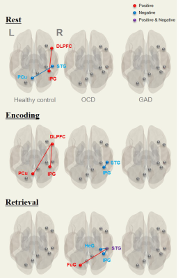 |
Differential task-induced brain activation and functional connectivity patterns between OCD and GAD: Preliminary study on verbal memory
Shin-Eui Park1, Gwang-Won Kim2, Yun-Hyeon Kim3, and Gwang-Woo Jeong3
11Advanced Institute of Aging Science, Chonnam National University, Gwangju, Korea, Republic of, 2Department of Psychiatry, Massachusetts General Hospital and Harvard Medical School, Boston, MA, United States, 3Department of Radiology, Chonnam National University Medical School, Gwangju, Korea, Republic of
This current study supports that abnormalities in task-induced functional connectivity and localized brain activation patterns in explicit, verbal short-term memory processing are potentially linked with cognitive and memory impairment. These findings will be helpful for understanding the differential neural mechanisms associated with the severity of clinical symptoms in OCD and GAD.
|
|||
1495.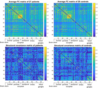 |
Functional and structural brain network features in Borderline Personality Disorder
Giovanni Sighinolfi1, Stefania Evangelisti2, Micaela Mitolo3, Claudio Bianchini2, Laura Ludovica Gramegna2,3, David Neil Manners2, Caterina Tonon2,3, Raffaele Lodi2,3, Francesca D'Adda2, Luca Pellegrini2, Marco Menchetti2, Domenico Berardi2, and Claudia Testa1
1Dipartimento di Fisica e Astronomia, Università di Bologna, Bologna, Italy, 2Dipartimento di Scienze Biomediche e Neuromotorie, Università di Bologna, Bologna, Italy, 3IRCCS Istituto delle Scienze Neurologiche di Bologna, Bologna, Italy
This study uses a graph-based approach to analyse the brain MRI data of early-stage Borderline Personality Disorder (BPD) patients. The topological properties of their Functional Connectivity and Structural Covariance weighted networks are explored. Compared to healthy controls, BPDs exhibit statistically significant alterations at global level, in terms of efficiency and modularity, and at local level, in centrality and efficiency, especially from the functional perspective. The nodal variations are mostly observed in the limbic system, in particular in those regions associated to emotion regulation. Such results may be anticipating structural alterations emerging in later stages of the disease.
|
|||
1496.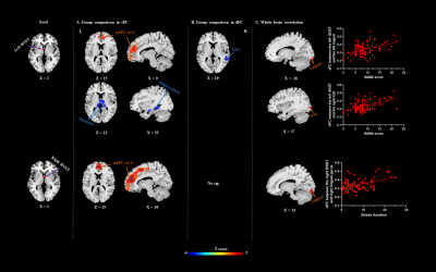 |
Static and dynamic functional connectivity in medication-free patients with obsessive compulsive disorder
Jing Liu1, Hailong Li1, Lingxiao Cao1, Xue Li2, Suming Zhang1, and Xiaoqi Huang1
1Huaxi MR Research Center (HMRRC), Functional and molecular imaging Key Laboratory of Sichuan Province, Department of Radiology, West China Hospital, Sichuan University, Chengdu, China, 2Sichuan University, Chengdu, China
In current study, we use static and dynamic functional connectivity (sFC/dFC) to examine the connectivity alternation of bed nucleus of the stria terminalis (BNST) in medication-free patients with obsessive-compulsive disorder (OCD) to clarify the neural underpinnings of OCD. We found that BNST demonstrated different connected regions in sFC and dFC, indicating that the combination between sFC and dFC can help to detect BNST network alternations in OCD in a more comprehensive way by considering both the static and time-varying aspects.
|
|||
1497.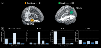 |
Meta-analytic investigation and funtional decoding on neural correlates of high familial risk for mood disorders
Kun Qin1, Nanfang Pan1, Ziyu Zhu1, Feifei Zhang1, Jing Yang1, Xueling Suo1, and Qiyong Gong1
1West China Hospital of Sichuan University, Chengdu, China
Despite the absence of positive clinical manifestations, there may be neurofunctional signatures of genetic vulnerability in the relatives of patients with mood disorders. We performed a meta-analysis integrating task-based fMRI studies comparing unaffected relatives of patients with mood disorders and healthy controls. Hyper-activation in the insula, as well as hypo-activation in the inferior parietal cortex and precuneus were identified in the high-risk relatives. Functional decoding further suggested emotion-related dysfunction might be primarily associated with abnormal activation pattern among deficits in multiple behavioral domains. These neurofunctional correlates may serve as high-risk biomarkers underlying illness onset and development in mood disorders.
|
|||
1498.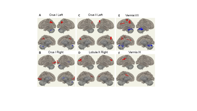 |
Aberrant Cerebellar Functional Connectivity and its Association with Motor and Non-motor Functions in de novo Drug Naïve Parkinson’s Disease
Li Jiang1,2, Brenda Hanna-Pladdy1,2, Jiachen Zhuo1,2, Paul Fishman3, and Rao Gullapalli1,2
1Center for Advanced Imaging Research, University of Maryland Baltimore, Baltimore, MD, United States, 2Department of Diagnostic Radiology & Nuclear Medicine, University of Maryland Baltimore, Baltimore, MD, United States, 3Department of Neurology, University of Maryland Baltimore, Baltimore, MD, United States
Parkinson’s disease (PD) is one of the most common neurodegenerative disorders characterized by progressive deterioration of motor function as well as a non-motor symptom complex. Resting-state fMRI (rs-fMRI) provides important insights on the pathophysiological mechanisms of PD. Here we used rs-fMRI to systematically investigate the cerebellar functional connectivity changes in de novo drug naïve PD patients compared with healthy controls, and the association between altered cerebellar connectivity and neuropsychological assessments. Our findings support that cerebellar connectivity changes while reflective of early symptoms of PD, also may suggest a possible compensatory mechanism prior to clinical presentation of non-motor features of the disease.
|
|||
1499.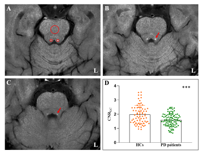 |
Locus coeruleus degeneration is associated with disorganized functional topology in Parkinson’s disease
Cheng Zhou1, Tao Guo1, Jingjing Wu1, Xueqin Bai1, Xiaojun Guan1, and Minming Zhang1
1Zhejiang University, Hangzhou, China
A major emphasis has been placed on the symptom related brain alterations caused by dopamine deficiency, the brain organization underlying the norepinephrine deficiency is largely unknown. We used the neuromelanin sensitive-magnetic resonance imaging for evaluating the degeneration of LC, and using graph theory-based network analysis for characterizing the brain functional topology in 94 PD patients and 68 healthy controls.Relationships between LC degeneration, network disruption and cognitive/motor manifestations in PD patients were assessed.An independent PD subgroup with MRI scanning before and after levodopa administration was enrolled to clarify whether LC degeneration related network disruption were independent of dopamine deficiency.
|
|||
1500.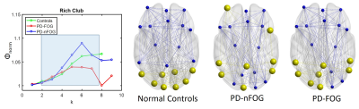 |
Reorganization of functional network topology in Parkinson’s disease patients with and without freezing of gait.
Karthik R Sreenivasan1, Xiaowei Zhuang1, Zhengshi Yang1, Dietmar Cordes1, Aaron Ritter1, Jessica Caldwell1, Zoltan Mari1, and Virendra Mishra1
1Cleveland Clinic Lou Ruvo Center for Brain Health, Las Vegas, NV, United States
Freezing-of-gait (FOG) can be attributed to overloading across neural networks in an attempt to compensate for reduced motor functions. Resting-state fMRI studies in Parkinson’s disease (PD) patients with FOG have implicated dysfunctional connectivity between cortical and subcortical regions over mainly the frontoparietal network, default mode, and visual networks. Our results suggest that despite not observing many global or local network differences there was a clear shift in the topological organization from occipital to frontal regions in both the PD-FOG patients and PD patients without FOG. Specifically, the PD-FOG groups showing significantly reduced rich-club connectivity when compared to the other groups.
|
|||
1501.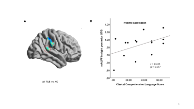 |
Altered Amplitude of Low Frequency Fluctuation in Language Eloquent Areas in Patients with Medically-refractory Temporal Lobe Epilepsy
Li Jiang1,2, Stephanie Chen3, Lorenna Vidal4, Jiachen Zhuo1,2, Rao Gullapalli1,2, and Prashant Raghavan2
1Center for Advanced Imaging Research, University of Maryland Baltimore, Baltimore, MD, United States, 2Department of Diagnostic Radiology & Nuclear Medicine, University of Maryland Baltimore, Baltimore, MD, United States, 3Department of Neurology, University of Maryland Baltimore, Baltimore, MD, United States, 4Department of Radiology, Children's Hospital of Philadelphia, Philadelphia, PA, United States Temporal lobe epilepsy (TLE) is the most common type of epilepsy in adults. Language impairment can result from both continued seizures and surgical attempts to treat it. Thus, accurate preoperative assessment of language function is essential. Here we used resting-state fMRI to investigate the altered amplitude of low frequency fluctuation (ALFF) in language eloquent areas in medically-refractory TLE patients and its relationship with clinical language test measures. Our findings suggest that left TLE disrupts language function more than right TLE and that intrinsic spontaneous brain activity is altered even in the absence of detectable clinical language impairment.
|
|||
1502.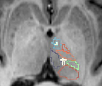 |
Volumetric and connectivity profile of regional thalamic abnormality in amyotrophic lateral sclerosis
Sicong Tu1, Marion Sourty2, Fernando Calamante1, Manojkumar Saranathan3, Ricarda Menke4, Kevin Talbot4, Matthew Kiernan1, and Martin Turner4
1The University of Sydney, Sydney, Australia, 2Université de Strasbourg, Strasbourg, France, 3University of Arizona, Tucson, AZ, United States, 4University of Oxford, Oxford, United Kingdom
Amyotrophic lateral sclerosis (ALS) is a rapidly progressive neurodegenerative disease with widespread extra-motor cortical and subcortical abnormality. The current findings highlight significant regional volumetric and connectivity abnormality in the thalamus associated with clinical features and may be a promising marker of disease burden.
|
|||
1503.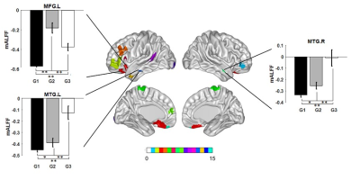 |
Brain amplitude of low frequency fluctuation alterations in optic neuritis patients: a 3-year follow-up study
Jing Huang1, Juan Wei2, and Jie Lu1
1Xuanwu Hospital, Capital Medical University, Beijng, China, 2GE Healthcare, MR Research, Beijng, China
The amplitude of low frequency fluctuation (ALFF) in middle temporal gyrus and middle frontal gyrus might play important role in the prognosis of visual acuity in optic neuritis, which might be used as a potential imaging marker to predict the outcome of visual acuity.
|
|||
1504.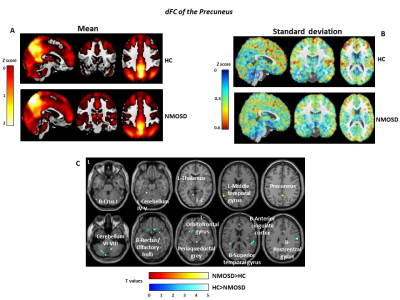 |
Altered Resting State Dynamic Functional Connectivity of the Precuneus Contributes to Cognition and Depression in Neuromyelitis Optica
Paola Valsasina1, Laura Cacciaguerra1,2,3, Damiano Mistri1, Vittorio Martinelli2, Massimo Filippi1,2,3,4,5, and Maria A. Rocca1,2,3
1Neuroimaging Research Unit, Division of Neuroscience, IRCCS San Raffaele Scientific Institute, Milan, Italy, 2Neurology Unit, IRCCS San Raffaele Scientific Institute, Milan, Italy, 3Vita-Salute San Raffaele University, Milan, Italy, 4Neurorehabilitation Unit, IRCCS San Raffaele Scientific Institute, Milan, Italy, 5Neurophysiology Service, IRCCS San Raffaele Scientific Institute, Milan, Italy
In this study, we used dynamic functional connectivity (dFC) to characterize time-varying connectivity abnormalities of the precuneus in 27 patients with neuromyelitis optica spectrum disorders (NMOSD) and test their correlation with cognitive impairment. Compared to controls, NMOSD patients showed reduced precuneus dFC with deep grey matter, temporal, occipital, frontal and cerebellar regions. Increased dFC within the precuneus, and between precuneus and thalamic, insular and temporal regions was also detected. In NMOSD, abnormally high dFC correlated with depression and cognitive deficits.
|
|||
1505.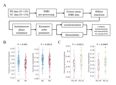 |
Pathological changes in subcortex disrupt cortical synchronization and metastability affecting cognitive function in Parkinson’s disease
Cheng Zhou1 and Minming Zhang1
1Zhejiang University, Hangzhou, China
In this paper, we investigated spatiotemporal dynamics of the phase interactions among resting-state blood oxygen level-dependent signals of Parkinson’s disease patients (n = 159) and normal controls (n = 152). We demonstrated that diminished dopaminergic function and the pathological changes in thalamus related structures responsible for decreased cortical synchronization and metastability, further affect cognitive function in Parkinson’s disease.
|
|||
1506. |
Aberrant spontaneous low-frequency brain activity in patients with subjective cognitive decline: A resting-state fMRI study
Yin Tang1, Ling Zhang1, Yi Zhu2, Hongyuan Ding1, Yaxin Gao2, Long Qian3, Weiqiang Dou3, and Ming Qi1
1Radiology department, The First Affiliated Hospital of Nanjing Medical University, Nanjing, China, 2Rehabilitation Department, The First Affiliated Hospital of Nanjing Medical University, Nanjing, China, 3MR Research China, GE Healthcare, Beijing, China
In this study, whole brain amplitude of low-frequency fluctuation (ALFF) changes have been respectively investigated for patients with subjective cognitive decline (SCD) and mild cognitive impairment (MCI) and healthy controls (HCs). Relative to HCs, significantly lower ALFF values have been separately found in the regions of right supramarginal gyrus, left precuneus and right supplementary motor area for SCD and MCI patients. Additionally, the ALFFs at these regions also showed strong correlations with multiple clinical scales. Therefore, ALFF might be considered an effective index in the early detection of SCD patients.
|
|||
1507.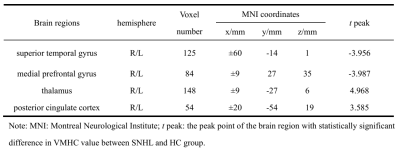 |
The changes of regional homogeneity and voxel-mirrored homotopic connectivity in Preschool Children with sensorineural hearing loss: a resting-state fMRI
Yi Yin1, Houyu Zhao2, Guiquan Shen1, Mingming Huang1, Xiaoxu Zhang1, Yawen Liu1, Lisha Nie3, and Hui Yu1
1Department of Imaging, Affiliated Hospital of Guizhou Medical University, Gui yang, China, 2Department of Otorhinolaryngology, Affiliated Hospital of Guizhou Medical University, Gui yang, China, 3GE Healthcare, MR Research, Beijing, China
Long-term hearing loss can lead to changes in brain structure and function in preschool children with sensorineural hearing loss(SNHL). we uses regional homogeneity(ReHo) and voxel mirror homology connectivity(VMHC) methods to evaluate the abnormal changes of functional activities in regional brain and interhemispheric functional connections in children after hearing deprivation during resting-state. The results of this study showed that the structure and function of auditory related brain regions were reorganized, and the interhemispheric information transfer was also changed, which would contribute to a better understanding the neuropathological mechanism of preschool deafness.
|
|||
1508.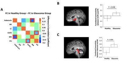 |
Alterations of the sleep-regulating systems in glaucoma
Ji Won Bang1, Carlos Parra1, Gadi Wollstein1, Joel S Schuman1, and Kevin C Chan1,2
1Department of Ophthalmology, New York University Grossman School of Medicine, New York, NY, United States, 2Department of Radiology, New York University Grossman School of Medicine, New York, NY, United States
Glaucoma patients have a high incidence of sleep disorders. This relationship implies that glaucoma may involve alterations in sleep-regulating systems. Here, we examined how glaucoma affects sleep-regulating subcortical systems. In particular, we focused on the ventrolateral preoptic nucleus (VLPO), a central sleep-inducing hub. We demonstrated that glaucoma patients had altered functional connectivity of VLPO with the subcortical arousal system and the occipital cortex. We also showed that glaucoma patients had reduced GABA levels in the occipital cortex. Overall, our study suggests that sleep-regulating subcortical structures involving VLPO and their inhibitory projections to the cortex are impaired in glaucoma.
|
|||
1509.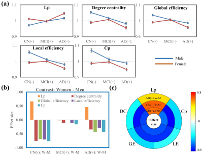 |
Multi-scale sex difference of brain function in Alzheimer’s disease
Zhengshi Yang1,2, Cieri Filippo1, Xiaowei Zhuang1,2, Marwan Sabbagh1, Jefferson W. Kinney2, Jeffrey L. Cummings2, Dietmar Cordes1,2,3, and Jessica Z.K. Caldwell1
1Cleveland Clinic Lou Ruvo Center for Brain Health, Las Vegas, NV, United States, 2University of Nevada Las Vegas, Las Vegas, NV, United States, 3University of Colorado Boulder, Boulder, CO, United States
Despite the prevalence of AD in women and the recognized sex-dependent genetic factors and male/female differences in cognitive measures in AD, how sex is related to AD phenotypic variability remains unclear. We demonstrated a varying spatial extent and magnitude of sex differences in brain function in an AD cohort, suggesting the dynamic contribution of sex in disease progression. Opposite network topological changes were observed from cognitively normal to MCI, and more rapid progression occurred in women than men from MCI to AD. The occipital lobe contributed more in men but frontal lobe contributed more in women in disease progression.
|
|||
1510.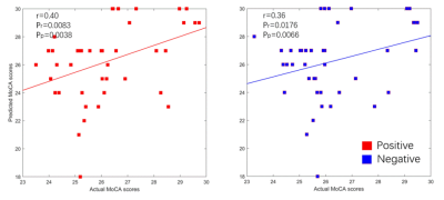 |
Resting-State Functional Connectivity Predicts Cognitive Impairment Related to Type 2 Diabetes Mellitus
An ping Shi1 and Xi yang Tang2
1Department of Radiology & Functional and Molecular Imaging Key Lab of Shaanxi Province, Department of Radiology & Functional and Molecular Imaging Key Lab of Shaanxi Province, Tangdu Hospital, Fourth Military Medical University (Air Force Medical University), Xi'an, Shaanxi, China, 2Department of thoracic surgery, Tangdu Hospital, Air Force Medical University., Xi'an, China
Resting-state functional connectivity (RSFC) patterns of the human brain show unique inherent or intrinsic characteristics, similar to a fingerprint. There is significant interest in using RSFC to predict human behavior. Inspired by previous RSFC fingerprinting studies, we adopted whole-brain RSFC as discriminative features to predicted the MoCA scores in 102 individuals with T2DM, using a connectome-based predictive modeling (CPM). We find that, the identified CPM, based on whole-brain RSFC patterns, are strong for predicting the MoCA scores in T2DM. The application of CPM to predict neurocognitive abilities can complement conventional neurocognitive assessments and aid the management of people with T2DM.
|
The International Society for Magnetic Resonance in Medicine is accredited by the Accreditation Council for Continuing Medical Education to provide continuing medical education for physicians.