Digital Posters
Diffusion: Across Age Spans
ISMRM & SMRT Annual Meeting • 15-20 May 2021

| Concurrent 5 | 19:00 - 20:00 |
 |
1690.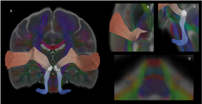 |
Microstructural characterization of auditory pathway developmental trajectory from infancy through adolescence
Kirsten Mary Lynch1, Ryan P Cabeen1, Stefanie C Bodison2,3, Arthur W Toga1, and Courtney C.J. Voelker4
1USC Mark and Mary Stevens Institute for Neuroimaging and Informatics, USC Keck School of Medicine, Los Angeles, CA, United States, 2Chan Division of Occupational Science and Occupational Therapy, USC Herman Ostrow School of Dentistry, Los Angeles, CA, United States, 3Division of Pediatrics, USC Keck School of Medicine, Los Angeles, CA, United States, 4USC Caruso Department of Otolaryngology – Head and Neck Surgery, USC Keck School of Medicine, Los Angeles, CA, United States
Auditory perception is established through experience-dependent stimuli during sensitive developmental periods; however, little is known regarding the structural development of the central auditory pathway. The present study quantified the magnitude and timing of regional microstructural development of the auditory pathway from the brainstem to the auditory cortex from infancy through adolescence using DTI and NODDI metrics. We found spatially varying white matter maturation along the length of the tract, with inferior brainstem regions developing earliest. These results help to characterize the processes that give rise to functional auditory processing and may provide a baseline for detecting abnormal development.
|
||
1691.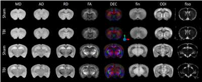 |
Assessment of microstructural changes following pediatric traumatic brain injury by advanced diffusion imaging
Yohan van de Looij1,2, Alice Jacquens3,4, Pierre Gressens4,5, Vincent Degos3,4, and Stéphane V Sizonenko1
1Division of Development and Growth, Department of Paediatrics and Gynaecology-Obstetrics, University of Geneva, Geneva, Switzerland, 2Center for Biomedical Imaging, Animal Imaging Technology section, Federal Institute of Technology of Lausanne, Lausanne, Switzerland, 3Department of Anesthesia and Intensive Care, Pitié-Salpêtrière Hospital, Paris, France, 4PROTECT, INSERM, Université Paris Diderot, Sorbonne Paris Cité, Paris, France, 5Centre for the Developing Brain, School of Biomedical Engineering and Imaging Sciences, King's College London, London, United Kingdom
Traumatic brain injury (TBI) on the immature brain can have dramatic consequences on cerebral development. Understanding the underlying lesions of this abnormal development is of high interest. In this work, we used a mouse model of pediatric TBI at Postnatal day 7 (P7 - impact acceleration model) and assessed the long-term subsequent microstructural damages (at P45) using DTI and NODDI at 9.4T. Severe changes in white matter and cortical developments were observed. In conclusion, DTI derived parameters as well as NODDI estimates allow an accurate determination of the location and extent of the brain lesions following TBI.
|
|||
1692. |
Characterization of individual DTI measurement age trajectories in a longitudinal study of autism spectrum disorder
Nagesh Adluru1, Douglas C Dean III1, Molly Prigge2, Jace B King2, Nicholas T Lange3, Erin D Bigler4, Brandon Zielinski2, Janet E Lainhart1, and Andrew L Alexander1
1UW-Madison, Madison, WI, United States, 2University of Utah, Salt Lake City, UT, United States, 3Harvard Medical School, Boston, MA, United States, 4Brigham Young University, Provo, UT, United States
The purpose of this analysis is to investigate the patterns of individual rates of change of diffusion tensor imaging (DTI) measures computed from a longitudinal study of autism spectrum disorder (ASD). The mean value and temporal rates of change of DTI measures were estimated for regions of white matter for individual subjects. The distributions of the individual longitudinal slopes versus mean measures for ASD and typically developing controls (TDC) were mapped and compared, revealing group differences in the distributions with generally greater heterogeneity in the ASD group.
|
|||
1693. |
Connectometry analysis of diffusion MRI in adolescents with sports-related concussion
Hon J Yu1, Mark Fisher2, and Min-Ying Su1
1Radiological Sciences, University of California, Irvine, CA, United States, 2Neurology, University of California, Irvine, Orange, CA, United States
This study evaluates a feasibility of diffusion MRI based connectometry in capturing sports-related concussion (SRC) induced effects in adolescents using high school football players with and without concussion history as cohorts. The results revealed that although there were some common tract bundles whose segments showed significant correlation with concussion history independent of the diffusion metrics used (QA, MD, or FA), there were also different tract bundles that showed either positive or negative correlation with concussion depending on the diffusion metric used. A further study would be necessary to fully examine clinical relevance of this study’s findings in adolescents with SRC.
|
|||
1694. |
Multimodality data improve diagnostic efficacy in brain injury of premature infants with necrotizing enterocolitis
Chunxiang Zhang1, Meiying Cheng1, Kaiyu Wang2, Xin Zhao1, and Xiaoan Zhang1
1Department of Radiology, The Third Affiliated Hospital of Zhengzhou University, Zhengzhou, China, 2GE Healthcare, MR Research China, Beijing, China
Diffusion tensor imaging (DTI) can noninvasively and quantitatively evaluate the development of brain white matter and myelination degree of damaged nerve fiber bundles. Serum C-reactive protein (CRP) and procalcitonin (PCT) as inflammatory indicators can easily reflect the systemic inflammation of necrotizing enterocolitis (NCE) in children. This study shows that the DTI combined with serum CRP and PCT was more effective than a single indicator in the diagnosis of brain developmental disorders in preterm infants with NEC.
|
|||
1695.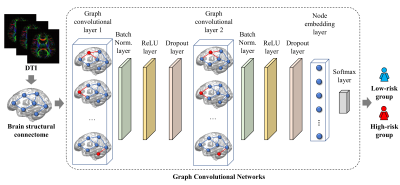 |
Early prediction of cognitive deficits in very preterm infants using graph convolutional networks with brain structural connectome
Hailong Li1, Ming Chen1,2, Jinghua Wang3, Nehal A. Parikh4,5, and Lili He1,5
1Imaging Research Center, Department of Radiology, Cincinnati Children's Hospital Medical Center, Cincinnati, OH, United States, 2Department of Electrical Engineering and Computer Science, University of Cincinnati, Cincinnati, OH, United States, 3Deep MRI Imaging Inc., Lewes, DE, United States, 4The Perinatal Institute and Section of Neonatology, Perinatal and Pulmonary Biology, Cincinnati Children's Hospital Medical Center, Cincinnati, OH, United States, 5Department of Pediatrics, University of Cincinnati College of Medicine, Cincinnati, OH, United States
Up to 40% of very preterm infants (≤32 weeks’ gestational age) are identified with cognitive deficits at 2 years of age. Reproducible approaches that serve as neonatal prognostic tools are urgently needed for early treatment decision. We developed a graph convolutional network model to learn the latent topological features of brain structural connectome obtained at term-equivalent age for predicting cognitive deficits at 2 years corrected age in very preterm infants. The proposed model was able to identify infants at high-risk of cognitive deficits with a balanced accuracy of 78.5% and an area under the receiver operating characteristic curve of 0.78.
|
|||
1696.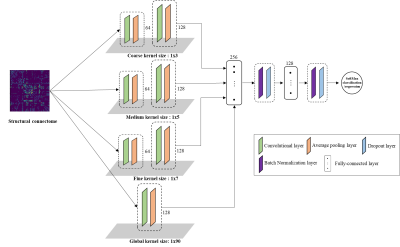 |
A novel multi-filter convolutional neural network for prediction of cognitive deficits using structural connectome in very preterm infants
Ming Chen1,2, Hailong Li1, Jinghua Wang3, Nehal A. Parikh4, and Lili He1
1Department of Radiology, Cincinnati Children's Hospital Medical Center, Cincinnati, OH, United States, 2Department of Electronic Engineering and Computing Science, University of Cincinnati, Cincinnati, OH, United States, 3Deep MRI Imaging Inc., Lewes, DE, United States, 4The Perinatal Institute, Cincinnati Children's Hospital Medical Center, Cincinnati, OH, United States
We proposed a novel multi-filter convolutional neural network for prediction of cognitive deficits using brain structural connectome data. In contrast to 2D grid convolutional filters in traditional convolutional neural networks, our proposed model contains multiple vector-shape convolutional filters that can better extract the topological information from brain connectome. We demonstrated the ability of our model to learn hidden patterns from brain connectome data for prediction tasks. Our proposed model was able to identify infants at a high risk of cognitive deficits with an area under the curve of 0.78, exceeding the performance of other existing peer convolutional neural network methods.
|
|||
 |
1697.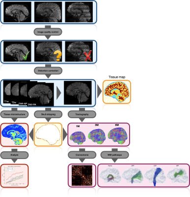 |
An Automated Processing Pipeline for Diffusion MRI in the Baby Connectome Project
Ye Wu1, Sahar Ahmad1, Khoi Minh Huynh1, Siyuan Liu1, Kim-Han Thung1, Weili Lin1, Pew-Thian Yap1, and UNC/UMN Baby Connectome Project Consortium1
1Biomedical Research Imaging Center, University of North Carolina, Chapel Hill, Chapel Hill, NC, United States
Processing baby diffusion MRI (dMRI) data is challenging due to the low and spatially-varying diffusion anisotropy, rendering standard analysis techniques developed for adult data inapplicable [1,2]. Here, we present a fully-automated processing pipeline for baby dMRI, tailored particularly to the data collected in the Baby Connectome Project (BCP).
|
||
1698.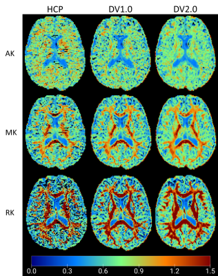 |
The effect of image pre-processing pipelines on age associations of diffusion and kurtosis in white matter
Jenny Chen1, Benjamin Ades-aron1, Hong-Hsi Lee1, Durga Kullakanda1, Saurabh Maithani1, Dmitry S. Novikov1, Jelle Veraart1, and Els Fieremans1
1Radiology, NYU School Of Medicine, New York, NY, United States
Diffusion MRI is prone to various artifacts such as noise, eddy current artifacts, and Gibbs ringing. This study compares diffusion tensor imaging (DTI) and diffusional kurtosis imaging (DKI) parameter estimates among healthy subjects in their 20s to 80s using a minimal diffusion pre-processing approach from Human Connectome Project (HCP) and two DESIGNER (Diffusion parameter EStImation with Gibbs and NoisE Removal) pipelines, which corrects for additional imaging artifacts HCP pipeline does not account for. Our results show that preprocessing quantitatively impacts parameter estimation as well as alters observed age correlations.
|
|||
1699.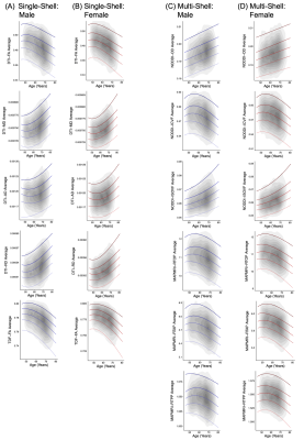 |
Age and Sex Effects on Brain White Matter Microstructure assessed with Advanced Single- and Multi-Shell Diffusion MRI Metrics
Katherine E Lawrence1, Leila Nabulsi1, Vigneshwaran Santhalingam1, Zvart Abaryan1, Julio E Villalon-Reina1, Talia M Nir1, Iyad Ba Gari1, Alyssa H Zhu1, Elizabeth Haddad1, Alexandra M Muir1, Neda Jahanshad1, and Paul M Thompson1
1University of Southern California, Marina del Rey, CA, United States
Characterizing the brain’s white matter microstructure is crucial for improving our understanding of healthy and diseased aging. Here we examined the ability of both traditional diffusion methods (diffusion tensor imaging) and advanced diffusion methods (tensor distribution function, neurite orientation dispersion and density imaging, mean apparent propagator MRI) to capture age and sex effects on white matter microstructure in a large sample of aging adults (15,628 UK Biobank participants; age range 45-80 years). Advanced diffusion models exhibited the greatest sensitivity to participant age and sex, suggesting that future aging studies may benefit from using advanced diffusion approaches.
|
|||
1700.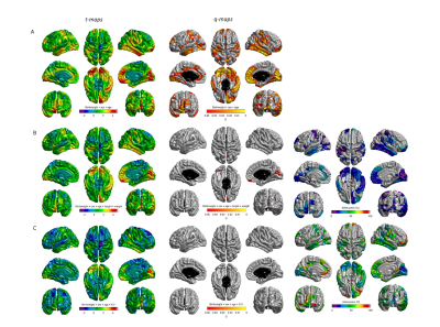 |
Birth weight is associated with brain tissue volumes seven decades later but not age-associated changes to brain structure
Emily Wheater1, Susan D Shenkin2,3, Susana Muñoz Maniega2,4, Maria Valdés Hernández2,4, Joanna M Wardlaw2,4, Ian J Deary4,5, Mark E Bastin2,4,6, James P Boardman1,2, and Simon R Cox4,5,6
1Centre for Reproductive Health, University of Edinburgh, Edinburgh, United Kingdom, 2Centre for Clinical Brain Sciences, University of Edinburgh, Edinburgh, United Kingdom, 3Geriatric Medicine, Usher Institute, University of Edinburgh, Edinburgh, United Kingdom, 4Lothian Birth Cohorts, University of Edinburgh, Edinburgh, United Kingdom, 5Psychology, University of Edinburgh, Edinburgh, United Kingdom, 6Scottish Imaging Network, A Platform for Scientific Excellence Collaboration (SINAPSE), Edinburgh, United Kingdom
Birthweight is a commonly used indicator of fetal growth weight and has been associated with neuropsychiatric and neurological sequalae. However, little is known about how birth weight impacts the brain in later life. We found positive associations with total brain, grey matter and normal appearing white matter volumes in later life, but not with white matter microstructure or hyperintensities. This relationship is explained by larger head size, rather than by age-associated tissue atrophy, and is furthermore independent of body size. This suggests that larger birthweight is linked to increased brain tissue reserve, but not age-associated brain features.
|
|||
1701.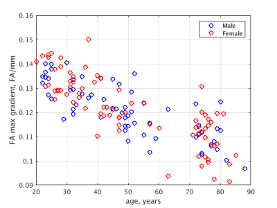 |
Profiling diffusion at the grey matter-white matter interface (GWI) to reveal unique microstructural features: proof of concept in aging
Roman Fleysher1 and Michael L Lipton1
1Radiology, Albert Einstein College of Medicine, Bronx, NY, United States
Microstructure differs greatly between cortical gray matter and adjacent white matter and this point of transition, the gray-white matter interface, is a predilection site for pathologies, including traumatic brain injury, small vessel vasculitis and microembolic ischemic injury. The interface region presents challenges to both voxel-wise and region of interest (ROI) analyses, because small registration or ROI placement errors may lead to large errors in extracted diffusion metrics. We propose an approach to assess alteration of the sharpness of the gray-white matter interface and illustrate its potential utility through the detection of age-related decline of the sharpness of this transition.
|
|||
1702.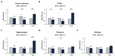 |
Longitudinal Apparent Diffusion Coefficient Trajectory in Different Severity Outcome Following Experimental Neonatal Hypoxic Ischemia
Yu-Chieh Jill Kao1, Chia-Feng Lu1, Bao-Yu Hsieh2, Cheng-Yu Chen3, and Chao-Ching Huang4
1National Yang Ming University, Taipei, Taiwan, 2Chang-Gung University, Taoyuan, Taiwan, 3Taipei Medical University, Taipei, Taiwan, 4National Cheng Kung University, Tainan, Taiwan
Temporal and regional profile of ADC-related MR characteristics after neonatal hypoxic ischemia between mild and severe outcome was delineated at 6 h, 24 h and 7 days after injury. Significant difference in ADC trajectories obtained early after HI are observed between outcome groups, suggesting that distinct ADC changes may be associated to tissue damage severity along the progress of HI.
|
|||
1703.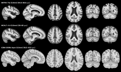 |
MIITRA atlas: Construction of high resolution T1w and DTI brain templates in a common space, based on 400 older adults
Yingjuan Wu1, Mohammad Rakeen Niaz1, Abdur Raquib Ridwan1, Xiaoxiao Qi1, David A. Bennett2, and Konstantinos Arfanakis1,2
1Illinois Institute of Technology, Chicago, IL, United States, 2Rush Alzheimer's Disease Center, Rush University, Chicago, IL, United States
As a critical step to establish the Multichannel Illinois Institute of Technology & Rush university Aging (MIITRA) atlas, the present work aimed to: a) develop high quality 0.5mm resolution T1-weighted (T1w) and diffusion tensor imaging (DTI) templates in a common space using data from a large, diverse, community cohort of non-demented older adults, and b) quantitatively compare the new templates to existing templates in terms of spatial normalization accuracy of external data. The new T1w and DTI templates allowed higher inter-subject and inter-modality spatial normalization of older adult data compared to other templates.
|
|||
1704.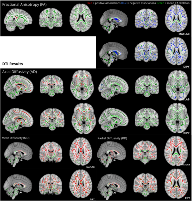 |
Using diffusional kurtosis imaging to capture white matter tissue complexity in aging: Does the choice of software package affect results?
Hiba Taha1,2, Jordan A. Chad2,3, and J. Jean Chen2,3
1Human Biology, University of Toronto, Toronto, ON, Canada, 2Rotman Research Institute, Baycrest Health Sciences, Toronto, ON, Canada, 3Medical Biophysics, University of Toronto, Toronto, ON, Canada
Studies in white matter (WM) aging have previously used diffusional kurtosis imaging (DKI), however it remains unclear if results have been affected by limited sample sizes or the choice of software package. In this study, we show positive age associations of diffusivity and negative age associations of kurtosis throughout WM tracts using two different DKI packages on a large sample of 700 adults. Unique regional specific changes were observed differentially using these two tools, suggesting that the choice of software package can influence the study of aging.
|
|||
|
1705.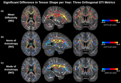 |
Orthogonal diffusion tensor decomposition reveals age-related degeneration patterns in complex fibre architecture
Jordan A. Chad1,2, Ofer Pasternak3, and J. Jean Chen1,2
1Rotman Research Institute, Baycrest Health Sciences, Toronto, ON, Canada, 2Department of Medical Biophysics, University of Toronto, Toronto, ON, Canada, 3Brigham and Women's Hospital, Harvard Medical School, Boston, MA, United States
Diffusion tensor imaging (DTI) consistently detects increased mean diffusivity (MD) and decreased fractional anisotropy (FA) with age in single white matter (WM) bundles, but findings have been inconsistent in more complex fibre architecture. In this study, an orthogonal moment tensor decomposition yields clear degeneration patterns across complex fibre architecture, indicating that apparent challenges of DTI in these regions were due to the choice of tensor decomposition rather than the DTI model itself. This study therefore presents a revised view of DTI of aging WM and indicates how degeneration in complex fibre architecture can manifest in forms other than decreased FA.
|
||
1706.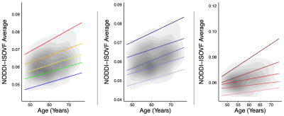 |
Exogenous sex hormone effects on brain microstructure in women: a diffusion MRI study in the UK Biobank
Leila Nabulsi1, Katherine E Lawrence1, Vigneshwaran Santhalingam1, Zvart Abaryan1, Christina P Boyle1, Julio E Villalon-Reina1, Talia M Nir1, Iyad Ba Gari1, Alyssa H Zhu1, Elizabeth Haddad1, Alexandra M Muir1, Neda Jahanshad1, and Paul M Thompson1
1Imaging Genetics Center, Mark and Mary Stevens Neuroimaging & Informatics Institute, University of Southern California, Marina del Rey, CA 90292, USA, Los Angeles, CA, United States
In women, higher levels of sex-related hormones have been associated with increased risk for age-related neurodegenerative diseases such as Alzheimer’s disease. We measured age-associated effects of exogenous sex hormones on brain white matter microstructure in pre- and post-menopausal women, using four diffusion-weighted MRI models (DTI, TDF, NODDI and MAPMRI). Estrogen therapy alone was associated with accelerated aging in white matter metrics, compared to hormone replacement therapy containing both estrogen and progestin. This effect was most evidenced by multi-shell diffusion method NODDI, compared to DTI and other advanced diffusion methods.
|
|||
1707.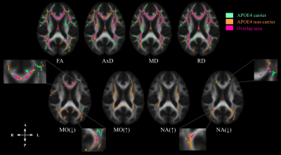 |
Effect of age on white matter microstructure in nondemented ApoE4 carriers and non-carriers
Patcharaporn Srisaikaew1,2, Jordan A. Chad3,4, Pasuk Mahakkanukrauh1,5, Nicole D. Anderson3,6, and J. Jean Chen3,4
1Department of Anatomy, Faculty of Medicine, Chiang Mai University, Chiang Mai, Thailand, 2PhD Program in Anatomy, Faculty of Medicine, Chiang Mai University, Chiang Mai, Thailand, 3Rotman Research Institute, Baycrest Health Centre, Toronto, ON, Canada, 4Department of Medical Biophysics, University of Toronto, Toronto, ON, Canada, 5Excellence in Osteology Research and Training Center (ORTC), Chiang Mai University, Chiang Mai, Thailand, 6Department of Psychology and Psychiatry, University of Toronto, Toronto, ON, Canada
With advancing age, ApoE4 increases the risk of developing AD compared to non-carriers. TBSS analysis and FSL’s randomise permutation tool were used to test diffusivity (FA, MD, RD, AxD, MO, and NA) differences in the effect of age in nondemented ApoE4+ compared to ApoE4- older adults. The difference between groups in the association of DTI metrics and age was most present in posterior WM regions. MO and NA were more sensitive to age-related effects than conventional DTI metrics in crossing fibres. We support the changes in DTI metrics due to the manifestation of the ApoE4 across whole-brain age-related WM microstructures.
|
|||
1708. |
Longitudinal Brain Atlases of Early Developing Cynomolgus Macaques from Birth to 48 Months of Age
Tao Zhong1,2, Liangjun Chen2, Fenqiang Zhao2, Zhengwang Wu2, Yuchen Pei2, Ya Wang2, Li Wang2, Yuyu Niu3, Yu Zhang1, and Gang Li2
1Southern Medical University, Guangzhou, China, 2University of North Carolina at Chapel Hill, Chapel Hill, NC, United States, 3Kunming University of Science and Technology, Kunming, China
In analyzing the early postnatal brain development featuring extremely dynamic imaging contrast, brain appearance, shape and size, longitudinal brain atlases with densely sampled time-points and ancillary anatomical information are of great importance, but remain absent in cynomolgus macaques, which is a highly valuable animal model for understanding human brains. To fill this critical gap, we construct the first set of spatiotemporal (4D) brain atlases and associated ancillary anatomical information with 12 time-points (i.e., 1, 2, 3, 4, 5, 6, 9, 12, 18, 24, 36, and 48 months of age) based on 175 longitudinal structural MRI scans from 46 cynomolgus macaques.
|
|||
1709.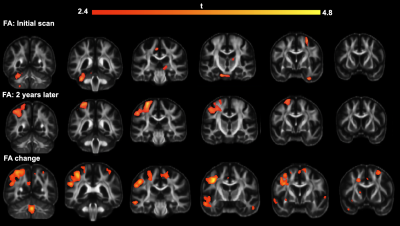 |
Widespread effect of age related macular degeneration on brain structural integrity.
Jacques Andrew Stout1, Robert BJ Anderson2, Simon Wilton Davis3, Jie Zhuang3,4, David Dunson5,6, Heather Elisabeth Whitson7, and Alexandra Badea1,2,3
1BIAC, Duke School of Medicine, Durham, NC, United States, 2Duke Radiology, Duke School of Medicine, Durham, NC, United States, 3Duke Neurology, Duke School of Medicine, Durham, NC, United States, 4School of Psychology, Shanghai University of Sport, Shanghai, China, 5Statistics, Duke University, Durham, NC, United States, 6Trinity College of Arts & Sciences, Duke University, Durham, NC, United States, 7Geriatrics, Duke School of Medicine, Durham, NC, United States
Age-related macular degeneration (AMD) has been associated with brain degeneration, particularly in the cortical regions of the brain. Our study involved the comparison of subjects with AMD to subjects, with both an initial scan at the beginning of the study, and a secondary one two years later. Using anatomical and diffusion acquisitions, we ascertained that there was a marked decrease in cortical volumetry and fractional anisotropy (FA), and that it evolved very rapidly in the case of FA. A PCA run on the connectivity matrices also identified the connections that were most affected.
|
The International Society for Magnetic Resonance in Medicine is accredited by the Accreditation Council for Continuing Medical Education to provide continuing medical education for physicians.