Digital Posters
Spine Imaging/Nervous System & More
ISMRM & SMRT Annual Meeting • 15-20 May 2021

Digital Posters
Spine Imaging/Nervous System & More
| Concurrent 5 | 19:00 - 20:00 |
 |
3461.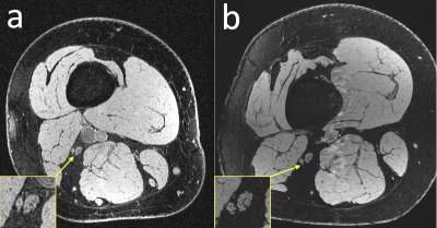 |
High-resolution anatomical and diffusion-weighted imaging in peripheral nerves at 7 Tesla using quantitative Double-Echo in Steady-State
Bragi Sveinsson1,2, Robert L Barry1,2,3, Olivia Rowe1,2, Jason Stockmann1,2, Daniel J Park1, Peter J Lally4, Matthew S Rosen1,2,5, and Reza Sadjadi6
1Athinoula A. Martinos Center for Biomedical Imaging, Massachusetts General Hospital, Boston, MA, United States, 2Radiology, Harvard Medical School, Boston, MA, United States, 3Harvard-Massachusetts Institute of Technology Health Sciences and Technology, Cambridge, MA, United States, 4Brain Sciences, Imperial College London, London, United Kingdom, 5Physics, Harvard University, Cambridge, MA, United States, 6Neurology, Massachusetts General Hospital, Boston, MA, United States
Research has suggested elevated diffusivity in nerves of subjects with peripheral nerve disease. Obtaining quantitative estimates of diffusivity at small scales within peripheral nerves could therefore provide important information about disease progression. However, diffusion weighted imaging at such small scales is challenging due to low signal to noise ratio (SNR) and distortion. Here, we demonstrate the feasibility of obtaining high-SNR, high-resolution, low-distortion anatomical images and quantitative diffusivity estimates in individual nerve fascicles with the quantitative Double-Echo in Steady-State sequence at 7 Tesla.
|
||
3462. |
Spinal cord perfusion mapping using Intra-Voxel Incoherent Motion at 3T in healthy individuals and Degenerative Cervical Myelopathy patients
Simon Lévy1,2,3, Patrick Freund4,5,6, Virginie Callot1,2,3, and Maryam Seif4,5
1CRMBM, Aix-Marseille University, CNRS, Marseille, France, 2CEMEREM, APHM, Hopital Universitaire Timone, Marseille, France, 3iLab-Spine International Research Laboratory, Marseille-Montreal, France, 4Spinal Cord Injury Center, University Hospital Balgrist, University of Zurich, Zurich, Switzerland, 5Max Planck Institute for Human Cognitive and Brain Sciences, Leipzig, Germany, 6Wellcome Trust Centre for Neuroimaging, UCL Institute of Neurology, London, United Kingdom
In the search for a technique to assess spinal cord perfusion, the Intra-Voxel Incoherent Motion technique, previously implemented at 7T, was adapted to 3T. B-values were optimized based on phantom acquisitions. The final protocol was applied within 11 healthy volunteers and 2 Degenerative Cervical Myelopathy patients. The technique demonstrated sensitivity to perfusion in healthy volunteers and to capillary network orientations, with a clear depiction of the gray matter perfusion and inter-slice reproducibility. No significant difference could be shown between healthy volunteers and patients given the small sample size but more patients will be included in the near future.
|
|||
3463. |
A Simulation Study on the Difference of PNS with Magnetic Fields and Electric Fields in an Arm Model
Yihe Hua1, Desmond TB Yeo1, and Thomas K Foo1
1GE Global Research, Niskayuna, NY, United States
PNS by the alternative magnetic field from the gradient coil is an important safety consideration in MRI scanning. Previous studies reported the chronaxie time of PNS in the arm by transcutaneous electrode stimulation (ES) might be very different from PNS by magnetic stimulation (MS). We simulated the electric field distributions and chronaxie of most sensitive nerve trajectories for both ES and MS cases in an anatomical real arm model and found the profile of the field distribution for both cases are very different but chronaxie values at the same order of magnitude.
|
|||
3464.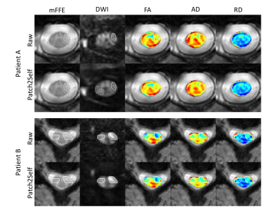 |
Patch2Self denoising of diffusion MRI in the cervical spinal cord improves repeatability and feature conspicuity
Kurt G. Schilling1,2, Shreyas Fadnavis3, Mereze Visagie2, Eleftherios Garyfallidis3, Bennett A. Landman2,4, Seth A. Smith1,2, and Kristin P. O'Grady1,2
1Radiology and Radiological Sciences, Vanderbilt University Medical Center, Nashville, TN, United States, 2Vanderbilt University Institute of Imaging Science, Vanderbilt University Medical Center, Nashville, TN, United States, 3Intelligent Systems Engineering, Indiana University Bloomington, Bloomington, IN, United States, 4Electrical Engineering and Computer Science, Vanderbilt University, Nashville, TN, United States
Diffusion MRI (dMRI) is a promising tool for evaluating the spinal cord in health and disease, however low SNR can impede accurate, repeatable, quantitative measurements. Here, we apply a recently proposed denoiser, Patch2Self, that strictly suppresses statistically independent random fluctuations in the signal originating from various sources of noise. Typical spinal cord dMRI scans have a smaller number of gradient directions (10-20) making PCA based 4D denoisers (require at least 30) inapplicable. Using self-supervised learning, Patch2Self addresses these issues which we quantitatively show with an improvement in repeatability and conspicuity of pathology in the spinal cord.
|
|||
3465.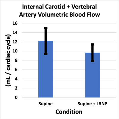 |
Spaceflight Associated Neuro-Ocular Syndrome: Quantitative MRI Evaluation of Lower Body Negative Pressure as a Potential Countermeasure
Larry A. Kramer1, Khader M. Hasan1, Brandon R. Macias2, Karina J. Marshall-Goebel3, Steven S. Laurie3, Refaat E. Gabr1, Leela Chaudhary1, and Alan R. Hargens4
1Diagnostic Imaging, UTHSC-Houston, Houston, TX, United States, 2NASA, Houston, TX, United States, 3KBR, Houston, TX, United States, 4Orthopedic Surgery, UCSD, La Jolla, CA, United States
The application of lower body negative pressure in the supine position significantly reduces bulk cerebral arterial flow and cross-sectional area of the internal jugular vein compared to the supine position alone. This is similar to the physiologic response expected with upright compared to supine positioning. Our results suggest that lower body negative pressure applied during spaceflight may help reduce headward fluid shift and venous congestion and mitigate the development of optic disc edema associated with long-duration spaceflight.
|
|||
3466.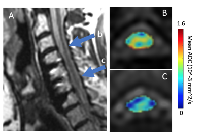 |
Assessment of Spinal Cord Diffusivity in Degenerative Cervical Myelopathy after Spinal Fusion Decompression
Kevin M Koch1, V. Emre Arpinar1, and Matthew Budde2
1Radiology, Medical College of Wisconsin, Milwaukee, WI, United States, 2Neurosurgery, Medical College of Wisconsin, Milwaukee, WI, United States
Metal artifacts from commonly-encountered spinal fusion stabilization hardware have historically confounded quantitative MRI (qMRI) research investigations of the injured and damaged spinal cord in human subjects. This report provides preliminary analysis of metal-artifact suppressed multi-spectral diffusion qMRI collected on instrumented degenerative cervical spondylotic myelopathy (CSM) subjects. The results of the study demonstrate group trends of reduced diffusivity in the CSM group at spinal cord levels fused by hardware. This is a unique finding in instrumented CSM subjects that may offer unique insight into the impact of spinal fusion interventions on conventional qMRI measures of the spinal cord.
|
|||
3467. |
Quantitative Multimodal Magnetic Resonance Imaging for the Evaluation of Dysthyroid Optic Neuropathy
Mengsha Zou1, Hongzhang Zhu1, Yunzhu Wu2, and Zhiyun Yang1
1Radiology, The first affiliated hospital of Sun Yet-sen University, Guangzhou, China, 2MR Sicentific Marketing, Siemens Healthineers, Shanghai, China
Dysthyroid optic neuropathy (DON) is one of the most severe types of Graves orbitopathy (GO) and requires urgent treatment to prevent blindness. However, DON is frequently difficult to diagnose clinically in its early stages because of confounding signs and symptoms of congestive orbitopathy. In our study, we evaluate orbital condition of DON patients by using quantitative multimodal MRI, which include anatomical and functional indicators. We found that mMI (at 21 mm) combined with the mean T2 value of the optical nerve can use as an effective imaging indicator for detecting DON.
|
|||
3468.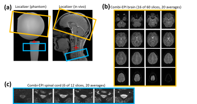 |
Partial Simultaneous Multi-Slice Acquisition of Combined T2*-Weighted Imaging of the Human Brain and Cervical Spinal Cord
Ying Chu1 and Jürgen Finsterbusch1
1Department of Systems Neuroscience, University Medical Center Hamburg-Eppendorf, Hamburg, Germany
Combined fMRI T2*-weighted imaging of the human brain and cervical spinal cord has been accelerated by simultaneous multi-slice (SMS) imaging of the brain volume. The implementation has been tested and evaluated in phantoms and in vivo and shows a very similar performance to non-accelerated and brain-only measurements. With the much shorter acquisition times achievable it could help to improve brain volume coverage of cortico-spinal fMRI.
|
|||
3469. |
A robust framework for characterising diffusion metrics of peripheral nerves: exploiting state of the art tracking methods
Arkiev D'Souza1, Chenyu Wang1,2, Sicong Tu1,2, Dominic Soligo3, Matthew Kiernan1,2,4, Michael Barnett1,4, and Fernando Calamante1,5,6
1Brain and Mind Centre, The University of Sydney, Sydney, Australia, 2Central Clinical School, The University of Sydney, Sydney, Australia, 3I-MED Radiology Network, Camperdown, Australia, 4Department of Neurology, Royal Prince Alfred Hospital, Camperdown, Australia, 5School of Biomedical Engineering, The University of Sydney, Sydney, Australia, 6Sydney Imaging, The University of Sydney, Sydney, Australia
Diffusion MRI has previously been used to quantify peripheral nerves; however, traditional post-processing techniques have several limitations. Here, we demonstrate the feasibility of using state-of-the-art diffusion analysis tools to reconstruct and quantify the ulnar nerve of the forearm. Constrained-spherical deconvolution was combined with probabilistic fibre-tracking to compute several track-weighted measurements in the ulnar nerve. The results suggest that a sample size of 22 would be sufficient to detect a 10% difference in any of the measured track-weighted metrics, and a sample size of 20 would be large enough to detect within-subject differences as small as 3%.
|
|||
3470. |
Investigating layer specific MR properties in the superior colliculus ex vivo at 14.1T
Ju Young Lee1, Andreas Mack2, Thomas Shiozawa-Bayer2, Marc Himmelbach3, Gisela Hagberg1,4, and Klaus Scheffler1,4
1Max Planck Institute for Biological Cybernetics, Tübingen, Germany, 2Eberhard Karls University Tuebingen, Tübingen, Germany, 3Hertie-Institue for Clinical Brain Research, Tübingen, Germany, 4University Hospital Tübingen, Tübingen, Germany
Layer specific MR properties have previously been observed in the superior colliculus in vivo at 9.4T. The present study investigates layer specific MR properties in R2* and R1 in more detail in post mortem brain stem samples at 14.1T. Similar to in vivo, one R2* maximum that likely corresponds to the superficial optic layer (layer III) was observed. Ex vivo we could observe high R2* and R1 in an area likely corresponding to the deep white layer (layer VII) of the superior colliculus. In the R1maps, this area showed a strong contrast towards the periaqueductal gray.
|
|||
3471. |
Visible “Butterfly” of the Cervical Spinal Cord: A Pilot study using high-resolution Phase-Sensitive and multiple Fast Field Echo MR imaging
bingbing gao1, Yanwei Miao1, Qingwei Song1, Ailian Liu1, and Jiazheng Wang2
1the First Affiliated hospital of Dalian Medical university, Dalian, China, 2Philips Healthcare, BeiJing, China
Discrimination of gray and white matters on the axial-spinal-marrow MRI remains a challenge, due to the small structure size and motion artifacts from such as swallowing and blood flow. The present study aims to visualize the cervical spinal marrow with high resolution axial phase-sensitive inversion recovery (PSIR) and multiple Fast Field Echo (FFE) images, and further to detect the image quality differentially on gray and white matters. The “butterfly” structure could be seen on both of the two contrast images, but the PSIR images were associated with significantly higher signal-to-noise ratios.
|
|||
3472.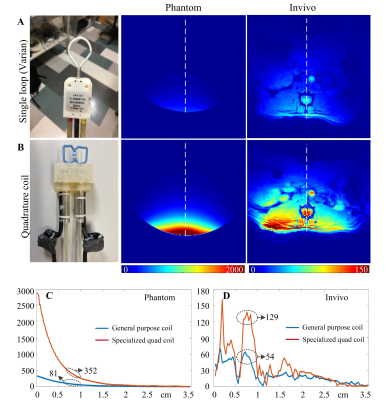 |
Design and Construction of an Interchangeable RF Coil System for Rodent Spinal Cord MR Imaging
Ming Lu1,2,3, Gary Drake1,2, Feng Wang1,2, Chaoqi Mu1,4, Limin Chen1,2, John C. Gore1,2,4, and Xinqiang Yan1,2
1Vanderbilt University Institute of Imaging Science, Vanderbilt University Medical Center, Nashville, TN, United States, 2Department of Radiology and Radiological Sciences, Vanderbilt University Medical Center, Nashville, TN, United States, 3College of nuclear equipment and nuclear engineering, Yantai University, Yantai, China, 4Department of Biomedical Engineering, Vanderbilt University, Nashville, TN, United States
Multiparametric MRI at high field provides comprehensive information to assess structural and functional changes that are important for clinical diagnosis and for evaluating therapies. Injuries may occur at different levels of the lumbar and thoracic cord, and the number of segments injured and their depths may vary along the spine, so it is challenging to build one universal RF coil that exhibits high-performance for all spinal cord imaging applications. We therefore developed an interchangeable RF coil system for a 9.4T small animal MRI scanner, and found the specialized coil has 2.4-fold SNR improvement compared to a commercial general-purpose sized coil.
|
|||
3473. |
Diffusion features in white and gray matter of healthy cervical spinal cord using PSIR imaging
Yunan Cui1, Yanwei Miao1, Ailian Liu1, Zhiwei Shen2, and Jiazheng Wang2
1THE FIRST AFFILIATED HOSPITAL OF DALIAN MEDICAL UNIVERSITY, DALIAN, China, 2Philips Healthcare, BEIJING, China
Due to the difficulty in distinguishing the structure of spinal by conventional MR sequence. In this study, the fusion images of Phase Sensitive Inversion Recovery sequence (PSIR) and zoomed Diffusion Tensor Imaging (DTI) was used to quantify the diffusion indicators, such as fractional anisotropy (FA), apparent diffusion coefficient (ADC), axial dispersion (AD) and relative anisotropy (RA) in the normal spinal cord. We found that there were significant differences of above indicators in different regions of cervical cord including the white columns of the cervical cord, the ipsilateral white matter and in the left and right sides of the cervical cord.
|
|||
3474.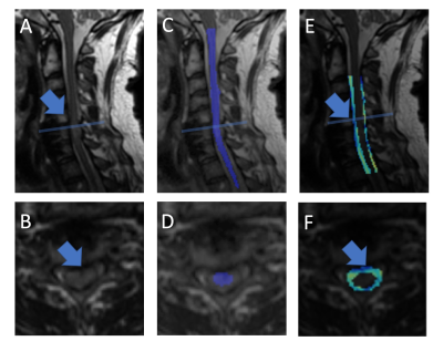 |
Morphological Assessment of the Instrumented Spinal Cord using Isotropic 3D-MSI MRI
Kevin M Koch1 and Andrew S Nencka1
1Radiology, Medical College of Wisconsin, Milwaukee, WI, United States
Morphological analysis of the spinal cord can provide valuable insight into injury and degeneration of damaged cord tissues. Accurate morphological analysis requires high resolution 3D anatomic imaging to be utilized as inputs into automated or semi-automated post-processing analysis algorithms. Historically, high resolution 3D imaging of the instrumented spinal cord has been difficult to achieve. As a result, the substantial damage and pathology near hardware cannot be analyzed using advanced morphological tools. In this technical feasibility study, isotropic 3D-MSI metal-artifact suppressed images are collected and utilized within a novel analysis framework constructed around the Spinal Cord Toolbox infrastructure.
|
|||
3475.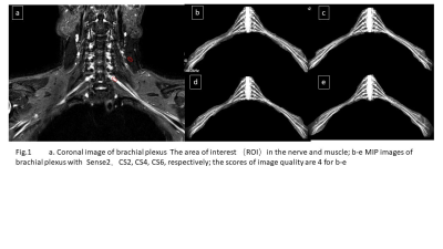 |
Optimization of Compressed SENSE accelerated Brachial Plexus MRI: Quality and Efficiency
Renwang PU1, Qingwei SONG1, Ailian LIU1, Zijing ZHANG1, Nan ZHANG1, Haonan ZHANG1, Bingbing GAO1, Lihua CHEN1, and Liangjie LIN2
1the First Affiliated Hospital of Dalian Medical Universityrsity, Dalian, China, 2Philips Healthcare, BEIJING, China
The 3D-NerveVIEW sequence with suppression of lipid and blood signals and a long echo time can obtain high-quality images for visualization of brachial plexus, while the long scan time may limit its clinical application. Compressed SENSE (CS) is a newly developed technique in MRI that enables accelerated acquisition with maintained image quality. By comparing results of 3D-NerveVIEW for brachial plexus imaging with acceleration by the conventional SENSE and the advanced CS with different acceleration factors. We found that the 3D-NerveVIEW for brachial plexus imaging with a CS acceleration factor of 4 can obtained favorable images within significantly reduced scan time.
|
|||
3476.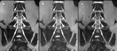 |
Application of 3D-FFE Based on Compressed Sensing for Lumbosacral Plexus Imaging: A Preliminary Study
Jian Wang1, Yanjun Chen1, Yingjie Mei2, Jialing Chen1, and Xiaodong Zhang1
1Department of Medical Imaging, The Third Affiliated Hospital of Southern Medical University, Guangzhou, China, 2China International Center, Philips Healthcare, Guangzhou, China
The anatomy of lumbosacral plexus (LSP) is complex, patients with trauma, neoplasms, or infection may experience motor weakness, sensory loss, and/or debilitating pain. Accurate and fast MRN scanning is an invaluable tool for evaluation of LSP diseases. In this study, the feasibility and performance of compressed sensing (CS) in scanning LSP was investigated and compared with conventional 3D-FFE with principles of the selective excitation technique. The results show that 3D-FFE with CS could reduce scan time obviously, influence image quality mildly, and meet clinical diagnosis.
|
|||
3477.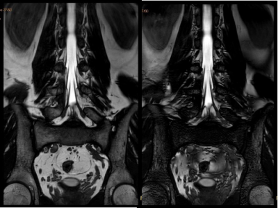 |
Banding artifacts reduction for lumbosacral plexus imaging with balanced FFE (b-FFE)
Geli Hu1, Jiazheng Wang1, Qingping Gu1, Yang Zhang1, and Jianxia Cao1
1Philips Healthcare, Beijing, China
The balanced FFE (b-FFE) sequence has the advantages of high resolution, SNR, CNR and superior differentiation between fluid and tissue. Therefore, it can be used for lumbosacral plexus imaging [1]. However, black band-like artifacts are frequently observed due to intra-voxel signal dephasing at the presence of magnetic field inhomogeneity. Here we proposed using a RF phase cycling technique (b-FFE-XD) to suppress the banding artifacts. The RF phase was set as [0,180+1*X, 2*X, 180+3*X,4*X,…] in consequence to limit the intravoxel dephasing, where X=n*360/NSA, and n is the nth average. Results demonstrated successful removal of banding artifacts with the proposed technique.
|
|||
| 3478. | WITHDRAWN | |||
3479.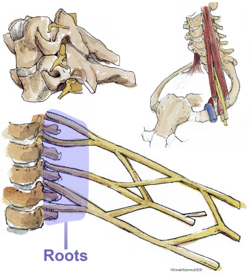 |
Diffusion Tensor Imaging of the Roots of the Brachial Plexus: A Systematic Review and Meta-Analysis of Normative Values
Ryckie George Wade1, Alexander Whittam2, Irvin Teh1, Gustav Andersson3, Fang-Cheng Yeh 4, Mikael Wiberg 3, and Grainne Bourke 1
1University of Leeds, Leeds, United Kingdom, 2Sheffield Teaching Hospitals, Sheffield, United Kingdom, 3Umeå University, Umeå, Sweden, 4University of Pittsburgh, Pittsburgh, PA, United States
In this systematic review and meta-analysis we summarise the normal diffusion tensor imaging (DTI) metrics for roots of the brachial plexus. We included 9 studies describing 316 adults (1:1 male:female) of mean age 35 years (SD 6). The normal fractional anisotropy was 0.36 (95% CI 0.34, 0.38; Figure 4). The normal mean diffusivity was 1.51 x10⁻³ mm2/s (95% CI 1.45, 1.56; Figure 5). DTI metrics varied according to experimental conditions and participant factors. Our summary estimates from different conditions which may be valuable to researchers and clinicians alike.
|
|||
3480.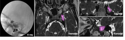 |
the value of preoperative MRI measurements of Meckel cavity volume in percutaneous balloon compression of the trigeminal nerve
Junjiao Hu1, Kai Deng1, Weijun Situ1, and Huiting Zhang2
1Department of Radiology, the Second Xiangya Hospital of Central South University, Changsha, China, 2MR Scientific Marketing, Siemens Healthcare Ltd., Wuhan, China This retrospective study aimed to investigate the feasibility of preoperative MRI measurements of Meckel cavity volume using 3D SPACE sequence and its values in guiding percutaneous balloon compression of the trigeminal nerve. Our results showed that there was no statistically significant difference between the volume of Meckel cavity using the axis, coronal and sagittal directions of MRI images and intraoperative balloon filling volume. 3D SPACE is helpful to guide the operation in clinical application. |
The International Society for Magnetic Resonance in Medicine is accredited by the Accreditation Council for Continuing Medical Education to provide continuing medical education for physicians.