Digital Posters
Psychiatry Neuroimaging
ISMRM & SMRT Annual Meeting • 15-20 May 2021

| Concurrent 4 | 19:00 - 20:00 |
1651.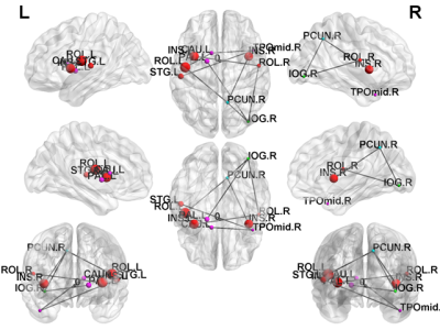 |
White matter connectome alterations in tuberous sclerosis complex patients with neuropsychiatric disorders revealed by DTI
Jheng-Yan Li1, Jeng-Dau Tsai2,3, Chao-Yu Shen4,5, and Jun-Cheng Weng1,6,7
1Department of Medical Imaging and Radiological Sciences, and Bachelor Program in Artificial Intelligence, Chang Gung University, Taoyuan, Taiwan, 2School of Medicine, Chung Shan Medical University, Taichung, Taiwan, 3Department of Pediatrics, Chung Shan Medical University Hospital, Taichung, Taiwan, 4Institute of Medicine, Chung Shan Medical University, Taichung, Taiwan, 5Department of Medical Imaging, Chung Shan Medical University Hospital, Taichung, Taiwan, 6Medical Imaging Research Center, Institute for Radiological Research, Chang Gung University and Chang Gung Memorial Hospital at Linkou, Taoyuan, Taiwan, 7Department of Psychiatry, Chang Gung Memorial Hospital, Chiayi, Taiwan
Previous studies indicate that the occurrence of tuberous sclerosis will cause neurotransmission in brain blocked by tumors and produce abnormal discharges. In the study we used diffusion tensor imaging (DTI), graph theoretical analysis (GTA) and network-based statistical (NBS) analysis to explain brain structural network alterations in TSC patients with different intellectual disability, seizure, and Neurological Severity Score.
|
|||
1652.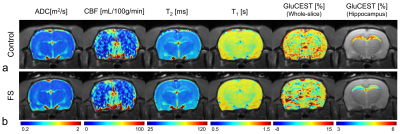 |
In vivo imaging of cerebral glutamate changes using chemical exchange saturation transfer MRI in a rat forced swimming test model of depression
Do-Wan Lee1, Hwon Heo2, Jae-Im Kwon3, Yeon Ji Chae2, Joongkee Min3, Monica Young Choi2, Chul‐Woong Woo3, Dong‐Cheol Woo2,3, Kyung Won Kim1, Jeong Kon Kim1, Hyo Jeong Chin4, and Dong‐Hoon Lee4
1Department of Radiology, Asan Medical Center, University of Ulsan College of Medicine, Seoul, Korea, Republic of, 2Department of Convergence Medicine, Asan Medical Center, University of Ulsan College of Medicine, Seoul, Korea, Republic of, 3Convergence Medicine Research Center, Asan Institute for Life Sciences, Asan Medical Center, Seoul, Korea, Republic of, 4Department of Radiological Science, College of Health Sciences, Yonsei University, Wonju, Korea, Republic of
Glutamate-weighted chemical exchange saturation transfer (GluCEST) imaging is a novel enhancement technique for the non-invasive detection and quantification of cerebral glutamate levels in neuro-molecular processes. The present study quantitatively evaluated glutamate signal changes in the hippocampal region of a rat forced swimming test model (FS) of depression. The GluCEST and proton magnetic resonance spectroscopy results showed that GluCEST values and glutamate concentrations were significantly lower in the FS rats than in the controls. These findings might provide a key marker to better understand the cerebral neurochemical responses in depressive disorders.
|
|||
 |
1653.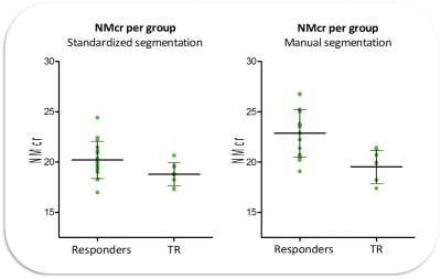 |
Neuromelanin MRI as biomarker for treatment resistance in first episode schizophrenia patients
Marieke Van der Pluijm1, Laura Meershoek1, Lieuwe De Haan2, Jan Booij1, and Elsmarieke Van de Giessen1
1Radiology and Nuclear Medicine, Amsterdam UMC, University of Amsterdam, Amsterdam, Netherlands, 2Psychiatry, Amsterdam UMC, University of Amsterdam, Amsterdam, Netherlands
The current study assessed whether neuromelanin sensitive MRI (NM-MRI) is a potential biomarker for treatment resistance (TR) in first episode schizophrenia patients. NM-MRI is a novel MRI sequence, which indirectly measures dopamine synthesis. Twenty-three first episodes schizophrenia patients underwent a NM-MRI scan, treatment response was determined during follow-up. Standardized and manual segmentation protocols of the NM-MRI data were used and compared. Both segmentation protocols showed significantly lower NM-MRI signal in TR compared to responders. These findings demonstrate the potential of NM-MRI as biomarker. The predictive value of NM-MRI for TR and optimal segmentation method still require further investigation
|
||
1654.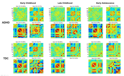 |
Developmental changes of functional network connectivity dynamics in typical development and ADHD youth
Yingxue Gao1, Xuan Bu1, Hailong Li1, Weijie Bao1, Kaili Liang1, and Xiaoqi Huang1
1Huaxi MR Research Center (HMRRC), Functional and Molecular Imaging Key Laboratory of Sichuan Province, Department of Radiology, West China Hospital, Sichuan University, Chengdu, China
We used dynamic functional network connectivity to explore the neurodevelopmental changes of whole-brain large scale intrinsic network connectivity dynamics from childhood to adolescence in attention deficit/hyperactivity disorder (ADHD) children and typical developing control (TDC) children. We found that the developmental changes of occurrence percentages, duration of stay and functional network connectivity patterns of states of internetwork hyperconnectivity and hypoconnectivity were different between ADHD and TDC children.
|
|||
1655.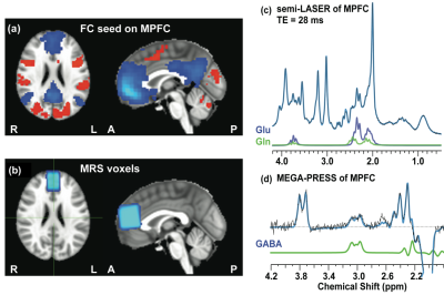 |
How neurotransmitter concentrations in default-mode network modulate brain functional activities and connectivities in psychosis
Xi Chen1, Dost Ongur1, and Fei Du1
1McLean Hospital; Harvard Medical School, Belmont, MA, United States
Failure to suppress default-mode network (DMN) activity during tasks and reduced anti-correlations between DMN and other brain networks at rest has been observed in various psychiatric disorders. However, the molecular mechanisms underlying this phenomenon are poorly understood. It has been shown that the neurotransmitter concentrations in DMN modulate the brain functional activities and connectivities in the healthy brain. In the current study, it was observed that the relationship between DMN neurotransmitter concentrations and the activities of brain functional network breaks down in first-episode psychosis patients. This finding provides opportunities for developing novel treatment strategies and earlier interventions for psychosis.
|
|||
1656.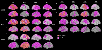 |
A Multi-Diffusion Model Investigation of White Matter Microstructure in Psychotic Spectrum Disorders
Faye McKenna1, Yu Veronica Sui2, Hillary Bertisch2, Donald Goff2, and Mariana Lazar2
1Radiology, New York University School of Medicine, New York, NY, United States, 2New York University School of Medicine, New York, NY, United States
We employed the recent Bingham neurite orientation dispersion and density imaging (NODDI-B) and diffusion kurtosis imaging (DKI) techniques, alongside classical diffusion tensor imaging (DTI) and found that psychotic spectrum disorders (PSD) and PSD sub-types had significantly increased mean and radial diffusion and orientation dispersion (MD, RD, ODI, ODIs) and significantly decreased mean and radial kurtosis (MK, RK), fractional anisotropy (FA) and neurite density index (NDI) in sub-cortical WM ROIs compared to healthy controls (HC). We also found significant relationships across PSD and HC groups between WM dMRI metrics and tests of episodic and working memory and several schizotypal traits.
|
|||
1657.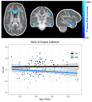 |
Investigation of Microstructural Alterations of the Corpus Callosum in Autism Using Multi-Shell Diffusion MRI and Quantitative Relaxometry
Douglas C Dean1,2,3, Nagesh Adluru3, Jace B King4, Molly B Prigge4, Carolyn King4, Erin D Bigler5,6,7,8, June Taylor4, Nick Lange9, Brandon A Zielinski4,5,10, Janet E Lainhart3,11, and Andrew L Alexander2,3,11
1Pediatrics, University of Wisconsin–Madison, Madison, WI, United States, 2Medical Physics, University of Wisconsin–Madison, Madison, WI, United States, 3Waisman Center, University of Wisconsin–Madison, Madison, WI, United States, 4Radiology, University of Utah, Salt Lake City, UT, United States, 5Neurology, University of Utah, Salt Lake City, UT, United States, 6Psychiatry, University of Utah, Salt Lake City, UT, United States, 7Psychology and Neuroscience Center, Brigham Young University, Provo, UT, United States, 8Neurology, University of California–Davis, Davis, CA, United States, 9Psychiatry, Harvard School of Medicine, Boston, MA, United States, 10Pediatrics, University of Utah, Salt Lake City, UT, United States, 11Psychiatry, University of Wisconsin–Madison, Madison, WI, United States
White matter microstructural alterations are consistently reported and believed to play a significant role in the defining characteristics of autism spectrum disorder. In this work, we utilized advanced diffusion imaging and quantitative relaxometry to examine microstructural differences in autism. We observe significant group and age-related deviations of the corpus callosum, as well as more widespread alterations of the neurite microstructure. These results are consistent with previous findings of the corpus callosum in ASD and suggest that while sensitive to underlying microstructural deviations, advanced diffusion imaging and relaxometry provide complementary information.
|
|||
1658.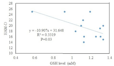 |
Association of anterior cingulate glutathione and degree of depression in unmedicated bipolar disorder – a 7T study
Pallab K Bhattacharyya1, Mark J Lowe1, and Amit Anand1
1Cleveland Clinic Foundation, CLEVELAND, OH, United States
Oxidative stress is a contributing factor in bipolar disorder (BD). Glutathione (GSH) acts as an antioxidant and reduces oxidative stress. GSH level at anterior cingulate cortex was measured in unmedicated patients with BD in depressed state and healthy controls at 7T using semi-LASER sequence. No significant difference in GSH level was observed; however, degree of depression was inversely correlated with GSH level.
|
|||
1659.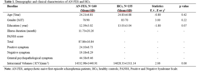 |
Morphological changes of the corpus callosum in antipsychotic-naive first-episode schizophrenia before and 1-year after treatment
Bo tao1, Yuan Xiao1, Wenjing Zhang1, Na Hu1, John A Sweeney1,2, and Su Lui1
1Huaxi MR Research Center (HMRRC), Functional and molecular imaging Key Laboratory of Sichuan Province, Department of Radiology, West China Hospital of Sichuan University, Chengdu, China, 2Department of Psychiatry and Behavioral Neuroscience, University of Cincinnati, Cincinnati, OH, United States
The present study demonstrates deficits of the callosal morphology in schizophrenia, which may reflect an neurodevelopment aberration. And short-term antipsychotics may not have a significant impact on the CC morphology in the early stage of illness.
|
|||
1660.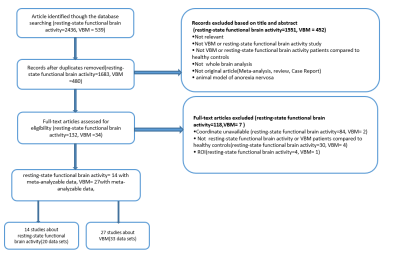 |
Functional and structural brain alterations in anorexia nervosa: a multimodal meta-analysis of neuroimaging studies
Ting Su1, Jia ying Gong2, Shao juan Qiu1, Pan Chen1, Guan mao Chen1, Jun jing Wang3, Li Huang1, and Ying Wang1
1Medical Imaging Center, First Affiliated Hospital of Jinan University, Guangzhou, China, 2Department of Radiology, Six Affiliated Hospital of Sun Yat-sen University, Guangzhou, China, 3Department of Applied Psychology, Guangdong University of Foreign Studies, Guangzhou, China Anorexia nervosa (AN) is a complex psychiatric disorder with poorly understood etiology. A whole-brain meta-analysis on resting-state functional imaging and VBM studies that measured differences in the intrinsic functional activity and gray matter volume (GMV) between AN patients and healthy controls. Overall, patients with AN displayed decreased resting-state functional activity in the bilateral ACC and MCC and increased in the right parahippocampal gyrus. In VBM studies, with AN patients displayed decreased GMV in the bilateral MCC , and left inferior parietal lobe. This multimodal meta-analysis identified functional activity and GMV reductions in the anterior and median cingulate in patients with AN. |
|||
1661.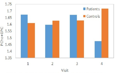 |
Changes in total choline level in left anterior cingulate during 26 weeks of Li treatment in patients with bipolar disorder
Pallab K Bhattacharyya1, Mark J Lowe1, and Amit Anand1
1Cleveland Clinic Foundation, CLEVELAND, OH, United States
Changes in total choline level in left anterior cingulate cortex (ACC) following lithium monotherapy of bipolar disorder in depressed state were studied at 7T. Patients were scanned with a semi LASER sequence at baseline and 2, 8 and 26 weeks from start of therapy. Healthy controls were also scanned at those 4 time points. A decrease in choline level at left dorsal/rostral ACC was observed in patients, and the reduction took place between 8 and 26 weeks after onset of therapy.
|
|||
1662.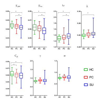 |
Disrupted Small-World Networks in Major Depressed Patients with Suicidality
Huiru Li1, Huawei Zhang1, Li Yin2, Zhiyun Jia3, and Qiyong Gong1
1Huaxi MR Research Center (HMRRC), Department of Radiology, West China Hospital of Sichuan University, Chengdu, China, 2Department of Psychiatry, West China Hospital of Sichuan University, Chengdu, China, 3Department of Nuclear Medicine, West China Hospital of Sichuan University, Chengdu, China
In this study, we investigated depressed suicidal brain from the level of network connection. We constructed brain structural networks using diffusion tensor imaging. Then all graph theoretical network parameters including small-world parameters (Cp, Lp, γ, λ and σ), network efficiency parameters (Eloc and Eglob) and nodal efficiency were analyzed. We found decreased Eloc/Eglob/Cp, increased Lp/λ and decreased nodal efficiency in fronto-striatal-limbic-thalamic circuit in depressed suicidal patients. In summary, suicidality involves complex neocortical network organization, which showed a weaker integration and disrupted fronto-striatal-limbic-thalamic circuit.
|
|||
1663.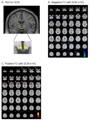 |
Circadian rhythm functional network in the central nervous system and its disruptions in chronic insomnia disorder
Ran Pang1,2, Jianli Wang3, Karunanayaka Prasanna3, Kuncheng Li4, and Qingxian Yang2
1Department of Radiology, Dongfang Hospital, Beijing University of Chinese Medicine, Beijing, China, 2Department of Neurosurgery, Pennsylvania State University College of Medicine, Hershey, PA, United States, 3Department of Radiology, Pennsylvania State University College of Medicine, Hershey, PA, United States, 4Department of Radiology, Xuanwu Hospital, Capital Medical University, Beijing, China
The etiology of chronic insomnia disease (CID) ultimately relates to the asynchrony of circadian rhythms. Using the anterior hypothalamus as a seed, functional connectivity (FC) of the circadian rhythm functional network (CRFN) during resting state was demonstrated in healthy subjects, consisting of both positive and negative FCs in the cerebrum and cerebellum. The CID patients exhibited an extensive weakening of FCs and abnormal local hyperactivities, reflecting underlying asynchrony in the CRFN.
|
|||
1664.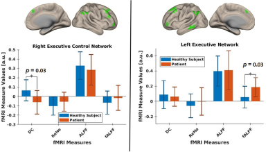 |
7T Mental health: Functional alterations in resting-state within the executive control network and its association with BDI-II and TMT-B in MDD
Ravichandran Rajkumar1,2,3, Gereon Johannes Schnellbächer2, Hasan Sbaihat1,2, N. Jon Shah1,4,5,6, Tanja Veselinović2, and Irene Neuner1,2,4
1Institute of Neuroscience and Medicine - 4 (Medical Imaging Physics), Forschungszentrum Juelich GmbH, Jülich, Germany, 2Department of Psychiatry, Psychotherapy and Psychosomatics, RWTH Aachen University, Aachen, Germany, 34JARA – BRAIN – Translational Medicine, Aachen, Germany, 4JARA – BRAIN – Translational Medicine, Aachen, Germany, 5Department of Neurology, RWTH Aachen University, Aachen, Germany, 6Institute of Neuroscience and Medicine, INM-11, Forschungszentrum Jülich GmbH, Jülich, Germany
Executive functioning is reported to be deficient in depression. In this pilot study, functional alterations in MDD patients and their association with mental flexibility and depression severity, particularly within the executive control network, are investigated. Our data contribute to a better understanding of the neurobiological signatures of depression. Further investigation in this area may lead to an improvement in the diagnosis and treatment of MDD patients.
|
|||
1665.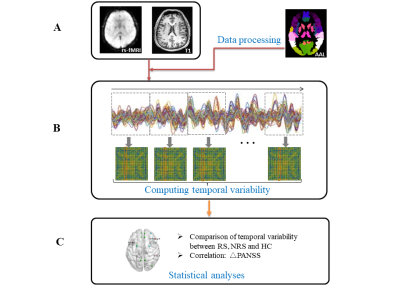 |
Different patterns of temporal variability of functional connectivity to predict response to electroconvulsive therapy in schizophrenia
Yunyun Jiao1, Jie Gong1, Hui Deng1, Dongchen Sun1, and Wei Qin1
1Engineering Research Center of Molecular and Neuro Imaging of the Ministry of Education, School of Life Science and Technology, Xidian University, Xi'an, China
We aimed to explore the relationship between the pre-treatment temporal variability of resting state functional connectivity and response to electroconvulsive therapy (ECT) in schizophrenia (SZ). Statistical analysis included grouping comparison and Pearson correlation analysis. The grouping comparison of temporal variability between responders to ECT with SZ, non-responders to ECT with SZ and healthy controls were conducted and Pearson correlation analysis was used to reveal the relationship between temporal variability and the response to ECT. The result showed that the temporal variability may serve as a promising indicator to predict the response to ECT in patients with SZ.
|
|||
1666.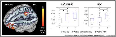 |
MRI measurements demonstrate gray matter increases induced by transcranial direct current stimulation treatment in depression
Mayank Jog1, Cole Anderson1, Elizabeth Kim1, Antoni Kubicki1, Michael Boucher1, Gerhard Hellemann2, Roger Woods1, and Katherine Narr1
1UCLA, Los Angeles, CA, United States, 2University of Alabama, Birmingham, Birmingham, AL, United States
Transcranial direct current stimulation (tDCS) is a low-cost and non-invasive neuromodulation technique. Although tDCS has been shown to improve symptoms in psychiatric disorders, the neurobiological effects of tDCS are not well-understood. In this study, we used MRI to investigate structural changes in the brain resulting from tDCS. T1-weighted MRI data from n=59 depressed participants was acquired pre/post tDCS treatment, and analysis revealed gray-matter increases near the stimulation-target and in a distant but functionally-connected region. These results indicate that tDCS treatments can elicit structural changes in the brain; both near the stimulation-target and in additional regions part of the same brain-network.
|
|||
1667.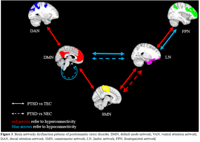 |
Alternations of functional networks in adult PTSD: a systematic review and meta-analysis of resting-state functional connectivity studies
WeiJie Bao1, YingXue Gao1, Hailong Li1, jing Liu1, Lingxiao Cao1, Xuan Bu1, and Xiaoqi Huang1
1Huaxi MR Research Center (HMRRC), Functional and Molecular Imaging Key Laboratory of Sichuan Province, Department of Radiology, West China Hospital, Sichuan University, Chengdu, China
We performed a systematic review of studies which explored network alterations by comparing PTSD and trauma-exposed controls (TEC) or nonexposed controls (NEC) using seed-based functional connectivity (SBFC) techniques. And quantitative meta-analysis was conducted when the number of studies reached an appropriate number. Our results supported that trauma may have long-lasting effects on the function of the default mode network (DMN) and limbic network (LN) regardless of whether it caused symptoms of PTSD. Moreover, the altered connectivity between the DMN and the somatomotor network (SMN) and between the LN and SMN may be underline neural mechanism specify to PTSD.
|
|||
1668. |
Effect of the outbreak of COVID-19 on college students with subthreshold depression:a resting-state functional MRI study
zhang zhang qi1, pan chen2, ZHEN YE LUO3, Long QIAN4, and YING WANG2
1JINAN UNIVERCITY, guang zhou, China, 2Jinan Univercity, guang zhou, China, 3lzy1735411016@163.com, guang zhou, China, 4MR Research, GE Healthcare, Beijing, China., BEI JING, China
Background: The current outbreak of the novel coronavirus disease 2019 (COVID-19), has rapidly spread across China and many other countries.Subthreshold depression (SD) is generally considered to present when individuals show depressive symptoms but diagnostic criteria for a major depressive disorder are not met. Methods: In total,49 SD individuals and 56 health controls(HC) were recruited from Jinan University. All subjects underwent MRI and MATRICS Consensus Cognitive Battery (MCCB) before COVID-19 outbreak. Conclusion: After the COVID-19 epidemic situation, changes of brain area and cognitive function of college students with significant stress symptoms were more obvious.
|
|||
|
1669.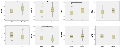 |
Brain volumetric measurements in children with ADHD:a comparative study between synthetic and conventional MRI
Yingqian Chen1, Shu Su1, Yan Dai1, Long Qian2, and Zhiyun Yang1
1First Affiliated Hospital, Sun Yat-sen University, Guangzhou, China, 2GE Healthcare, Beijing, China
The developmental mechanism underlying the brain development abnormalities in attention deficit hyperactivity disorder (ADHD) is still unclear. The currently developed synthetic MRI (SyMRI) offers a novel approach for brain segmentation and myelin volume estimation. Our study investigated the profiles of brain volumetric measurements in children with ADHD with both SyMRI and conventional T1 weighted(cT1w) MRI. And the results proved the global brain development retardation but normal whole brain myelination of children with ADHD. Besides, the high consistency of brain segmentation with cT1w image and SyMRI has also been proved, which indicate the clinical feasibility of SyMRI in children.
|
The International Society for Magnetic Resonance in Medicine is accredited by the Accreditation Council for Continuing Medical Education to provide continuing medical education for physicians.