Digital Posters
MS: White Matter & Other Structures
ISMRM & SMRT Annual Meeting • 15-20 May 2021

Digital Posters
MS: White Matter & Other Structures
| Concurrent 6 | 13:00 - 14:00 |
2790.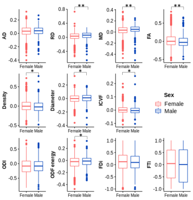 |
Sex-based differences in tissue microstructure in multiple sclerosis detected using multi-model diffusion MRI
Olayinka Adeoluwa Oladosu1, Cayden Murray2, Syed Rizvi2, Mariana Bento3,4, G Bruce Pike3,4, and Yunyan Zhang3,4
1Neuroscience, University of Calgary, Calgary, AB, Canada, 2University of Calgary, Calgary, AB, Canada, 3Radiology, University of Calgary, Calgary, AB, Canada, 4Clinical Neurosciences, University of Calgary, Calgary, AB, Canada
Sex difference in multiple sclerosis (MS) prevalence is well recognized but impacts on tissue microstructure and its variations with respect to disease modifying therapies (DMTs) remains unclear. Here we investigated sex-based differences in lesion microstructure and its variations with therapeutic approaches in 52 relapsing-remitting MS patients using diffusion tensor, compartment, and orientation models. We found that men had greater myelin and axonal damage than women in MS lesions, and there was less axonal damage in women taking oral versus injectable DMTs. These results may support further investigations of sex-driven differences in disease monitoring and treatment choices to promote personalized medicine.
|
|||
2791.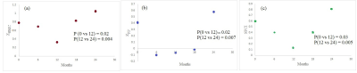 |
Tracking longitudinal disease progression of MS during fingolimod therapy using SFCI, a combined structural and functional connectivity metric
Pallab K Bhattacharyya1, Robert Fox1, Jian Lin1, Paola Raska1, Ken Sakaie1, and Mark J Lowe1
1Cleveland Clinic Foundation, CLEVELAND, OH, United States
It has been reported that structural and functional connectivity impairment of motor and cognitive network in multiple sclerosis (MS), as measured by diffusion tensor imaging (DTI) and functional connectivity (fcMRI) respectively, stabilize after one year of fingolimod treatment. Structural and functional connectivity index (SFCI), a combined metric of DTI and fcMRI has previously been demonstrated as a sensitive imaging-based measure of progression of MS. Change of (i) motor (ii) cognitive and (iii) pathway combined SFCI were tracked over 2-year fingolimod therapy of MS. SFCI stabilized after one year, which shows its effectiveness to measure disease progression following therapy .
|
|||
2792.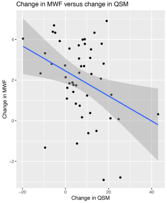 |
Greater increase in magnetic susceptibility following acute MS lesion formation is associated with reduced myelin repair
Lily Zexter1, Thanh D. Nguyen2, Elizabeth M. Sweeney3, Yi Wang2, and Susan A. Gauthier1
1Department of Neurology, Weill Cornell Medicine, New York, NY, United States, 2Department of Radiology, Weill Cornell Medicine, New York, NY, United States, 3Department of Population Health Sciences, Weill Cornell Medicine, New York, NY, United States
Quantitative susceptibility mapping (QSM) provides an effective means to directly map the distribution of magnetic susceptibility sources, such as brain iron. In this study, we examined the relationship between initial QSM change and short-term myelin recovery, measured by myelin water fraction (MWF). The results showed that change in lesion susceptibility at three months was significantly and negatively associated with change in MWF at one year. This significant association provides evidence of the damaging effect of brain iron during early stages of lesion development.
|
|||
2793.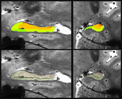 |
Hippocampal subfield volumes relate to future cognitive performance in Multiple Sclerosis
Katherine A Koenig1, Jian Lin1, Daniel Ontaneda1, Kedar Mahajan1, Jenny Feng1, Stephen Rao1, Sanghoon Kim1, Stephen Jones1, and Mark J Lowe1
1The Cleveland Clinic, Cleveland, OH, United States
Cognitive dysfunction is a common symptom of Multiple Sclerosis (MS), and patients would benefit from a measure that estimates their risk of future decline. Previous work suggests that hippocampal subfield volumes may have value as a predictive measure. Using 7 tesla MRI, we measured hippocampal volumes in 77 participants with MS. We found relationships between subfield volumes and future cognitive performance. These relationships were driven by relapse remitting MS patients, although the secondary progressive MS group did not show clear differences in the relationship patterns.
|
|||
2794.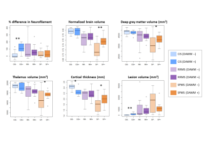 |
Diffusely abnormal white matter in clinically isolated syndrome is associated with parenchymal loss and elevated neurofilament levels
Irene Margaret Vavasour1, Jackie T Yik2,3, Pierre Becquart4, Jasmine Gill4, Shannon H Kolind1,2,3,5, Alice J Schabas5, Ana-Luiza Sayao5, Virginia Devonshire5, Robert Carruthers5, Anthony Traboulsee5, GR Wayne Moore3,4,5, Sophie Stukas4, Cheryl Wellington4,
Jacqueline Quandt4, David KB Li1, and Cornelia Laule1,2,3,4
1Radiology, University of British Columbia, Vancouver, BC, Canada, 2Physics and Astronomy, University of British Columbia, Vancouver, BC, Canada, 3International Collaboration on Repair Discoveries (ICORD), University of British Columbia, Vancouver, BC, Canada, 4Pathology and Laboratory Medicine, University of British Columbia, Vancouver, BC, Canada, 5Medicine, University of British Columbia, Vancouver, BC, Canada
We characterized the frequency of diffusely abnormal white matter (DAWM) in a broad cohort of multiple sclerosis (MS) participants. 35% of clinically isolated syndrome (CIS) and ~60% of MS participants had DAWM. CIS with DAWM had smaller cortical thickness, higher lesion load and higher concentration of neurofilament light chain compared to CIS without DAWM. DAWM may be useful in identifying brains at risk of injury but only in the CIS population when lesion load is low. Longitudinal studies are warranted.
|
|||
2795.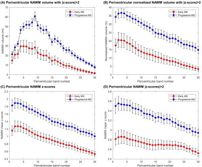 |
Periventricular gradients of brain pathology in early and progressive MS revealed by qMRI
Gian Franco Piredda1,2,3, Tom Hilbert1,2,3, Manuela Vaneckova4, Jan Krasensky4, Michaela Andelova5, Tomas Uher5, Barbora Srpova5, Eva Kubala Havrdova5, Karolina Vodehnalova5, Dana Horakova5, Veronica Ravano1,2,3, Bénédicte Maréchal1,2,3, Jean-Philippe Thiran2,3,
and Tobias Kober1,2,3
1Advanced Clinical Imaging Technology, Siemens Healthcare AG, Lausanne, Switzerland, 2Department of Radiology, Lausanne University Hospital and University of Lausanne, Lausanne, Switzerland, 3LTS5, École Polytechnique Fédérale de Lausanne (EPFL), Lausanne, Switzerland, 4Department of Radiology, First Faculty of Medicine, Charles University and General University Hospital, Prague, Poland, 5Department of Neurology and Center of Clinical Neuroscience, First Faculty of Medicine, Charles University and General University Hospital, Prague, Poland
In multiple sclerosis, gradients of tissue damage severity were found in periventricular areas and proved to change in response to treatment. This work investigates whether these gradual microstructural changes can be detected with high-resolution, whole-brain quantitative T1 mapping and how they compare between two cohorts of early and progressive multiple sclerosis patients. T1 deviations were computed in patients from normative atlases established in a healthy cohort and corrected for age and gender. Results show that a periventricular gradient of abnormal T1 values in normal-appearing white matter is indeed present and increases from early to progressive stages of the disease.
|
|||
2796.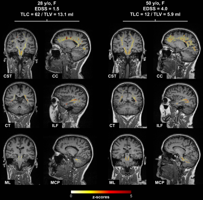 |
T1 abnormalities in atlas-based white matter tracts: reducing the clinico-radiological paradox in multiple sclerosis using qMRI
Veronica Ravano1,2,3, Gian Franco Piredda1,2,3, Manuela Vaneckova4, Jan Krasensky4, Michaela Andelova5, Tomas Uher5, Barbora Srpova5, Eva Kubala Havrdova5, Karolina Vodehnalova5, Dana Horakova5, Tom Hilbert1,2,3, Bénédicte Maréchal1,2,3, Reto Meuli2,
Jean-Philippe Thiran2,3, Tobias Kober1,2,3, and Jonas Richiardi2
1Advanced Clinical Imaging Technology, Siemens Healthineers, Lausanne, Switzerland, 2Department of Radiology, Lausanne University Hospital and University of Lausanne, Lausanne, Switzerland, 3LTS5, Ecole Polytechnique Fédérale de Lausanne, Lausanne, Switzerland, 4Department of Radiology, First Faculty of Medicine, Charles University and General University Hospital, Prague, Czech Republic, 5Department of Neurology and Center of Clinical Neuroscience, First Faculty of Medicine, Charles University and General University Hospital, Prague, Czech Republic
In multiple sclerosis, standard radiological metrics (i.e. lesion load) correlate poorly with clinical outcomes. To overcome this limitation, we propose a novel method to evaluate T1 relaxometry abnormalities along thirty-seven major white matter pathways extracted from a tractography atlas, i.e. without needing a diffusion scan. Evaluating T1 z-scores along WM tracts strongly improved correlation with disability compared to lesion load. The strongest correlations were found for T1 abnormalities in normal-appearing white matter, especially in infratentorial tracts. These results suggest that diffuse pathological changes in normal-appearing WM measured along atlas-based tracts using T1 relaxometry could aid clinical evaluation of multiple sclerosis.
|
|||
2797.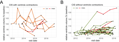 |
3.0 T MRI detects brain ventricle oscillations in patients with clinically-isolated syndrome
Jason Michael Millward1, Claudia Chien2, Joseph Kuchling2, Friedemann Paul2, Thoralf Niendorf1, and Sonia Waiczies1
1Berlin Ultrahigh Field Facility (B.U.F.F.), Max Delbrück Center for Molecular Medicine in the Helmholtz Association, Berlin, Germany, 2NeuroCure Clinical Research Center, Charité - Universitätsmedizin Berlin, Berlin, Germany
We used clinical high resolution 3T MRI to investigate brain ventricle changes over time in patients with clinically isolated syndrome (CIS) who are at the early stages of disease, which may eventually become multiple sclerosis. While most patients showed an overall increase in ventricle volume, 23% also showed contractions greater than the range of variation in healthy subjects, even over a period of years. Patients with contracting ventricles were significantly younger than those without. This suggests that, in addition to neurodegeneration, other processes are implicated that lead to a transient enlargement of the ventricles, which then later resolves.
|
|||
2798.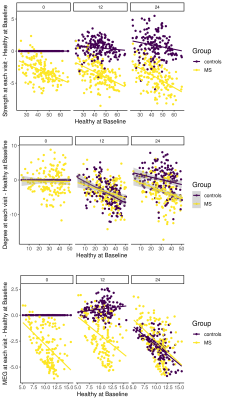 |
Structural connectivity is more sensitive to track cognition progression individual level than fMRI and MEG over 2 years in mildly disabled RRMS
Arzu Ceylan Has Silemek1, Guido Nolte2, Jana Pöttgen1,3, Andreas K. Engel4, Christoph Heesen1,3, Stefan M. Gold1,5, and Jan-Patrick Stellmann6,7
1Institute of Neuroimmunology and Multiple Sclerosis (INIMS), University Medical Center Eppendorf, Hamburg, Germany, 2Department of Neurophysiology and Pathophysiology, University Medical Center Eppendorf, Hamburg, Germany, 3Department of Neurology, University Medical Center Eppendorf, Hamburg, Germany, 4Institute of Neurophysiology and Pathophysiology, University Medical Center Eppendorf, Hamburg, Germany, 5Department of Psychiatry and Psychotherapy, Charité University Medical Center, Campus Benjamin Franklin, Hindenburgdamm 30, Berlin, Germany, 6CRMBM AMU-CNRS, Aix-Marseille Université, Marseille, France, 7CEMEREM, APHM, CHU Timone, Marseille, France
There is still substantial inconsistency between clinical disabilities and findings on brain networks and lack of longitudinal study in Multiple Sclerosis (MS). We aimed to elucidate how topology alters and how disability progression affects the structural and functional organizations over 2-years using graph theory approach in relapsing-remitting MS (RRMS). RRMS patients have encountered with lack of improvement in PASAT over 2-years. Structural connectivity was more sensitive to show a relationship with a cognitive function over 2-years than the rs-fMRI and MEG functional metrics in RRMS patients. These findings underline the difficulties associated with functional imaging studies in MS.
|
|||
2799.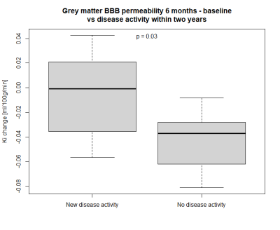 |
Blood-brain barrier permeability changes in multiple sclerosis during alemtuzumab treatment
Maria Højberg Knudsen1,2, Helle Juhl Simonsen1, Jette Lautrup Battistini Frederiksen2,3, Ulrich Lindberg1, Mark Bitsch Vestergaard1, Henrik Bo Wiberg Larsson1,2, and Stig Præstekjær Cramer1
1Dept. Clinical Physiology, Rigshospitalet, Glostrup, Denmark, 2Department of Clinical Medicine, University of Copenhagen, Copenhagen, Denmark, 3Dept. of Neurology, Rigshospitalet, Glostrup, Denmark
Ineffective disease control in multiple sclerosis leads to permanent disability, and biomarkers for early detection of inadequate treatment response are needed. The potential of the patlak derived influx constant (Ki) as a biomarker was investigated in fifteen relapsing-remitting multiple sclerosis patients undergoing alemtuzumab treatment. The subjects underwent dynamic contrast enhanced MRI and 3DT1-weighted scans to assess Ki in grey and white matter before and after treatment. The treatment associated change in Ki in grey matter predicted disease activity within two years. Treatment associated changes in Ki may be used as a biomarker of treatment efficacy.
|
|||
2800.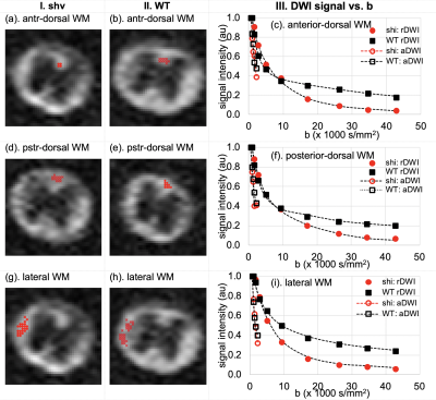 |
Ultrahigh-b radial Diffusion Weighted Imaging (UHb-rDWI) of Wild Type and Shiverer Mouse Spines
Kyle Jeong1, You-Jung Lee1, Suk-Keu Yeom2, Noel Carlson3, Lubdha Shah4, John Rose5, and Eun-Kee Jeong4
1Utah Center for Advanced Imaging research, University of Utah, Salt Lake City, UT, United States, 2Radiology, Korea University Ansan Hospital, Ansan, Korea, Republic of, 3GRECC, Veteran Affairs, Salt Lake City, UT, United States, 4Radiology and Imaging Sciences, University of Utah, Salt Lake City, UT, United States, 5Neuroimmunology Division, University of Utah, Salt Lake City, UT, United States
Ultrahigh-b radial DWI (UHb-rDWI) is an MRI technique that can quantitatively evaluate the MS lesions with respect to degrees of demyelination and axonal damage and/or loss. With the current lack of sensitive diagnostic imaging for grading cervical spinal cord (CSC) injury and repair, our UHb-rDWI can potentially serve as a powerful tool to observe cervical spinal cord once validated through animal studies. Therefore, the main objective of this study was to evaluate mouse spinal cords with and without demyelination using UHb-rDWI and immunohistochemical analyses to authenticate the reliability and reproducibility of UHb-rDWI.
|
|||
2801.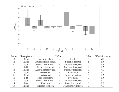 |
Evaluation of PASAT test performance and diffusivity indices in U-fiber regions in healthy subjects and RRMS patients.
Cristian Andrés Montalba1,2,3, Tomás Labbe4,5, Marcelo Andia1,2,3, Miguel Guevara6, Jean-François Mangin7, Juan Pablo Cruz2, Ethel Ciampi8,9, Claudia Carcamo5,8, Pamela Guevara6, and Sergio Uribe1,2,3
1Biomedical Imaging Center, Pontificia Universidad Católica de Chile, Santiago, Chile, 2Radiology Department, School of Medicine, Pontificia Universidad Católica de Chile, Santiago, Chile, 3Millennium Nucleus for Cardiovascular Magnetic Resonance, Santiago, Chile, 4School of Medicine, Pontificia Universidad Católica de Chile, Santiago, Chile, 5Interdisciplinary Center of Neurosciences, Pontificia Universidad Católica de Chile, Santiago, Chile, 6Faculty of Engineering, Universidad de Concepción, Concepción, Chile, 7UNATI, Neurospin, CEA, Université Paris-Saclay, Gif-sur-Yvette, France, 8Neurology Department, School of Medicine, Pontificia Universidad Católica de Chile, Santiago, Chile, 9Neurology Service, Hospital Dr. Sótero del Río, Santiago, Chile
Multiple Sclerosis patients develop cognitive impairment at the earliest stages of the disease, with changes in cortical recruitment related to cognitive tasks. We evaluated the relation between information-processing speed performance with different diffusivity measurements in subcortical regions between healthy subjects and RRMS patients. We evaluated the FA, MD, RD, and AD diffusivity maps of U-fibers with PASAT test scores in healthy subjects and RRMS patients. PASAT test scores are significantly linear related to Frontal, Temporal, and Parietal cortical areas in healthy subjects. There is no linear relationship between PASAT test scores and diffusivity maps in RRMS patients.
|
|||
2802.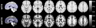 |
Assessment of white matter damages in Multiple Sclerosis using normative templates of conventional and inhomogeneous Magnetization Transfer
Lucas Soustelle1,2, Andreea Hertanu1,2, Arnaud Le Troter1,2, Soraya Gherib1,2, Samira Mchinda1,2, Patrick Viout1,2, Lauriane Pini1,2, Claire Costes1,2, Sylviane Confort-Gouny1,2, Adil Maarouf1,2,3, Bertrand Audoin1,2,3, Audrey Rico1,2,3, Clémence Boutière1,2,3,
Maxime Guye1,2, Jean-Philippe Ranjeva1,2, Gopal Varma4, David C. Alsop4, Jean Pelletier1,2,3, Guillaume Duhamel1,2, and Olivier M. Girard1,2
1Aix Marseille Univ, CNRS, CRMBM, Marseille, France, 2APHM, Hôpital Universitaire Timone, CEMEREM, Marseille, France, 3APHM, Hôpital Universitaire Timone, Service de neurologie, Marseille, France, 4Division of MR Research, Radiology, Beth Israel Deaconess Medical Center, Harvard Medical School, Boston, MA, United States
Characterization of Multiple Sclerosis (MS) is of paramount importance for patient care. In this work, we compare inhomogeneous (ihMT) and conventional magnetization transfer (MT) MRI techniques for white matter (WM) evaluation in a group comparison analysis after normalization in a standard space (templates of MS patients and healthy controls). Regions of interest, voxel-based morphometry and histogram analyses revealed that signal changes in pathological WM are not equivalent for ihMT and MT, highlighting that both metrics provide complementary information.
|
|||
2803.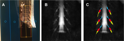 |
Assessing proximal and distal peripheral nerve damage in relapsing-remitting multiple sclerosis using magnetisation transfer ratio
Marios C. Yiannakas1, Ratthaporn Boonsuth1, Carmen Tur1,2, Marco Battiston1, Francesco Grussu1,3, Rebecca S. Samson1, Torben Schneider4, Masami Yoneyama5, Ferran Prados1,6,7, Sara Collorone1, Rosanna Cortese1, Olga Ciccarelli1, and Claudia A. M. Gandini Wheeler-Kingshott1,8,9
1NMR Research Unit, Queen Square MS Centre, Department of Neuroinflammation, UCL Queen Square Institute of Neurology, Fuculty of Brain Sciences, University College London, London, United Kingdom, 2Multiple Sclerosis Centre of Catalonia (Cemcat), Vall d’Hebron Institute of Research, Vall d'Hebron Barcelona Hospital Campus, Barcelona, Spain, 3Radiomics Group, Vall d’Hebron Institute of Oncology, Vall d’Hebron Barcelona Hospital Campus, Barcelona, Spain, 4Philips Healthcare, Surrey, Guildford, United Kingdom, 5Philips Japan, Minatoku, Tokyo, Japan, 6Centre for Medical Image Computing, Medical Physics and Biomedical Engineering, University College London, London, United Kingdom, 7Universitat Oberta de Catalunya, Barcelona, Spain, 8Department of Brain and Behavioural Sciences, University of Pavia, Pavia, Italy, 9Brain Connectivity Research Centre, IRCCS Mondino Foundation, Pavia, Italy
Whilst multiple sclerosis (MS) is thought to be a disease of the central nervous system, evidence from neuropathological investigations has demonstrated that the peripheral nervous system (PNS) can also be affected in MS, with demyelination and axonal degeneration being the main pathophysiological mechanisms involved. In this study, PNS damage is assessed in vivo at proximal (lumbar plexus) and distal (sciatic nerve) anatomical locations in people with relapsing-remitting MS and healthy controls using magnetisation transfer ratio (MTR). Results demonstrate significantly reduced MTR values at distal anatomical locations, however no relationship is identified between these changes and clinical scores of disability.
|
|||
2804.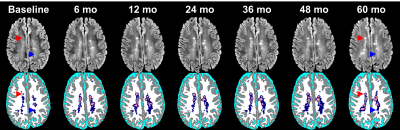 |
Automatic Segmentation of Diffusely Abnormal White Matter in MS Using Deep Neural Network
Refaat E Gabr1 and Ponnada A Narayana1
1Diagnostic and Interventional Imaging, University of Texas Health Science Center at Houston, Houston, TX, United States
Deep neural network was used to automatically segment diffusely abnormal white matter (DAWM) in 100 relapsing remitting multiple sclerosis patients (RRMS). Our calculated DAWM prevalence of 32% is comparable to ~ 25% reported elsewhere. Based on our studies, only 13% of T2 lesions at baseline converted into DAWM by 60 months. Of the DAWM detected at baseline, only 15% converted to lesions, 45% persisted, and 40% resolved (converted to NAWM). These initial results suggest that DAWM may present a significant disease burden by itself.
|
|||
2805.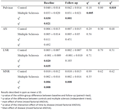 |
Decline of both iron concentration and iron content within the thalamus of patients with multiple sclerosis over two years
Fahad Salman1, Niels Bergsland1,2, Michael G Dwyer1,3, Bianca Weinstock-Guttman4, Robert Zivadinov1,3, and Ferdinand Schweser1,3
1Buffalo Neuroimaging Analysis Center, Department of Neurology at the Jacobs School of Medicine and Biomedical Sciences, University at Buffalo, The State University of New York, Buffalo, NY, United States, 2IRCCS, Fondazione Don Carlo Gnocchi ONLUS, Milan, Italy, 3Center for Biomedical Imaging, Clinical and Translational Science Institute, University at Buffalo, The State University of New York, Buffalo, NY, United States, 4Jacobs Multiple Sclerosis Center, Department of Neurology, Jacobs School of Medicine and Biomedical Sciences, University at Buffalo, The State University of New York, Buffalo, NY, United States
This study investigates the temporal evolution of iron content and concentration in thalamic nuclei of patients with multiple sclerosis over two years. Both metrics declined significantly relative to controls in the pulvinar. Pulvinar iron may potentially serve as an imaging biomarker for oligodendroglial vitality in MS.
|
|||
2806.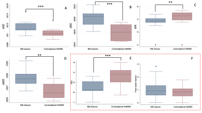 |
Diffusion Compartment Imaging Characterization of White Matter Microstructural Changes in Pediatric Onset Multiple Sclerosis
Fedel Machado-Rivas1,2, Camilo Jaimes1,2, Benoit Scherrer1,2, Mark Gorman1,2, Simon K Warfield1,2, and Onur Afacan1,2
1Radiology, Boston Children's Hospital, Boston, MA, United States, 2Radiology, Harvard Medical School, Boston, MA, United States
DTI has been used to evaluate brain microstructure in MS lesions and normal appearing white matter (NAWM). A DIAMOND model with an isotropic compartment + an anisotropic compartment may help disentangle the individual contribution of the compartments arising within a voxel, more accurately reflecting changes in the WM microstructure. In addition to finding differences in cAD, cRD, cMD and cFA, compartment diffusion model heterogeneity index was found to be significantly different in lesions. Our results support that a DIAMOND analysis can provide insights to MS lesion microstructure beyond conventional DTI metrics.
|
|||
2807.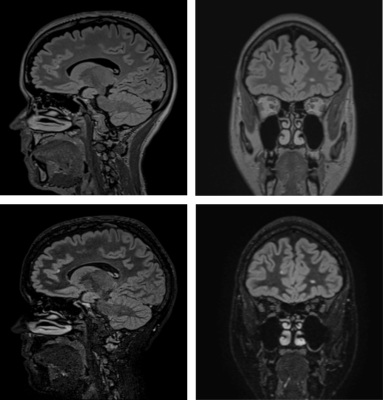 |
MR Inversion Recovery Simulation and Scanning of Subjects with Focus on White Matter Lesion Contrast Optimization
Øystein Bech Gadmar1, Anne-Hilde Farstad2, Berit Elstad2, Piotr Sowa2, and Wibeke Nordhøy1
1Diagnostic Physics, Oslo University Hospital, Oslo, Norway, 2Radiology, Oslo University Hospital, Oslo, Norway
A MatLab-based inversion recovery sequence simulator/calculator was developed with the purpose of determining and testing optimal parameters for 3D IR acquisitions with the purpose of detecting Multiple Sclerosis lesions in brains. Single inversion FLAIR and dual inversion DIR sequences were studied including a “True-T2” DIR sequence removing the undesired T1 weighting inherent in IR sequences to improve lesion-WM contrast. A T2 preparation phase further helps facilitate T1 suppression. Optimized IR sequences were tested on healthy volunteers and some MS patients, on 1.5 T and 3.0 T MR scanners. Good lesion contrast efficiency with high SNR was found for True-T2 DIR.
|
|||
2808.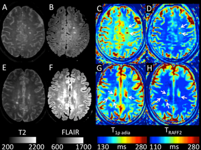 |
Prediction of multiple sclerosis clinical progression using whole brain adiabatic T1rho and Relaxation Along a Fictitious Field imaging
Ivan Jambor1, Aida Steiner2, Marko Pesola3, Timo Liimatainen4, Marcus Sucksdorff3, Eero Rissanen 2, Laura Airas2, Hannu Aronen3, and Harri Merisaari3
1Icahn School of Medicine at Mount Sinai, New York, NY, United States, 2Turku University Hospital, Turku, Finland, 3University of Turku, Turku, Finland, 4University of Oulu, Oulu, Finland
In this single center prospective clinical trial, we have shown feasibility of whole brain of T1ρadiab and TRAFF2 at 3T. Both of these methods provided higher lesion-to-normal appearing white matter contrast compared with conventional used T1-weighted imaging, and demonstrated potential to predict multiple sclerosis disease severity scores (EDSS, MSSS) at the time of imaging and 1-year follow-up. These encouraging findings stimulate further application of T1ρadiab and TRAFF2 at 3T in patients with multiple sclerosis.
|
|||
2809.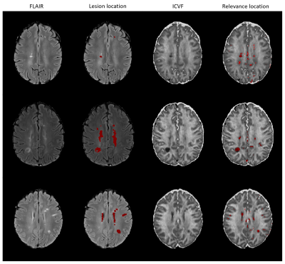 |
Relevance analysis of identifying multiple sclerosis patients based on diffusion imaging data using CNN
Alina Lopatina1,2, Stefan Ropele3, Renat Sibgatulin1, Jürgen R Reichenbach1,2,4, and Daniel Güllmar1
1Medical Physics Group / IDIR, Jena University Hospital, Jena, Germany, 2Michael-Stifel-Center for Data-Driven and Simulation Science, Jena, Germany, 3Department of Neurology, Medical University of Graz, Graz, Austria, 4Center of Medical Optics and Photonics Jena, Jena, Germany
To analyze the classification procedure of identifying multiple sclerosis (MS) based on diffusion-weighted imaging data by using convolutional neural networks (CNNs), we generated relevance maps. The relevance maps indicate the contribution of each input voxel to the final classification score and may facilitate new findings regarding MS-specific biomarkers. The study showed that voxels in the central brain area including some of the lesion voxels are important for correct classification. This information may be used in the future to perform a more detailed analysis in order to classify different MS-phenotypes or predict disease progression.
|
The International Society for Magnetic Resonance in Medicine is accredited by the Accreditation Council for Continuing Medical Education to provide continuing medical education for physicians.