Digital Posters
MS: Myelin & Lesion Characterization
ISMRM & SMRT Annual Meeting • 15-20 May 2021

Digital Posters
MS: Myelin & Lesion Characterization
| Concurrent 6 | 13:00 - 14:00 |
2810.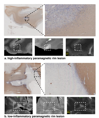 |
Paramagnetic Rim Lesions in MS are characterized by heterogeneous damage and inflammatory activity: a combined T1 relaxometry-diffusion study
Muhamed Barakovic1,2,3, Riccardo Galbusera1,2,3, Reza Rahmanzadeh1,2,3, Matthias Weigel1,2,3, Po-Jui Lu1,2,3, Erik Bahn4, Simona Schiavi5, Alessandro Daducci5, Pascal Sati6,7, Pietro Maggi8,9, Ludwig Kappos2,3, Jens Kuhle2,3, Laura Gaetano10,
Stefano Magon11, and Cristina Granziera1,2,3
1Translational Imaging in Neurology (ThINk) Basel, University Hospital Basel and University of Basel, Basel, Switzerland, 2Neurologic Clinic and Policlinic, University Hospital Basel and University of Basel, Basel, Switzerland, 3Research Center for Clinical Neuroimmunology and Neuroscience (RC2NB) Basel, University Hospital Basel and University of Basel, Basel, Switzerland, 4Institute of Neuropathology, University Medical Center, Göttingen, Germany, 5Department of Computer Science, University of Verona, Verona, Italy, 6Translational Neuroradiology Section, National Institute of Neurological Disorders and Stroke, Bethesda, MD, United States, 7Department of Neurology, Cedars-Sinai Medical Center, Los Angeles, CA, United States, 8Department of Neurology, Lausanne University Hospital, Lausanne, Switzerland, 9Cliniques universitaires Saint Luc, Université catholique de Louvain, Louvain, Belgium, 10F. Hoffmann-La Roche Ltd., Basel, Switzerland, 11Pharmaceutical Research and Early Development, Roche Innovation Center Basel F. Hoffmann-La Roche Ltd., Basel, Switzerland
Paramagnetic rim lesions (PRL) are characterized by heterogeneous damage in multiple sclerosis (MS) patients. In this study we acquired in vivo, postmortem and histopathology with the aim to further characterize the microstructural properties of these lesions. Furthermore, we disentangled the heterogeneity of PRL by combining a stick-ball-sphere diffusion model with the quantification of mean T1 relaxation times.
|
|||
2811. |
Deep Multiple Sclerosis Lesion Segmentation with Anatomical Convolution and Lesion-wise Loss
Hang Zhang1, Jinwei Zhang1, Pascal Spincemaille1, Thanh D. Nguyen1, and Yi Wang1
1Cornell University, New York, NY, United States
We propose an anatomical convolutional module to couple anatomical information into deep neural network. We further develop a loss function based on the mass center of individual lesions called lesion-wise loss, which can regularize the network training, thereby improving the performance of lesion localization and segmentation. We validate our methods on a public dataset, ISBI-15 Multiple Sclerosis Lesion Segmentation Challenge [1], where the results showed that we achieved the best performance on all published methods.
|
|||
2812. |
Characterization of multiple sclerosis lesion types with texture analysis of advanced and conventional MRI
Zahra Hosseinpour1, Olayinka Oladosu2, Mahshid Soleymani1, G Bruce Pike2, and Yunyan Zhang2
1Biomedical Engineering program, Schulish School of Engineering, University of Calgary, Calgary, AB, Canada, 2Department of Radiology and Clinical Neurosciences, Hotchkiss Brain Institute, University of Calgary, Calgary, AB, Canada
Multiple sclerosis (MS) lesion characterization using MRI is valuable for monitoring MS and probing remyelination therapies. However, there is a lack of in-vivo MRI metrics in clinical studies. We assessed MS lesion types using statistical texture measures of advanced and conventional MRIs, accompanied by random forest classification and percentile statistics, to differentiate MS lesions based on myelination. The best texture parameters and percentiles that classified re- and de-myelinated lesions were derived from histology-verified MRI. Applying the established parameters from postmortem to in-vivo MRI identified two types of lesions: highly demyelinated and potentially remyelinated.
|
|||
2813.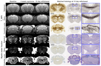 |
MRI reveals inflammation-mediated demyelination in a cuprizone toxin model of multiple sclerosis
Stephen T. Vito1, Kai H. Barck2, Rohan S. Virgincar2, Amy Easton1, Robby M. Weimer2, Tracy J. Yuen1, and Luke Xie2
1Neuroscience, Genentech, South San Francisco, CA, United States, 2Biomedical Imaging, Genentech, South San Francisco, CA, United States
Multiple sclerosis (MS) is the most common disease of the CNS that causes neurodegeneration and demyelination. MRI can track changes in MS lesions, but how imaging monitors inflammatory cell response or myelin content is not well characterized. In this study, we use a cuprizone toxin mouse model to determine how T2 relaxation time, myelin water fraction (MWF), diffusion tensor imaging (DTI), and gRatio MRI relate to changes with microglia, astrocytes, oligodendrocytes, and myelin. Using longitudinally imaging, MRI was able to detect inflammation-mediated demyelination. T2 reflected changes in inflammation (microglia and astrocytes) while MWF and gRatio suggested demyelination.
|
|||
2814.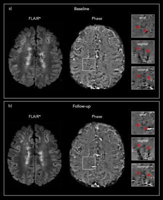 |
Longitudinal automated assessment of paramagnetic rim lesions in multiple sclerosis using RimNet
Maxence Wynen1, Francesco La Rosa2,3,4, Amina Sellimi5, Germán Barquero2,3,4, Gaetano Perrotta6, Valentina Lolli7, Vincent Van Pesch5, Cristina Granziera8,9, Tobias Kober10, Pascal Sati11,12, Benoît Macq13, Daniel S. Reich11, Martina Absinta11,14,
Meritxell Bach Cuadra2,3,4, and Pietro Maggi5,15
1Ecole Polytechnique de Louvain, Université Catholique de Louvain, Louvain-la-Neuve, Belgium, 2Signal Processing Laboratory (LTS5), École Polytechnique Fédérale de Lausanne, Lausanne, Switzerland, 3Medical Image Analysis Laboratory, Center for Biomedical Imaging (CIBM), University of Lausanne, Lausanne, Switzerland, 4Radiology Department, Lausanne University Hospital and University of Lausanne, Lausanne, Switzerland, 5Department of Neurology, Cliniques Universitaires Saint-Luc, Université Catholique de Louvain, Brussels, Belgium, 6Department of Neurology, Erasme University Hospital, Université Libre de Bruxelles, Brussels, Belgium, 7Department of Radiology, Erasme University Hospital, Université Libre de Bruxelles, Brussels, Belgium, 8Neurologic Clinic and Policlinic, Departments of Medicine, Clinical Research and Biomedical Engineering, University Hospital Basel and University of Basel, Basel, Switzerland, 9Translational Imaging in Neurology (ThINk) Basel, Department of Medicine and Biomedical Engineering, University Hospital Basel and University of Basel, Basel, Switzerland, 10Advanced Clinical Imaging Technology, Siemens Healthcare AG, Lausanne, Switzerland, 11Translational Neuroradiology Section, National Institute of Neurological Disorders and Stroke, National Institutes of Health, Bethesda, MD, United States, 12Department of Neurology, Cedars-Sinai Medical Center, Los Angeles, CA, United States, 13ICTEAM Institute, Université catholique de Louvain, Louvain-la-Neuve, Belgium, 14Department of Neurology, Johns Hopkins University, Baltimore, MD, United States, 15Department of Neurology, Lausanne University Hospital and University of Lausanne, Lausanne, Switzerland
The automatic assessment of paramagnetic rim lesions in multiple sclerosis is important, and a deep learning-based algorithm called RimNet has recently been proposed. This work evaluates the generalizability of RimNet and its longitudinal performance on MRI data acquired at different clinical centers. We found that RimNet’s performance was nearly as good on totally unseen data as in the original paper (receiver-operating-characteristic area-under-the-curve (AUC) 0.88 vs. 0.94, precision-recall AUC 0.69 vs. 0.70), and it made consistent predictions on longitudinal data (binary consistency 82%, probability consistency 93%).
|
|||
2815.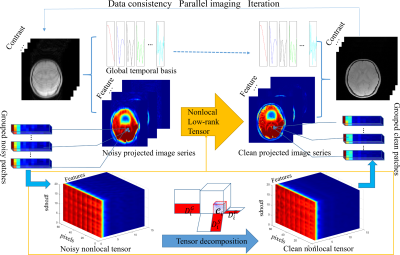 |
Accelerating Quantification of Myelin Water Fraction with Nonlocal Low-Rank Tensor in the Feature Domain
Quan Chen1, Huajun She1, and Yiping P. Du1
1School of Biomedical Engineering, Shanghai Jiao Tong University, Shanghai, China
Acceleration of myelin water fraction (MWF) mapping using the Feature domain nonlocal Low-Rank Tensor based (FnLRT) algorithm is investigated in this study. The global temporal information of the whole images is used to project the T2* weighted images (T2*WIs) into the feature domain. The nonlocal and local spatial redundancies in the feature domain are further exploited. The tensor-based decomposition is used to explore the multi-dimensional redundancies. The human brain experiments demonstrate the outperformance of the FnLRT algorithm over the state-of-the-art reconstructions at R=6. The FnLRT algorithm provides the potential to obtain the whole brain MWF mapping in 1 minute.
|
|||
2816.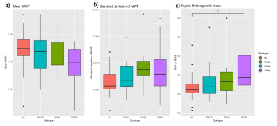 |
Beyond the Mean: Myelin Heterogeneity Index as a Sensitive Metric for Assessing Myelin Damage in Multiple Sclerosis
Poljanka Johnson1, Irene M Vavasour2, Shawna Abel1, Lisa E Lee3, Stephen Ristow1, Cornelia Laule4, Roger Tam2, David K.B. Li2, Nathalie Ackermans1, Alice Schabas1, Jillian Chan1, Helen Cross1, Ana-Luiza Sayao1, Virginia
Devonshire1, Robert Carruthers1, Anthony Traboulsee3, and Shannon Kolind5
1Medicine, University of British Columbia, Vancouver, BC, Canada, 2Radiology, University of British Columbia, Vancouver, BC, Canada, 3University of British Columbia, Vancouver, BC, Canada, 4Radiology, Physics and Astronomy, Pathology, University of British Columbia, Vancouver, BC, Canada, 5Medicine, Physics & Astronomy, University of British Columbia, Vancouver, BC, Canada
Myelin water imaging is a quantitative MRI technique that has been used to investigate myelin content in multiple sclerosis (MS). Three metrics derived from myelin water imaging were compared in the normal appearing white matter of different MS subtypes and correlations with disability were investigated. The myelin heterogeneity index was best able to distinguish between MS subtypes and demonstrated the strongest correlation with disability.
|
|||
2817. |
Application of an exponential recovery model to multiparametric 3D MRI to characterize the evolution of active lesions in Multiple Sclerosis
Lucas Soustelle1,2, Andreea Hertanu1,2, Arnaud Le Troter1,2, Soraya Gherib1,2, Samira Mchinda1,2, Patrick Viout1,2, Lauriane Pini1,2, Claire Costes1,2, Sylviane Confort-Gouny1,2, Adil Maarouf1,2,3, Bertrand Audoin1,2,3, Audrey Rico1,2,3, Clémence Boutière1,2,3,
Maxime Guye1,2, Jean-Philippe Ranjeva1,2, Gopal Varma4, David C. Alsop4, Jean Pelletier1,2,3, Olivier M. Girard1,2, and Guillaume Duhamel1,2
1Aix Marseille Univ, CNRS, CRMBM, Marseille, France, 2APHM, Hôpital Universitaire Timone, CEMEREM, Marseille, France, 3APHM, Hôpital Universitaire Timone, Service de neurologie, Marseille, France, 4Division of MR Research, Radiology, Beth Israel Deaconess Medical Center, Harvard Medical School, Boston, MA, United States
Active lesions in Multiple Sclerosis (MS) present successive phases, from formation through their chronic stage. Characterizing these phases may help us better comprehend the disease evolution. In this work, multiparametric MRI was performed in a 12-month longitudinal study of MS patients presenting new active lesions. An exponential recovery model was proposed to characterize the evolution of MR metrics in these lesions, allowing derivation of the recovery rates of inhomogeneous MTR, conventional MTR, DTI and T1-weighted images. Results show that recovery capacities are patient-dependent and that metrics differ in performance, presumably due to their respective sensitivity to the underlying MS mechanisms.
|
|||
2818.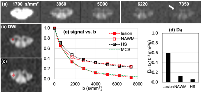 |
High-b Diffusivity of MS Lesion in Cervical Spinal Cord using Ultrahigh-b DWI (UHb-DWI)
Kyle Jeong1, Lubdha Shah2, You-Jung Lee1, Bijaya Thapa1, Nabraj Sapkota1, Erica Bisson3, Noel Carlson4, Eun-Kee Jeong2, and John Rose5
1Utah Center for Advanced Imaging Research, University of Utah, Salt Lake City, UT, United States, 2Radiology and Imaging Sciences, University of Utah, Salt Lake City, UT, United States, 3Neurosurgery, University of Utah, Salt Lake City, UT, United States, 4GRECC, Veterans Affairs, Salt Lake City, UT, United States, 5Neuroimmunology and Neurovirology Division, University of Utah, Salt Lake City, UT, United States
Ultrahigh-b radial DWI (UHb-rDWI) is a technique that can quantitatively evaluate MS lesions with respect to degrees of demyelination and axonal damage and/or loss. Currently, there is a lack of sensitive diagnostic technique to quantitatively and non-invasively grade cervical spinal cord (CSC) injury and repair. Our UHb-rDWI can potentially be a powerful tool to quantitatively observe disease progression and monitor treatment responses in MS CSC. Therefore, the main objective of this study was to investigate UHb-rDWI signal in white matter tracts of the CSC and compare quantitative values between healthy control WM with both MS NAWM and WM lesions.
|
|||
2819.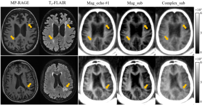 |
Brain Ultrashort T2 Component imaging using a STAIR Prepared Dual-Echo UTE Sequence with Complex Echo Subtraction
Ya-Jun Ma1, Hyungseok Jang1, Zhao Wei1, Mei Wu1, Saeed Jerban1, Eric Y Chang1,2, Jody Corey-Bloom1, Graeme M Bydder1, and Jiang Du1
1UC San Diego, San Diego, CA, United States, 2VA Health system, San Diego, CA, United States
We combine a Short TR Adiabatic Inversion Recovery preparation and dual-echo Ultrashort TE data collection with complex Echo Subtraction (ES) (STAIR-dUTE-ES) for both morphological and quantitative imaging of ultrashort T2 components in the whole brain. The 3D STAIR-dUTE-ES technique provides robust water suppression within the whole brain and allows accurate ultrashort T2 proton quantification. UltraShort T2 Proton Fraction (USPF) reduction in multiple sclerosis (MS) lesions suggests that the STAIR-dUTE-ES technique has potential for evaluation of demyelination and remyelination in the diagnosis and treatment of patients with MS.
|
|||
2820.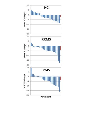 |
Myelin Water Imaging Demonstrates Myelin Loss in Multiple Sclerosis Normal Appearing White Matter over Two Years
Irene Margaret Vavasour1, Poljanka Johnson2, Shawna Abel3, Stephen Ristow3, Jared Splinter3, Cornelia Laule1,4,5,6, Roger Tam1, David KB Li1, Nathalie Ackermans3, Alice J Schabas3, Jillian Chan3, Helen Cross3, Ana-Luiza Sayao3,
Virginia Devonshire3, Robert Carruthers3, Anthony Traboulsee3, and Shannon H Kolind1,3,4,6
1Radiology, University of British Columbia, Vancouver, BC, Canada, 2Neuroscience, University of British Columbia, Vancouver, BC, Canada, 3Medicine, University of British Columbia, Vancouver, BC, Canada, 4Physics and Astronomy, University of British Columbia, Vancouver, BC, Canada, 5Pathology and Laboratory Medicine, University of British Columbia, Vancouver, BC, Canada, 6International Collaboration on Repair Discoveries (ICORD), University of British Columbia, Vancouver, BC, Canada
Therapies that target remyelination are under development and a non-invasive specific and sensitive imaging biomarker to evaluate their efficacy within the timescale of a clinical trial is vital. Using myelin water imaging, we followed a group of relapsing-remitting and progressive multiple sclerosis (MS) participants and healthy controls over approximately 2 years to compare their rate of change in myelin water fraction. A significant decrease in mean myelin water fraction was found over 2 years in the normal-appearing white matter of participants with MS, with a larger decrease in relapsing-remitting MS than progressive MS.
|
|||
2821.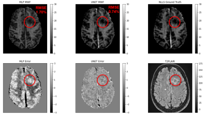 |
Sub-second Accurate Myelin Water Fraction Reconstruction with UNET
Jeremy Kim1,2, Thanh Nguyen2, Jinwei Zhang2, and Yi Wang2
1Stanford University, New York, NY, United States, 2Weill Cornell, New York, NY, United States
We developed UNET neural network for rapid, accurate and reproducible extraction of myelin water fraction map from FAST-T2 multi-echo T2 decay data. Testing results on 109 MS brains showed that UNET shortens post-processing time to less than a second and outperforms existing multi-layer perceptron algorithm.
|
|||
2822.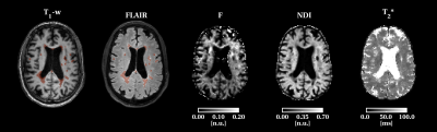 |
Quantitative multi-modal MRI shows correlations between lesion iron deposition and neuro-axonal density in progressive multiple sclerosis
Sara Collorone1, Marco Battiston1, Ferran Prados 1,2,3, Alberto Calvi1, Baris Kanber4, Francesco Grussu1,5, Marios Yiannakas1, Carmen Tur1,6, Rebecca Samson1, Olga Ciccarelli1,7, and Claudia A.M. Gandini Wheeler-Kingshott1,8,9
1NMR Research Unit, Queen Square MS Centre, Department of Neuroinflammation, UCL Queen Square Institute of Neurology, Faculty of Brain Sciences, University College London (UCL), London, United Kingdom, 2Centre for Medical Image Computing (CMIC), Department of Medical Physics and Biomedical Engineering, University College London (UCL), London, United Kingdom, 3Universitat Oberta de Catalunya, Barcelona, Spain, 4Centre for Medical Image Computing (CMIC), Department of Medical Physics and Biomedical Engineering, University College London(UCL), London, United Kingdom, 5Radiomics Group, Vall d’Hebron Institute of Oncology, Vall d’Hebron Barcelona Hospital Campus, Barcelona, Spain, 6Multiple Sclerosis Centre of Catalonia (Cemcat), Vall d’Hebron Institute of Research, Vall d’Hebron Barcelona Hospital Campus, Barcelona, Spain, 7University College London Hospitals, Biomedical Research Centre, National Institute for Health Research, London, United Kingdom, 8Department of Brain & Behavioural Sciences, University of Pavia, Pavia, Italy, 9Brain Connectivity Center Research Department, IRCCS Mondino Foundation, Pavia, Italy
We present preliminary results on ten patients with progressive multiple sclerosis(PMS) who underwent multi-modal MRI including quantitative magnetisation transfer (qMT), relaxometry and neurite orientation dispersion and density imaging (NODDI). We obtained metrics sensitive to iron deposition (T2*), myelin (restricted proton fraction (F)), and neuro-axonal density (neurite density index (NDI)) in lesions and brain tissues. Increased lesion T2* was related to decreased NDI in lesions and cortical grey matter. The lesion high T2* and low NDI were related to worse cognitive performance. In PMS, iron deposition from innate immune activity may lead to axonal loss and represent a future therapeutic target.
|
|||
2823.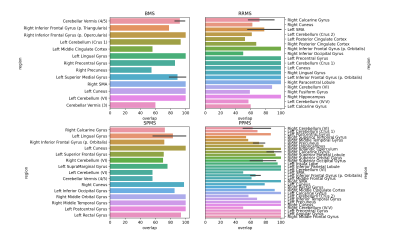 |
A Comparison of Brain Iron Accumulation Patterns Between Subtypes of Multiple Sclerosis
Aly Khalifa1 and Michael D Noseworthy2,3
1Department of Medical Biophysics, University of Toronto, Toronto, ON, Canada, 2Electrical and Computer Engineering, McMaster University, Hamilton, ON, Canada, 3School of Biomedical Engineering, McMaster University, Hamilton, ON, Canada
A data analysis pipeline was developed to correlate multiple sclerosis (MS) disease severity with regional susceptibility weighted imaging (SWI) intensity scores. It was assumed that reduced brain SWI signal correlated with elevated iron accumulation. Susceptibility weighted imaging (SWI) was used to map regional variations in brain iron content within each MS subtype. Number of brain areas with abnormal brain iron (relative to healthy age/sex matched controls), and the size of abnormal SWI image clusters, correlated with disease severity, based on subtype.
|
|||
2824.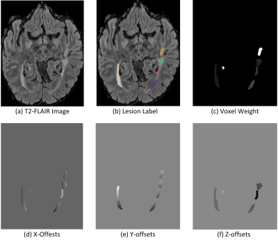 |
MS-Voter: Learning Where to Vote for Confluent Multiple Sclerosis Lesion Separation
Hang Zhang1, Jinwei Zhang1, Junghun Cho1, Susan A. Gauthier1, Pascal Spincemaille1, Thanh D. Nguyen1, and Yi Wang1
1Cornell University, New York, NY, United States
Lesion count, which encodes the lesion historical information, is an important biomarker for diagnosis and treatment of multiple sclerosis. Confluent lesions pose a great challenge to traditional automated methods, as these lesions are connected spatially, which requires expert experience to separate them. In this abstract, we propose a Hough voting method based on deep neural networks to resolve the issue. Experimental results on an in-house dataset demonstrates the superiority of our approach.
|
|||
2825.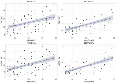 |
Increasing age is independently associated with higher free water in non-active MS brain - A multi-compartment analysis using FAST-T2
Liangdong Zhou1, Yi Li1, Xiuyuan Hugh Wang1, Elizabeth Sweeney1, Hang Zhang1,2, Emily B Tanzi1, Jennette Prince2, Victor Antonio Su-Ortiz2, Susan A Gauthier1, and Thanh D Nguyen1
1Radiology, Weill Cornell Medicine, New York, NY, United States, 2Cornell University, Ithaca, NY, United States
Multiple Sclerosis (MS) includes both inflammatory demyelination and axonal degeneration, which may affect the tissue water fractions of brain parenchyma in both myelin and the CSF water compartments. Quantification of brain parenchyma CSF fraction (CSFF) may help us better understanding of this complicated neurodegenerative disease. Aging is the most common cause of neurodegenerations. Here we tested CSFF in a 111 non-active MS cohort (age:45-79 years) by using the multi-echo T2 relaxometry-based multiple water-compartment method. Multivariate multiple regression shows that in cortical gray matter CSFF increases with age by controlling other factors including gender, disease duration, and MS lesion burden.
|
|||
2826.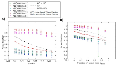 |
Impact of the Acquisition Protocol on the Sensitivity to Demyelination and Axonal Loss of DKI and NODDI: A Simulation Study
Stefania Oliviero1 and Cosimo Del Gratta1
1Department of Neuroscience, Imaging, and Clinical Sciences, University of Chieti Pescara G. D'Annunzio, Chieti, Italy
There is a general lack of agreed-upon guidelines regarding the DWI sequences to use, particularly to optimize the microstructural characterization of lesions. We evaluated the effects of demyelination and axonal loss on DKI and NODDI metrics and the impact of the sequence on the sensitivity to damage by performing a Monte-Carlo diffusion simulation inside a novel model of damaged WM. The sequence strongly affected the means and sensitivities of the metrics. Consequently, comparing DKI and NODDI analysis employing different sequences could lead to erroneous conclusions regarding the damage assessment: further investigations are needed for a community consensus on acquisition details.
|
|||
2827.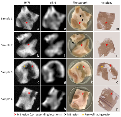 |
Mapping myelin content in ex-vivo MS brain tissue using short-T2 MRI of the lipid-protein bilayer
Emily Louise Baadsvik1, Markus Weiger1, Romain Froidevaux1, Wolfgang Faigle2, Benjamin Victor Ineichen2, and Klaas Paul Pruessmann1
1ETH Zurich and University of Zurich, Zurich, Switzerland, 2University Hospital Zurich and University of Zurich, Zurich, Switzerland
Ultrashort-T2 signal from white matter can be captured for imaging using dedicated short-T2 techniques. In this work, such methods were applied to D2O-exchanged human brain tissue from four donors diagnosed with multiple sclerosis to obtain 0.39 mm isotropic resolution MR images of the ultrashort-T2 tissue components and a lower-resolution multi-TE image series which, via a model fitting procedure, was used to produce maps of the relative content of myelin lipid-protein bilayer. As proof-of-principle, the contrast in these images was compared with corresponding myelin-stained cryosections and sample photographs, yielding promising correlation for white matter, grey matter and multiple sclerosis lesions.
|
|||
2828.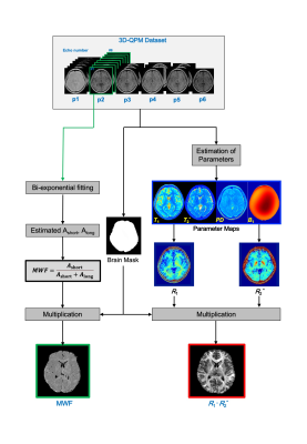 |
Myelin imaging derived quantitative parameter mapping compared to myelin water fraction
Yuki Kanazawa1, Masafumi Harada1, Yo Taniguchi2, Syun Kitano3, Nagomi Fukuda3, Yuki Matsumoto1, Hiroaki Hayashi4, Kosuke Ito2, Yoshitaka Bito2, and Akihiro Haga1
1Graduate School of Biomedical Sciences, Tokushima University, Tokushima, Japan, 2Healthcare Business Unit, Hitachi, Ltd., Tokyo, Japan, 3Faculty of Medicine, Tokushima University, Tokushima, Japan, 4Faculty of Health Sciences, Institute of Medical, Pharmaceutical and Health Sciences, Kanazawa University, Kanazawa, Japan
We evaluated myelin imaging R1·R2* derived from QPM-relaxation parameters compared with the MWF derived from the same QPM gradient-echo dataset. Linear regression analysis indicated a correlation between R1·R2* products values and MWF values derived from same QPM dataset (R = 0.53, P < 0.0001).The R1·R2* products map derived from QPM offers as follow advantage; quantitative analysis; artifact free; and
post-processing without fitting algorithm after QPM estimation. Myelin map derived from QPM can be applied for the quantitative assessment of white matter structure as substitute for MWF.
|
|||
2829.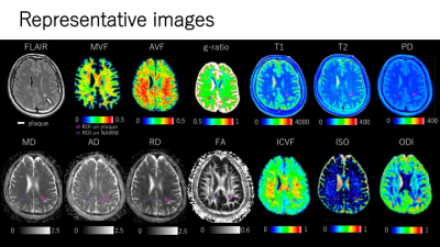 |
Association of estimated time from the onset of multiple sclerosis plaques with myelin and axon-related quantitative MRI measurement
Tomoko Maekawa1,2, Akifumi Hagiwara1,3, Masaaki Hori1,4, Christina Andica1, Shohei Fujita1,5, Toshiaki Akashi1, Koji Kamagata1, Akihiko Wada1, and Shigeki Aoki1
1Department of Radiology, Juntendo University School of Medicine, Tokyo, Japan, 2Division of Regenerative Medicine, The Jikei University School of Medicine, Tokyo, Japan, 3Department of Radiological Science, David Geffen School of Medicine, University of California Los Angeles, Los Angeles, CA, United States, 4Department of Diagnostic Radiology, Toho University Omori Medical Center, Tokyo, Japan, 5Department of Radiology, Graduate School of Medicine, The University of Tokyo, Tokyo, Japan
We investigated the association between estimated time from the onset of multiple sclerosis (MS) plaques and myelin- and axon-related quantitative MRI measurements using synthetic MRI (SyMRI) and neurite orientation dispersion and density imaging (NODDI). We retrospectively analyzed 32 MS patients with 73 newly appearing plaques. MS plaques with longer estimated time from the onset had significantly lower myelin volume fraction and higher g-ratio. It is suggested that myelin in plaques is under ongoing damage more than axons. SyMRI and NODDI may be useful for the quantitative assessment of temporal change in MS plaques.
|
The International Society for Magnetic Resonance in Medicine is accredited by the Accreditation Council for Continuing Medical Education to provide continuing medical education for physicians.