Digital Posters
Epilepsy & TBI: Damaged by Trauma, Etc.
ISMRM & SMRT Annual Meeting • 15-20 May 2021

Digital Posters
Epilepsy & TBI: Damaged by Trauma, Etc.
| Concurrent 5 | 13:00 - 14:00 |
| 1053. | WITHDRAWN | |||
1054.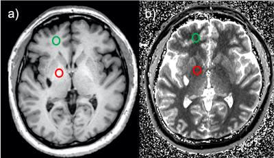 |
Sensitivity and Specificity of MRI Markers of Excess Manganese Brain Deposition
Humberto Monsivais1, Grace Francis2, Sandy Snyder1, Jonathan Kuhn3, and Ulrike Dydak1,4
1School of Health Sciences, Purdue University, West Lafayette, IN, United States, 2Department of Physics and Astronomy, Purdue University, West Lafayette, IN, United States, 3Mathematics and Statistics, Purdue University Northwest, Westville, IN, United States, 4Radiology and Imaging Scienes, Indiana University School of Medicine, Indianapolis, IN, United States
The pallidal index (PI) and the R1 relaxation rate are two commonly used MRI markers to diagnose manganese (Mn) neurotoxicity caused by excess Mn accumulation in the brain. While it has been hypothesized that the R1 relaxation rate is more sensitive and specific to Mn accumulation than the PI, a formal comparison is still missing. This study aimed to compare these two MRI markers' sensitivity and specificity to distinguish different levels of exposure to Mn by performing a receiver operating characteristic curve (ROC) analysis on a cohort of welders, occupationally exposed to Mn in welding fumes.
|
|||
1055.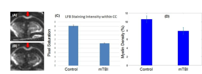 |
UTE MRI can detect myelin loss in mice using an open field low intensity blast injury model of mild traumatic brain injury (mTBI)
Ya-Jun Ma1, Catherine E Johnson2, Jonathan Wong1,3, Hyungseok Jang1, Roland Lee1, Eric Y Chang1,3, Zezong Gu2, and Jiang Du1
1UC San Diego, San Diego, CA, United States, 2Missouri University of Science and Technology, Rolla, MO, United States, 3VA Health System, San Diego, CA, United States
Mild traumatic brain injury (mTBI) may cause significant myelin damage, leading to significant degradation of elaborate cognitive functions. However, conventional neuroimaging techniques are unable to accurately assess myelin, and fail to show abnormalities in the majority of mTBI cases. UTE MRI sequences with echo times (TEs) <0.1 ms allow direct imaging and quantitative assessment of myelin density. Here we aim to investigate whether the 3D IR-UTE sequence can detect myelin loss in mice using an open field low intensity blast injury model of mTBI. This technique provides a new approach for potentially more accurate diagnosis and treatment monitoring of mTBI.
|
|||
1056.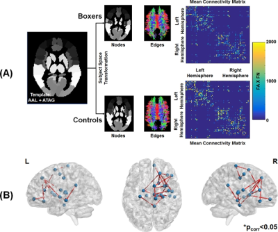 |
Topological reorganization due to repetitive head impacts: Insights from Professional Fighters Brain Health Study
Virendra R Mishra1, Karthik Sreenivasan1, Dietmar Cordes1, Aaron Ritter1, and Charles Bernick2
1Cleveland Clinic Lou Ruvo Center for Brain Health, Las Vegas, NV, United States, 2University of Washington - Seattle, Seattle, WA, United States
Repetitive head impact (RHI) is thought to induce robust white-matter (WM) damage which may be the risk factor for various disorders. Though single-tensor diffusion MRI (dMRI) studies have improved our understanding of WM abnormalities at a regional level in participants exposed to RHI, there are no studies that attempt to understand topological WM structural connectivity changes due to RHI. Utilizing a high spatial resolution (1.5mm3) dMRI from 12 active male boxers and 10 demographically matched healthy controls (HC), we showed that RHI induces a topological shift as compared to HC, and this shift is correlated with neuropsychological scores.
|
|||
1057.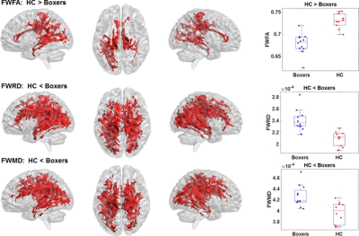 |
Investigating the extent of difference in single tensor and beyond single tensor diffusion MRI-derived voxelwise measures
Virendra R Mishra1, Karthik Sreenivasan1, Dietmar Cordes1, Aaron Ritter1, and Charles Bernick2
1Cleveland Clinic Lou Ruvo Center for Brain Health, Las Vegas, NV, United States, 2University of Washington - Seattle, Seattle, WA, United States
In this study, we investigated the spatial extent and location of white-matter (WM) disorganization due to repeated head impacts (RHI) by estimating single-tensor (ST) diffusion MRI (dMRI)-measures along with free-water (FW)-corrected ST measures, diffusion kurtosis imaging (DKI), and Neurite Orientation Dispersion and Density Imaging (NODDI) measures, along with understanding the correlation of such voxelwise measures with exposure to fighting and neuropsychological scores. Overall, our findings suggest that WM disorganization is prevalent in thalamocortical and corpus-callosum fibers due to RHI, although, the spatial extent and location of these differences are heavily dependent on the dMRI-models utilized in the study.
|
|||
1058.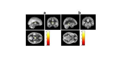 |
Hippocampal and Anterior Cingulate Blood Flow is Associated with Affective Symptoms in Chronic Traumatic Brain Injury
Binu P. Thomas1,2, Takashi Tarumi3,4, Ciwen Wang3, David C. Zhu5, Tsubasa Tomoto4, C. Munro Cullum3,6,7, Marisara Dieppa3, Ramon Diaz-Arrastia8, Kathleen Bell9, Christopher Madden6, Rong Zhang3,4, and Kan Ding3
1Advanced Imaging Research Center, University of Texas Southwestern Medical Center, Dallas, TX, United States, 2Department of Bioengineering, University of Texas at Arlington, Arlington, TX, United States, 3Department of Neurology, University of Texas Southwestern Medical Center, Dallas, TX, United States, 4Institute for Exercise and Environmental Medicine, Texas Health Presbyterian Hospital, Dallas, TX, United States, 5Department of Radiology and Cognitive Imaging Research Center, Michigan State University, East Lansing, MI, United States, 6Department of Psychiatry, University of Texas Southwestern Medical Center, Dallas, TX, United States, 7Department of Neurological Surgery, University of Texas Southwestern Medical Center, Dallas, TX, United States, 8Department of Neurology, University of Pennsylvania Perelman School of Medicine, Philadelphia, PA, United States, 9Department of Physical Medicine and Rehabilitation, University of Texas Southwestern Medical Center, Dallas, TX, United States
Chronic Traumatic-brain-injury (TBI) has lifelong implications on brain function. It is characterized by cerebral-blood-flow (CBF) deficits, often accompanied by TBI-related symptoms. It is crucial that we understand mechanisms of CBF alterations and its association with TBI-symptoms. We observed CBF deficits in patients with TBI in the thalamus, hippocampus and other subcortical structures compared to a group of normal control participants. Furthermore, CBF in the hippocampus and anterior cingulate were negatively associated with TBI-related symptoms of anxiety, depression, fatigue and sleep issues. Our results suggest that regional CBF deficits may be useful biomarkers for perfusion-targeted therapies to ameliorate TBI-related symptoms.
|
|||
1059.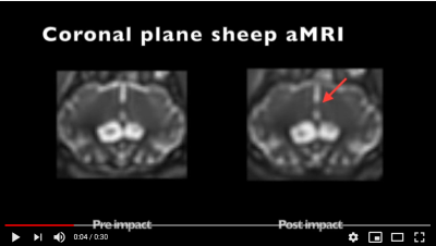 |
Analysis and visualisation of physiological changes before and after a mild Traumatic Brain injury
Catherine Emata*1, Eryn Kwon*2,3, Maryam Tayebi3, Leo Dang2,4, Adam Donaldson5, Vickie Shim3, Allen Champagne6, Itamar Terem7,8, Alan Wang2,3,4, David Dubowitz 2,9,10, Sarah-Jane Guild11, Miriam Scadeng 2,4,10, and Samantha Holdsworth2,4
1University of Auckland, Auckland, New Zealand, 2Anatomy and Medical Imaging, Faculty of Medical and Health Sciences, University of Auckland, Auckland, New Zealand, 3Auckland Bioengineering Institute, University of Auckland, Auckland, New Zealand, 4Centre for Brain Research, University of Auckland, Auckland, New Zealand, 5Mechatronics Engineering, University of Canterbury, Christchurch, New Zealand, 6Centre for Neuroscience Studies, Queen’s University, Kingston, ON, Canada, 7Electrical Engineering, Stanford University, Stanford, CA, United States, 8Structural Biology, Stanford University, Stanford, CA, United States, 9Centre for Advanced MRI, University of Auckland, Auckland, New Zealand, 10Center for functional MRI, University of California, San Diego, CA, United States, 11Physiology, Faculty of Medical and Health Science, University of Auckland, Auckland, New Zealand
Three advanced sequences were used in conjunction to characterize the physiological changes associated with a mild traumatic brain injury. Amplified Magnetic Resonance Imaging (aMRI) is a motion detection and visualization technique, and is used to amplify pulsatile brain motion. Four-dimensional Flow Magnetic Resonance Imaging (4D flow MRI) is a sequence utilised to analyse and visualise blood flow, while diffusion MRI (dMRI) delineates features of tissue microstructure. Findings included diffusion changes, an increase in blood velocity, and a change in the profile of blood flow to the brain in the carotid arteries, along with increased parenchymal micro-displacements within the brain.
|
|||
1060.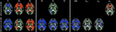 |
White Matter Microstructural Alterations In Contact-Sport Athletes With And Without Concussion: A Multi-Shell Diffusion Study
Sohae Chung1,2, Junbo Chen3, Tianhao Li3, Yao Wang3, and Yvonne W. Lui1,2
1Center for Advanced Imaging Innovation and Research (CAI2R), Department of Radiology, New York University Grossman School of Medicine, New York, NY, United States, 2Bernard and Irene Schwartz Center for Biomedical Imaging, New York University Grossman School of Medicine, New York, NY, United States, 3Department of Electrical and Computer Engineering, NYU Tandon School of Engineering, Brooklyn, NY, United States
There is growing concern that there may be negative effects on the brain from repetitive head impacts as well as sport-related concussion (SRC). Here, we demonstrate widespread white matter microstructural differences between contact-sport athletes and non-contact sport controls using multi-shell diffusion MRI. Importantly, microstructure differences were present in contact-sport athletes both with and without concussion, suggesting that exposure to multiple head impacts effects brain microstructure. Decreased radial diffusivity (RD) and extra-axonal radial diffusivity (D$$$e,\perp$$$) and increased fractional anisotropy (FA) suggest pathologies such as cytotoxic edema may be present acutely after injury.
|
|||
1061. |
Radio-chemotherapy induced toxic leukoencephalopathy: ultra-high field MR findings
Laura Biagi1,2, Rosa Pasquariello1, Raffaello Canapicchi 1, Chiara Ticci3, Claudia Dosi3, Graziella Donatelli2,4, Mauro Costagli1,2, Mirco Cosottini2,4,5, Roberta Battini3,6, Michela Tosetti1,2, and Network IDEA7
1Laboratory of Medical Physics and Magnetic Resonance, IRCCS Fondazione Stella Maris, Pisa, Italy, 2Imago7 Research Foundation, Pisa, Italy, 3Department of Developmental Neuroscience, IRCCS Fondazione Stella Maris, Pisa, Italy, 4Neuroradiology Unit, Azienda Ospedaliero-Universitaria Pisana, Pisa, Italy, 5Department of Translational Research and New Technologies in Medicine and Surgery, University of Pisa, Pisa, Italy, 6Department of Clinical and Experimental Medicine, University of Pisa, Pisa, Italy, 7Italian DEvelopmental Age Health Network (IDEA Network), Rome, Italy
The affirmation of the role of UHF MRI in the clinical setting requires the demonstration of its diagnostic and prognostic power, especially in the context of fields not yet sufficiently explored, such as diseases of pediatric age. This study presents a single case of radio-chemotherapy induced leukoencephalopathy in which UHF-MRI demonstrated its added value compared to traditional MR imaging in the characterization of lesions and in the detailed delineation of their localization.
|
|||
1062.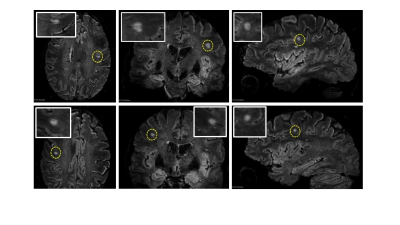 |
Central vein sign occurrence on FLAIR* in neuropsychiatric systemic lupus erythematosus patients
Francesca Inglese1, Kenneth Hergaarden1, Pierre-Louis Bazin2, Gerda M. Steup-Beekman3, Tom J.W. Huizinga3, Jeroen de Bresser1, and Itamar Ronen1
1Department of radiology, Leiden University Medical Center, Leiden, Netherlands, 2Faculty of Social and Behavioural Sciences, University of Amsterdam, Amsterdam, Netherlands, 3Department of Rheumatology, Leiden University Medical Center, Leiden, Netherlands
The central vein sign (CVS) is an imaging biomarker useful in multiple sclerosis (MS) diagnosis and to differentiate MS from other autoimmune diseases with inflammatory lesions, such as systemic lupus erythematosus with neuropsychiatric events (NP). The prevalence of CVS in SLE patients experiencing NP is unknown. We examined 16 NPSLE patients for presence of CVS using FLAIR* images, generated from FLAIR images acquired at 3T and T2*-weighted images acquired at 7T. Out of 491 white matter lesions, we found 4 confirmed and 1 suspected CVS lesions. Our data suggests that CVS may be present in random occurrence in NPSLE.
|
|||
1063.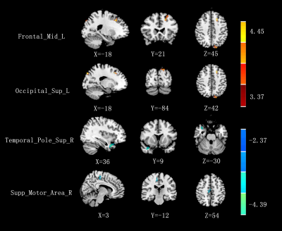 |
Parahippocampal gyrus neuroanatomical and functional connectivity alterations in mTBI at the acute stage:a VBM and rsfMRI study
wenjing huang1, jing zhang2, wanjun hu2, guangyao liu2, yanli jiang2, and shaoyu wang3
1Second Clinical School, Lanzhou University, Lanzhou, China, 2Lanzhou University Second Hospital, Lanzhou, China, 3Siemens Healthineers, Shanghai, China
mTBI is not a static event, but a progressive injury. The neuroanatomical and functional alterations of mild traumatic brain injury (mTBI) at the acute stage can be an initial step of damages leading to cognitive and emotional deficit which can be developed in future in long-term period of injury. We analyzed early alterations in the gray matter volume (GMV) and functional connectivity (FC) alterations of mTBI patients within 7 days after injury, and found that early disruptions in left parahippocampal gyrus may appear in mTBI, which might be potentially related to the cognitive and emotional impairments.
|
|||
1064.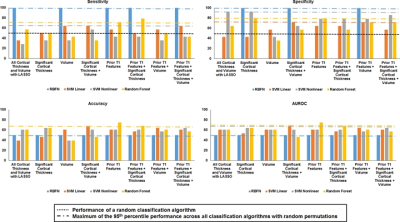 |
Hypothesis-driven or regression-driven machine learning? What technique to choose? Insights from Professional Fighters Brain Health Study
Virendra R Mishra1, Xiaowei Zhuang1, Dietmar Cordes1, Aaron Ritter1, and Charles Bernick2
1Cleveland Clinic Lou Ruvo Center for Brain Health, Las Vegas, NV, United States, 2University of Washington - Seattle, Seattle, WA, United States
Whether routinely obtained T1-derived volumetric and cortical thickness measures can identify boxers with neuropsychological impairment using machine-learning (ML) techniques in active male boxers is currently unknown. We utilized conventionally acquired MPRAGE data from 72 impaired and 72 nonimpaired boxers, and identified regions that have significantly different cortical thickness, volumetric differences, and cortical thickness and brain volumes correlated with exposure to fighting and neuropsychological scores. Further, we investigated whether these regression-defined regions or prior hypothesis-defined brain regions can identify boxers with neuropsychological deficits. Hypothesis-driven regions with random forest algorithm outperformed other ML techniques with either regression of hypothesis-driven feature selection.
|
|||
1065. |
An application of BOLD-fMRI on functional changes in shenmen and neiguan electroacpunctured population with chronic partial sleep-deprivation
Wang Chunyan1, Qiu Ganbin1, Weiyin Vivian Liu2, and Ma Liheng1
1Medical Imaging, the first affiliated hospital of Guangdong pharmaceutical university, Guangzhou, China, 2MR Research, GE Healthcare, Beijing, Beijing, China
An application of BOLD-fMRI on functional changes in shenmen and neiguan electroacpunctured population with chronic partial sleep-deprivation
|
|||
1066.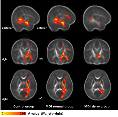 |
Altered structural asymmetries of mild white matter injury in preterm infants with different cognitive outcomes
Miaomiao Wang1, Xianjun Li1, Congcong Liu1, Xiaoyu Wang1, Mengxuan Li1, Cong Tian1, Peiyao Chen1, Chao Jin1, Xiaocheng Wei2, and Jian Yang1
1Department of Diagnostic Radiology, the First Affiliated Hospital of Xi’an Jiaotong University, Xi‘an, China, 2MR Research China, GE Healthcare, Beijing, China
Punctate white matter lesions (PWMLs) are common in the preterm. The mild PWMLs may result in cognitive impairments and lesion location is closely associated with neurodevelopmental outcomes. Cognition is a high-order function with multiregional information integration and functional hemispherical lateralization. Therefore, this study aims to investigate the white matter (WM) asymmetries of mild PWMLs in preterm infants with different cognitive outcomes at a corrected age of 6 months by diffusion tensor imaging. Left lateralization of WM development is obviously reduced in mild PWMLs with cognitive delay, while WM asymmetries of PWMLs with normal cognition score are similar with controls.
|
|||
1067.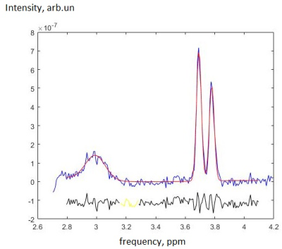 |
GABA and Glu in posterior cingulate cortex of mild TBI patients: MEGA-PRESS and TE averaged PRESS study with spectral averaging
Andrei V. Manzhurtsev1,2,3, Peter Bulanov3, Maxim Ublinskiy1,2, Petr E. Menshchikov2,4, Alexey Yakovlev1,2, Tolib Akhadov1,3, and Natalia Semenova1,2,3,5
1Radiology, Clinical and Research Institute of Emergency Pediatric Surgery and Trauma, Moscow, Russian Federation, 2Emanuel Institute of Biochemical Physics of the Russian Academy of Sciences, Moscow, Russian Federation, 3Moscow State University, Moscow, Russian Federation, 4Philips Healthcare, Russia, Moscow, Russian Federation, 5Semenov Institute of Chemical Physics of the Russian Academy of Sciences, Moscow, Russian Federation
In this study, GABA and Glu are measured in the posterior cingulate cortex (PCC) of children with acute mTBI using MEGA-PRESS and TE averaged PRESS. Spectra were processed individually and after between-subject group averaging. The results demonstrate the increase in Glu without glutamine contribution and the stability of GABA without macromolecule contamination which means that the excitatory-inhibitory neurotransmitter balance is shifted towards excitation in the cerebral region studied. Spectral averaging over groups of participants recommends itself as a powerful tool, if used with proper preprocessing steps.
|
|||
1068.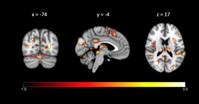 |
Blast-Induced Neurotrauma Results in Spatially Distinct Gray Matter Alteration alongside Hormonal Alteration
Sarah Hellewell1 and Ibolja Cernak2
1Curtin University, Nedlands, Australia, 2Mercer University, Macon, GA, United States
Blast-induced neurotrauma (BINT) occurs frequently in military personnel, who are also vulnerable to occupational stress. This study compared Canadian Armed Forces members/Veterans with a history of BINT to emergency first responders, performing voxel-based morphometry on T1-weighted images to examine gray matter alteration, and assessment of stress-related hormones to delineate BINT effects from stress. We found widespread, symmetric loci of reduced gray matter volume specific to BINT, which occurred alongside significant increases in testosterone, cortisol and the testosterone/cortisol ratio. These results indicate that BINT may cause structural and endocrine alterations unseen in emergency service workers who experienced occupational stress alone.
|
|||
1069.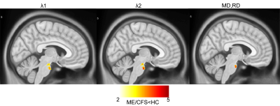 |
Study of brain abnormalities in Myalgic Encephalomyelitis /Chronic Fatigue Syndrome patients using diffusion tensor imaging
Kiran Thapaliya1,2, Donald Staines1, Sonya Marshall-Gradisnik1, and Leighton Barnden1
1Griffith University, Gold Coast, Australia, 2Centre for Advanced Imaging, The University of Queensland, Brisbane, Australia
Myalgic Encephalomyelitis (ME)/Chronic fatigue syndrome (CFS) patients suffer from a variety of physical and neurological complaints indicating the central nervous system plays a role in ME/CFS pathophysiology. Structural and functional magnetic resonance imaging have been used to identify the pathomechanism of ME/CFS However, changes in tissue microstructure to understand the pathomechanism of ME/CFS using diffusion tensor imaging (DTI) have not been fully investigated. Our DTI study showed abnormality of the brain stem in ME/CFS patients relative to healthy controls.
|
|||
1070.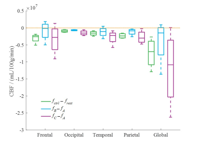 |
Effects of vibration on cerebral blood flow using 3D arterial spin labeling imaging
Linghan Kong1, Suhao Qiu1, Zhao He1, RunKe Wang1, Yu Chen1, Qiang He2, and Yuan Feng1
1Institute for Medical Imaging Technology, School of Biomedical Engineering, Shanghai Jiao Tong University, Shanghai, China, 2Shanghai United Imaging Healthcare Co Ltd, Shanghai, China
The relation between vibration and brain injury is still largely unknown. We applied a 30Hz vibration to brain and measured the cerebral blood flow (CBF) change using 3D arterial spin labeling perfusion imaging (3D-ASL). Results showed a decrease of CBF both regionally and globally after the vibration. This provided clues to understand the mechanism of traumatic brain injury.
|
|||
1071.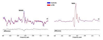 |
Separate NAAG and NAA quantification after pediatric mild traumatic brain injury in the acute phase.
Anna Ivantsova1, Petr Menshchikov1,2,3, Andrei Manzhurtsev1,3, Maxim Ublinskii1,3, Alexey Yakovlev1,3,4, Ilya Melnikov1, Dmitrii Kupriyanov2, Tolib Akhadov1, and Natalia Semenova1,3,4
1Clinical and Research Institute of Emergency Paediatric Surgery and Traumatology, Moscow, Russian Federation, 2Clinical Science, LLC Philips Healthcare, Moscow, Russian Federation, 3Emanuel Institute of Biochemical Physics, Russian Academy of Sciences, Moscow, Russian Federation, 4Semenov Institute of Chemical Physics, Russian Academy of Sciences, Moscow, Russian Federation
The main finding of the study is that the tNAA signal reduction in WM after mTBI is associated with a decrease in the NAAG concentration rather than a decrease in the NAA concentration, as was thought previously. This finding highlights the importance of separating these signals, at least for WM studies, to avoid misinterpretation of the results. NAAG plays an important role in selectively activating mGluR3 receptors, thus providing neuroprotective and neuroreparative functions immediately after mTBI. NAAG shows potential for the development of new therapeutic strategies for patients with injuries of varying severity
|
|||
1072.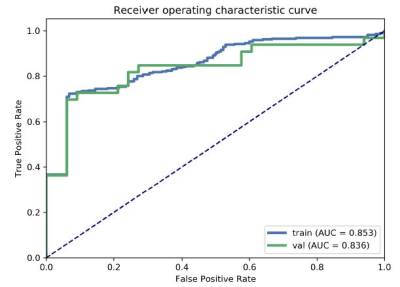 |
Exploring the Application of an Advanced-Diffusion-Model-Based Radiomics Model in detecting radiation-induced brain injury
Mengzhu Wang1 and Weike Zeng2
1MR Scientific Marketing, Siemens Healthcare, Guangzhou, China, 2Sun Yat-sen Memorial Hospital, Sun Yat-sen University, Guangzhou, China
The aim of this study was to elucidate a multiple diffusion-model-based radiomics model in detecting radiation-induced brain injury (RI). We used diffusion features derived from DTI, DKI, NODDI and MAP-MRI models and chose LR as the classifier to construct a prediction model. The results showed that the most accurate prediction was achieved by incorporating the DKI-AD and RTPP into a nomogram, with AUC and accuracy reached 0.8356 and 0.8182.
|
The International Society for Magnetic Resonance in Medicine is accredited by the Accreditation Council for Continuing Medical Education to provide continuing medical education for physicians.