Digital Posters
Oxygen, Reactivity & Tissue Function in Stroke Imaging
ISMRM & SMRT Annual Meeting • 15-20 May 2021

Digital Posters
Oxygen, Reactivity & Tissue Function in Stroke Imaging
| Concurrent 6 | 19:00 - 20:00 |
4322.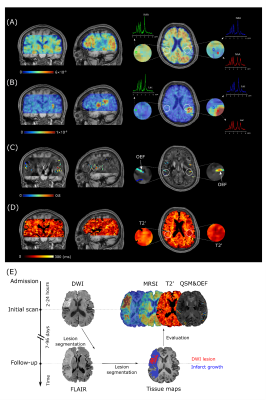 |
Simultaneous Oxygenation and Metabolic Imaging Relates Oxygen and Neuronal Metabolism in Acute Stroke
Tianxiao Zhang1, Rong Guo2,3, Tianyao Wang4, Zengping Lin1, Yudu Li2,3, Yibo Zhao2,3, Jun Liu4, Danhong Wu5, Zheng Jin6, Xin Yu7, Zhi-Pei Liang2,3, and Yao Li1
1School of Biomedical Engineering, Shanghai Jiao Tong University, Shanghai, China, 2Beckman Institute for Advanced Science and Technology, University of Illinois at Urbana-Champaign, Urbana, IL, United States, 3Department of Electrical and Computer Engineering, University of Illinois at Urbana-Champaign, Urbana, IL, United States, 4Radiology Department, The Fifth People's Hospital of Shanghai, Fudan University, Shanghai, China, 5Neurology Department, The Fifth People's Hospital of Shanghai, Fudan University, Shanghai, China, 6Shanghai Minhang Hospital of Integrated Traditional Chinese and Western Medicine Hospital, Shanghai, China, 7Department of Biomedical Engineering, Case Western Reserve University, Cleveland, OH, United States
Elevated oxygen extraction fraction and impaired neurometabolic metabolism are hallmarks of at-risk tissue in acute ischemic stroke. This study investigated the concurrent changes of oxygen and neuronal metabolisms. In an 8-min scan using SPICE, we simultaneously obtained 3D maps of neurometabolites (2.0 x 3.0 x 3.0 mm3 nominal spatial resolution) and T2'/oxygen extraction fraction (1.0 x 1.0 x 1.9 mm3 nominal resolution). Our results showed the expected changes in oxygenation and neurometabolite markers individually, and also their coupling. This study may lay a foundation for prediction of tissue viability in acute stroke using noninvasive multimodal high-resolution metabolic imaging.
|
|||
| 4323. | WITHDRAWN | |||
4324.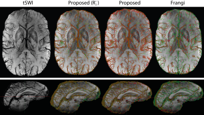 |
A Shearlet-based whole brain vein segmentation algorithm and its application for the detection of regional differences in venous oxygenation
Sina Straub1, Janis Stiegeler1,2, Edris El-Sanosy3, and Till M. Schneider4
1Division of Medical Physics in Radiology, German Cancer Research Center (DKFZ), Heidelberg, Germany, 2Faculty of Physics and Astronomy, University of Heidelberg, Heidelberg, Germany, 3Division of Radiology, German Cancer Research Center (DKFZ), Heidelberg, Germany, 44Department of Neuroradiology, University of Heidelberg, Heidelberg, Germany
Generating whole-brain vein segmentations can be very time-consuming. In this abstract, a method is proposed that can segment brain veins from a single-echo or multi-echo gradient echo scan. The segmentation algorithm combines classical vessel enhancement filtering and local thresholding methods with a shearlet-based multi-scale approach. Compared with a ground truth, the algorithm performs better for multi-echo data when R2* information is included in the segmentation. Moreover, the combination of venous segmentation with masks for deep and superficial venous territories yields higher susceptibility values for the superficial venous vasculature which is in accordance with a higher oxygen consumption of the cortex.
|
|||
4325.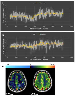 |
A Flexible Computational Framework for Characterization of Dynamic Cerebrovascular Response to Global Hemodynamic Stimuli
Siddhant Dogra1, Xiuyuan Wang2, Jelle Veraart3, Alejandro Gupta3, Koto Ishida3, Deqiang Qiu4, and Seena Dehkharghani3
1Radiology, New York University Langone Health, New York, NY, United States, 2Weill Cornell, New York, NY, United States, 3New York University Langone Health, New York, NY, United States, 4Emory University, Atlanta, GA, United States
Cerebrovascular reactivity (CVR) is conventionally assessed by comparing discrete cerebral blood flow measurements obtained prior to and following a hemodynamic stimulus. Blood-oxygen-level-dependent (BOLD)-MRI provides potentially continuous, dynamic CVR characterization when coupled to stimuli such as acetazolamide (ACZ), but poor signal-to-noise generally limits its analysis to an analogous comparison of arbitrarily defined terminal BOLD signals relative to baseline. We present a novel framework incorporating temporal and spatial denoising to pre-condition the dynamic time-signal course for robust voxel-level maximal and terminal CVR, and novel CVR time-to-peak and related kinetic features, disambiguating confounders related to potentially non-optimal terminal CVR assignment in conventional use.
|
|||
4326.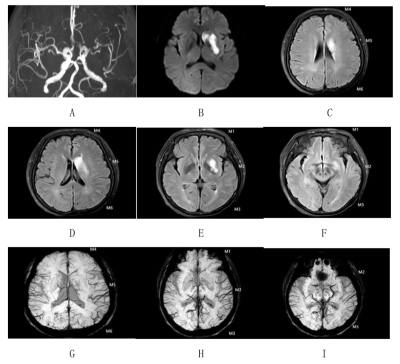 |
Relationship of FLIAR vascular hyperintensity territory with APCVs and venous oxygen saturation in patients with cerebral infarction
Pei-pei chang1, Yan-wei Miao1, Yu-han Jiang1, Yi-wei Che1, Bing-bing Gao1, Li-hua Chen1, Qing-wei Song1, Ren-wang Pu1, Ai-lian Liu1, Liang-jie Lin2, and Jia-zheng Wang2
1The First Affiliated Hospital of Dalian Medical University, Da Lian, China, 2Philips Healthcare, Bei Jing, China
Previous studies have suggested that fluid-attenuated inversion recovery vascular hyperintensity (FVH) and asymmetrical prominent cortical veins (APCVs) may be related with collateral flow in cerebral ischemia. Some studies showed susceptibility-weighted imaging (SWI) can reflect oxygenation fraction (OEF) in venous blood. Here, we prospectively compared the differences of FVH and APCVs scores in different types of vascular stenosis, and analyzed the relationships of the FVH score with the APCVs score, the venous-oxygen-saturation related MR measurements as well as the NIHSS score in patients with cerebral infarction. It was found that FVH and APCVs scores in severe-occlusion group were significantly higher than those in mild-to-moderate group. In addition, FVH score was positively correlated with APCVs score, rΔφ (phase value) and NIHSSadmission. It suggests that the occurrence of FVH is related to cerebral oxygen metabolism.
|
|||
4327.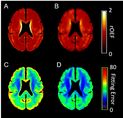 |
A Local Linear Regression Algorithm for Partial Volume Correction in Brain Oxygen Extraction Fraction Estimation
Yasheng Chen1, Chunwei Ying2, Peter Kang1, Slim Fellah1, Amy Mirro3, Melanie Fields3, Kristin Guilliams1, Jin-Moo Lee1, Andria Ford1, and Hongyu An4
1Neurology, Washington University School of Medicine, St. Louis, MO, United States, 2Biomedical Engineering, Washington University in St. Louis, St. Louis, MO, United States, 3Pediatrics Hematology, Washington University School of Medicine, St. Louis, MO, United States, 4Washington University School of Medicine, St. Louis, MO, United States
Measurement of oxygen extraction fraction (OEF) provides important information about tissue oxygen utilization. OEF maps can be obtained using a double echo asymmetric spin echo (ASE) sequence. Partial volume effect may lead to inaccurate OEF measurements in regions with mixed tissue types. We introduced a local linear regression algorithm to correct for partial volume effect. The PVC OEF method reduced model fitting errors and signal contamination of CSF, and it improved the association between OEF and WMH lesion burden.
|
|||
4328.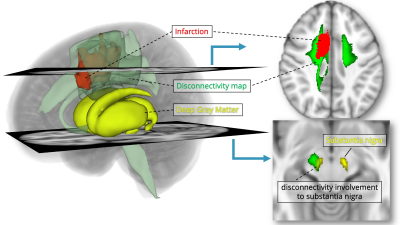 |
Predicting the Neurodegeneration after Stroke using Disconnectivity Map
Takayuki YAMAMOTO1, Hikaru FUKUTOMI1, Vincent DOUSSET1,2, Igor SIBON3, and Thomas TOURDIAS1,2
1Institut de Bio-imagerie IBIO, Université de Bordeaux, Bordeaux, France, 2Neuroimagerie diagnostique et thérapeutique, CHU de Bordeaux, Bordeaux, France, 3Unité de soins intensifs neurovasculaires, CHU de Bordeaux, Bordeaux, France
We investigated remote effects of brain infarction. We previously demonstrated higher iron content (higher R2* values) in deep nuclei as a consequence of their disconnection. Here, we aimed at predicting such long term remote degeneration from the acute stage. Through a disconnectivity approach, we mapped the fibers that were likely to be disconnected by projecting the acute stroke masks on tractograms from 180 healthy volunteers. We showed that disconnected areas based on this approach were likely to show significantly higher iron content at follow-up. This validates the prediction of deep nuclei disconnection from acute stage through a disconnectivity approach.
|
|||
4329.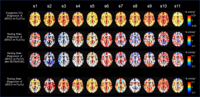 |
Revisit on the cerebrovascular responses to end-tidal CO2 fluctuations during spontaneous breathing as a surrogate of regional cerebrovascular reactivity assessment under hypercapnic challenge
Suk-tak Chan1, Karleyton C. Evans2, Tian-yue Song1, Andre van der Kouwe1, Bruce R. Rosen1, Yong-ping Zheng3, and Kenneth K. Kwong1
1Athinoula A. Martinos Center for Biomedical Imaging, Department of Radiology, Massachusetts General Hospital, Charlestown, MA, United States, 2Biogen, Inc., Cambridge, MA, United States, 3Department of Biomedical Engineering, Hong Kong Polytechnic University, Kowloon, Hong Kong
Regional cerebraovascular reactivity (CVR) to endogenous carbon dioxide (CO2) during spontaneous breathing (i.e. end-tidal partial pressure of CO2 (PETCO2) measured at rest), was substantially different from that obtained under externally applied CO2 challenge. Individual maps of CVR to endogenous CO2 showed significant inter-subject variability while CVR maps under hypercapnic challenge did not. Such inter-subject variability was not reduced by correction of respiratory effects. In addition, the CVR to end-tidal partial pressure of oxygen (PETO2) during spontaneous breathing shows less inter-subject variability. Our findings question the compatibility of using CVR during spontaneous breathing as a surrogate of CVR under hypercapnic challenge.
|
|||
4330.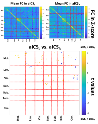 |
Distinct Effects on Cognition Caused by the Side of Asymptomatic Carotid Artery Stenosis
Jyun-Ru Chen1, Chun-Jen Lin2,3, I-Hui Lee2,3,4, and Chia-Feng Lu1
1National Yang-Ming University, Taipei, Taiwan, 2School of Medicine, National Yang-Ming University, Taipei, Taiwan, 3Neurological Institute, Taipei Veterans General Hospital, Taipei, Taiwan, 4Institute of Brain Science, National Yang-Ming University, Taipei, Taiwan
Previous studies reported that the patients with asymptomatic internal carotid stenosis (aICS) had a higher risk of stroke and neurocognitive impairment. However, whether the side of aICS could cause different neurocognitive outcomes was less explored. In this study, significant difference of functional connectivity (FC) was found between the left and right aICS groups. The association between FC and neuropsychological measures (dizziness, immediate and delayed recall verbal memory) revealed by the correlation analysis also presented different profiles between two aICS groups.
|
|||
4331.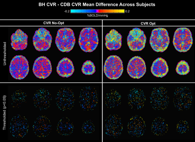 |
Agreement of CVR maps acquired using hypocapnic and hypercapnic breathing tasks
Kristina M. Zvolanek1,2, Rachael C. Stickland2, Stefano Moia3,4, Apoorva Ayyagari1,2, César Caballero-Gaudes3, and Molly G. Bright1,2
1Biomedical Engineering, Northwestern University, Evanston, IL, United States, 2Physical Therapy and Human Movement Sciences, Northwestern University, Chicago, IL, United States, 3Basque Center on Cognition, Brain and Language, Donostia, [Gipuzkoa], Spain, 4University of the Basque Country EHU/UPV, Donostia, [Gipuzkoa], Spain
The addition of a breath-hold or deep breathing task to the beginning of a resting state fMRI scan is a feasible method for cerebrovascular reactivity (CVR) mapping. However, these two tasks rely on different physiological mechanisms with different temporal properties. We used a bespoke analysis method to account for voxelwise hemodynamic lag and assessed its impact on agreement between CVR measurements from each breathing task. Good agreement was observed both across the brain and across subjects, regardless of hemodynamic lag correction, indicating both tasks are comparable options for BOLD CVR measurements in healthy subjects.
|
|||
4332. |
Effect of Myelin Content on Cognitive Outcomes in Cerebral Small Vessel Disease
Elizabeth Dao1, Roger Tam1, Ging-Yuek R Hsiung1, Lisanne ten Brinke1, Rachel Crockett1, Cindy K Barha1, Youngjin Yoo1, Walid al Keridy2, Stephanie H Doherty1, Alex L MacKay1, Cornelia Laule1, and Teresa Liu-Ambrose1
1University of British Columbia, Vancouver, BC, Canada, 2King Saud University, Riyadh, Saudi Arabia
Pathology studies report myelin damage as a salient feature in cerebral small vessel disease (cSVD). Currently, the role of myelin content in-vivo on cognition is poorly understood. Thus, the main purpose of this study was to investigate the association between myelin content and cognitive function in cSVD. Normal appearing white matter (NAWM) myelin water fraction (MWF) was quantified in 55 people with cSVD with spin-echo myelin water imaging. After accounting for age, education, and white matter hyperintensity volume, lower NAWM MWF was significantly associated with slower processing speed and poorer working memory, but not with set shifting or inhibitory control.
|
|||
4333.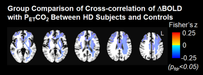 |
Impaired cerebrovascular reactivity in patients with Huntington's disease
Suk-tak Chan1, Nathaniel Mercaldo2, Kenneth K. Kwong1, Steven M. Hersch3, and Herminia D. Rosas3
1Athinoula A. Martinos Center for Biomedical Imaging, Department of Radiology, Massachusetts General Hospital, Charlestown, MA, United States, 2Department of Radiology, Massachusetts General Hospital, Boston, MA, United States, 3Department of Neurology, Massachusetts General Hospital, Boston, MA, United States
Multiple subcortical white matter areas showing impaired cerebrovascular reactivity (CVR) in our pre-symptomatic and early Huntington Disease (HD) subjects overlap with the white matter areas and tracts that have been reported to demonstrate altered microstructural integrity. They are also adjacent to cortical brain regions that atrophy as disease progresses in HD. Our findings support that abnormal cerebrovascular function contributes to the neuropathology of HD.
|
|||
4334. |
Quantification of Relative Blood Volume in Squirrel Monkey Brain in vivo using an MRI-based template
Zhangyan yang1,2, Feng Wang1,3, Chaohui Tang1, Li Min Chen1,3, and Gore C. John1,2,3
1Institute of Imaging Science, Vanderbilt University, Nashiville, TN, United States, 2Biomedical Engineering, Vanderbilt University, Nashiville, TN, United States, 3Department of Radiology and Radiological Science, Vanderbilt University, Nashiville, TN, United States
Cerebral blood volume (CBV) is a fundamental hemodynamic characteristic of brain related to blood flow and metabolism. High resolution (sub-millimeter) maps of relative values of CBV (rCBV) provide important information for studies of brain function and changes that occur in brain structure and organization. In this study, by using an intravascular superparamagnetic contrast agent in non-human primates, we quantified high-resolution rCBV maps across subjects, identified alterations of rCBV across regions, and created an rCBV atlas using a brain template.
|
|||
4335.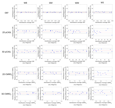 |
Test–Retest Repeatability of Brain Oxygen Metabolism Measurement using MRI
Chunwei Ying1, Michael M. Binkley2, Peter Kang2, Yasheng Chen2, Jin-Moo Lee2,3, Andria L. Ford2, and Hongyu An3
1Department of Biomedical Engineering, Washington University in St. Louis, St. Louis, MO, United States, 2Department of Neurology, Washington University School of Medicine, St. Louis, MO, United States, 3Mallinckrodt Institute of Radiology, Washington University School of Medicine, St. Louis, MO, United States
We evaluated the test-retest repeatability of MR-measurement of CBF, OEF and CMRO2 in whole brain, gray matter, white matter, and watershed regions in healthy subjects. The inter-session wCV was 2.20% in white matter and 2.76% in watershed region for OEF measured with ASE, 15.02% in white matter and 28.73% in watershed region for CBF measured with 2D multi-slice pCASL, and 9.11% in white matter and 17.14% in watershed region for CBF measured with background suppressed 3D pCASL. CMRO2 had similar test-retest repeatability to CBF.
|
|||
4336. |
Detecting Magnetic Resonance Changes in Brain Structure and Function During Stroke Rehabilitation
Jonathan Taylor1, Oun Al-iedani2,3, Saadallah Ramadan3,4, Neil Spratt1, and Sarah Valkenborghs5
1School of Biomedical Sciences and Pharmacy, University of Newcastle, Newcastle, Australia, 2School of Health Sciences, University of Newcastle, Newcastle, Australia, 3Hunter Medical Research Institute, Newcastle, Australia, 4Faculty of Health and Medicine, University of Newcastle, Newcastle, Australia, 5University of Newcastle, Newcastle, Australia
Using Magnetic Resonance (MR) data acquired as part of a feasibility study in stroke rehabilitation, a novel post-processing pipeline was designed and implemented to explore metabolic factors with MR Spectroscopy (MRS). The stroke study looked at the effect of aerobic exercise performed immediately prior to the usual task-specific rehabilitation training. Examining the clinical motor function results in relation to the metabolic data revealed by the MRS pipeline showed some interesting correlations among metabolites and rehabilitation outcomes.
|
|||
4337.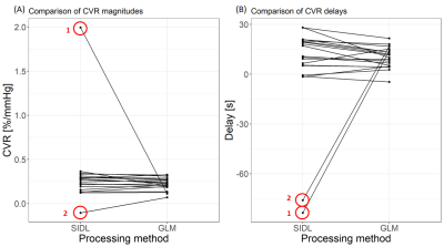 |
Processing cerebrovascular reactivity data using shift-invariant dictionary learning
Emilie Sleight1,2, Michael S Stringer1,2, Ian Marshall1,2, Joanna M Wardlaw1,2, Sotirios A Tsaftaris3, and Michael J Thrippleton1,2
1Centre for Clinical Brain Sciences, University of Edinburgh, Edinburgh, United Kingdom, 2UK Dementia Research Institute, Edinburgh, United Kingdom, 3Institute for Digital Communications, University of Edinburgh, Edinburgh, United Kingdom
The BOLD response to a hypercapnic challenge, i.e. cerebrovascular reactivity (CVR), may vary between individuals and tissue types. Linear regression (GLM) between the BOLD signal and the end-tidal CO2 is the most common CVR processing method but does not allow for different haemodynamic responses across the brain. We propose to use shift-invariant dictionary learning (SIDL) as a promising method to enable data-driven extraction of BOLD response(s). We show that CVR and delay estimates from SIDL are comparable to estimates from GLM. Future work will focus on reducing the effect of drift on SIDL estimates.
|
|||
4338.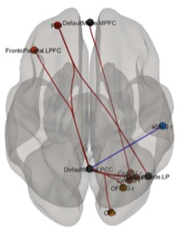 |
Cerebral circulation time related DMN connectivity in intracranial dural arteriovenous fistula before and after treatment
Bejoy Thomas1, Jithin S S1, Sabarish S Sekar1, Santhosh Kannath1, and Ramshekhar N Menon2
1Imaging Sciences and Interventional Radiology, Sree Chitra Tirunal Institute for Medical Sciences and Technology, Thiruvananthapuram, India, 2Neurology, Sree Chitra Tirunal Institute for Medical Sciences and Technology, Thiruvananthapuram, India
Cognitive decline is a common non-hemorrhagic complication of intracranial dural arteriovenous fistula (DAVF). Increase in cerebral circulation time (CCT) can be related to significant resting-state functional changes in the brain. 30 DAVF patients and a similar cohort of age-matched controls underwent resting-state functional MRI (rsfMRI) and the results were correlated with CCT and neuropsychological profile. Patients with high CCT showed decreased connectivity at DMN seed regions and less compensatory pre-frontal connectivity (pFDR < 0.05), which showed a trend in reversal after a month of embolisation therapy. These findings may be of significance in formulating treatment strategies in DAVF.
|
|||
4339.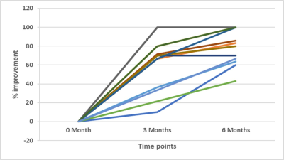 |
fMRI based evaluation of yoga-induced changes in ischemic post-stroke patients
Rama Jayasundar1, Dushyant Kumar1, Rajesh Mishra1, Priyanka Jain2, Jaideep Sachdeva3, Chahat Kumar1, Priyanka Bhagat4, and Padma Srivastava4
1Department of NMR, All India Institute of Medical Sciences, New Delhi, India, 2Centre for Development of Advanced Computing, New Delhi, India, 3Manipal University, Jaipur, India, 4Department of Neurology, All India Institute of Medical Sciences, New Delhi, India
This fMRI study has evaluated for the first time, the functional changes effected by yoga in post-stroke (ischemic) recovery. Left hemisphere stroke patients (n=13) with motor deficits practiced Hatha yoga for one hour daily for six months under the supervision of certified yoga trainers. Pre-intervention, and 3 and 6 months post-yoga intervention assessments were carried out using fMRI at 3T and also clinically evaluated using NIHSS score. Results show positive response to yoga, reflected both in the significantly reduced NIHSS scores and the increased BOLD activity in the left pre-central gyrus region in the stroke patients.
|
|||
4340.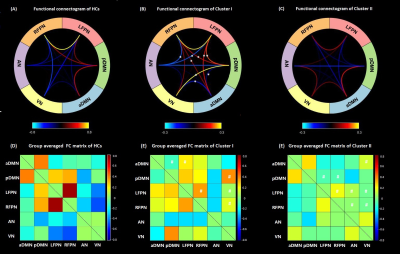 |
Clinical Phenotype Polymorphism of Ischemic Stroke Underpinned by Inter-network Functional Connectivity
Lijuan Zhang1, Siqi Cai1,2, Chunxiang Jiang1,2, Shihui Zhou1,2, and Li Yi3
1Shenzhen Institutes of Advanced Technology, Chinese Academy of Sciences, Shenzhen, China, 2University of Chinese Academy of Sciences, Beijing, China, 3Peking University Shenzhen Hospital, Shenzhen, China
The clinical profile of motor deficit after ischemic stroke may vary greatly, which are not fully attributable to the lesion topography. Investigation of the clinical phenotype polymorphism is promising to provide new insights to infer the relevance of functional remodeling to the refinement of the disease characterization and management of ischemic stroke.
|
|||
4341.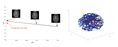 |
Monte Carlo Simulation Study of Asymmetric Spin Echo and Preliminary Estimation of Vessel Size Index
Jian Shen1 and John Wood1,2
1University of Southern California, Los Angeles, CA, United States, 2Children's Hospital Los Angeles, Los Angeles, CA, United States
Asymmetric spin echo (ASE) sequence is a technique for mapping oxygen extraction fraction (OEF) and venous cerebral blood volume (vCBV). However, there might be an overestimation of vCBV due to the diffusion effect related to vessel size. In this study, we use Monte Carlo simulation to study this effect and investigate the feasibility of estimating vessel size index from ASE data.
|
The International Society for Magnetic Resonance in Medicine is accredited by the Accreditation Council for Continuing Medical Education to provide continuing medical education for physicians.