Digital Posters
Head, Neck & Other Brain Tumor
ISMRM & SMRT Annual Meeting • 15-20 May 2021

| Concurrent 6 | 15:00 - 16:00 |
 |
3927.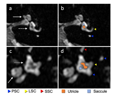 |
The visualization of the saccule and utricle with non-contrast-enhanced FLAIR sequence.
Hikaru Fukutomi1, Xavier Barreau2, Lydia Hamitouche2, Takayuki Yamamoto1, Laurent Denat1, Bei Zhang3, Lijun Zhang4, Bruno Triaire5, Valentin Prevost5, Vincent Dousset1,2, and Thomas Tourdias1,2
1Institut de Bio-imagerie IBIO, Université de Bordeaux, Bordeaux, France, 2CHU de Bordeaux, Neuroimagerie diagnostique et thérapeutique, Bordeaux, France, 3Canon Medical Systems Europe, Zoetermeer, Netherlands, 4Canon Medical Systems China, Beijing, China, 5Canon Medical Systems Corporation, Tochigi, Japan
Delayed post-gadolinium FLAIR can delineate peri- and endo-lymphatic spaces to capture endolymphatic hydrops, the pathological counterpart of Ménière’s disease. However, the four hours delay between injection and acquisition is a burden for patients and staff. No previous work has proposed a satisfying delineation of inner ear internal anatomy without injection yet. This study presents step-by-step 3D-FLAIR optimizations with specific T2 preparation and inversion time to maximize the contrast between peri- and endo-lymphatic spaces. We showed that optimized 3D-FLAIR sequence allowed delineating saccule and utricle in healthy volunteers without injection, paving the way toward future application to diagnose Meniere disease.
|
||
3928.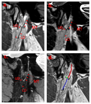 |
Negative-contrast neurography of the extracranial facial nerve and branches based on variable flip angle turbo spin echo imaging
Timothy Bray1,2, Alan Bainbridge1,3, Susan Jawad2, Sofia Otero2, Timothy J Beale2, Sumandeep Kaur2, Mark McGurk4, Margaret A Hall-Craggs1,2, and Simon Morley2
1Centre for Medical Imaging, University College London, London, United Kingdom, 2Department of Imaging, University College London Hospital, London, United Kingdom, 3Medical Physics, University College London Hospital, London, United Kingdom, 4Head and Neck Academic Centre, University College London, London, United Kingdom
Injury to the facial nerve is a major risk during parotid surgery; pre-operative identification of its course is key to minimising complications. Unfortunately, conventional MRI sequences offer insufficient resolution whilst conventional ‘positive-contrast’ neurographic methods suffer from artifacts and inconsistent quality. We propose an approach to imaging the extracranial facial nerve at high resolution using variable flip angle turbo spin echo imaging. This method depicts the nerve as a low-signal structure (‘black nerve’) against the high-signal parotid parenchyma (‘white parotid’). We show that this ‘negative-contrast’ method outperforms a widely-used positive-contrast neurographic sequence and might be further improved using gadolinium-based contrast agent.
|
|||
3929.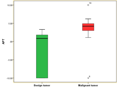 |
Differentiation of benign and malignant neck tumors by APT
Xiaohan Song1, Lijun Wang1, Ailian Liu1, Jiazheng Wang1, and Lihua Chen1
1the First Affiliated Hospital of Dalian Medical University, Dalian, China Neck malignant tumors is the seventh most common cancer in the world and the ninth most common cause of cancer death. Meanwhile, challenges remain in the differential diagnosis of benign and malignant tumors in the neck. In this study, we explored the value of APTw imaging in the differential diagnosis between malignant and benign neck tumors. Results showed significantly higher APTw values in the malignant tumors than in benign tumors (AUC=0.882, P=0.006), suggesting APT a promising non-invasive method in the differentiation of the neck malignant and benign tumors. |
|||
3930.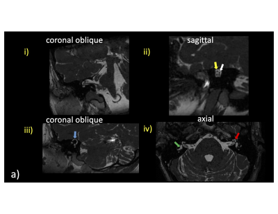 |
3D bSSFP Imaging performance using high performance gradients at 0.5T is comparable to standard field strengths: Clinical use in Temporal Bone MRI
David Volders1, James Rioux1, Steven Beyea1, Chris V Bowen2, and Elena Adela Cora1
1Diagnostic Radiology, Nova Scotia Health, Halifax, NS, Canada, 2Diagnostic Imaging, Nova Scotia Health, Halifax, NS, Canada
Evaluation of temporal bone MRI scans using a 0.5T high performance gradient head only system reveals high quality visualization of clinically relevant structures using 3D bSSFP imaging.
|
|||
3931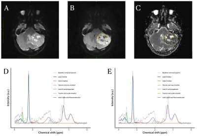 |
Childhood Brain Tumour Classification Through Proton Magnetic Resonance Spectroscopy and Diffusion Weighted Imaging Video Permission Withheld
Dadi Zhao1,2, James T. Grist1,2, Heather E.L. Rose1,2, Huijun Li2, Lesley MacPherson2, Yu Sun1,2, and Andrew C. Peet1,2
1Institute of Cancer and Genomic Sciences, University of Birmingham, Birmingham, United Kingdom, 2Department of Oncology, Birmingham Children's Hospital, Birmingham, United Kingdom
Multi-modal functional imaging is expected to improve the classification of childhood brain tumours. Forty-three patients with a confirmed childhood brain tumour were enrolled in this 1.5T multi-modal functional imaging study. Short-echo proton magnetic resonance spectroscopy (1H-MRS) and diffusion weighted imaging (DWI) were acquired and analysed through multi-class receiver operating characteristics for feature selection and a wavelet-based data-driven framework for 1H-MRS noise suppression. The balanced classification accuracy across the three tumour types was improved to 95% through linear discriminant analysis by combining DWI and noise-suppressed 1H-MRS, showing improved from 84% through only DWI and 88% through only noise-suppressed 1H-MRS.
|
|||
3932.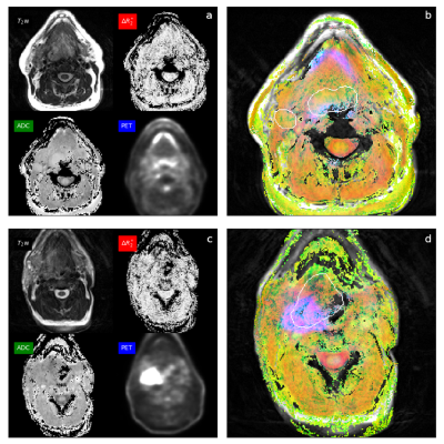 |
Multiparametric Physiologic MR Imaging of Head and Neck Cancer: Imaging Biomarker for Tumor Hypoxia and Heterogeneity
Yoshimi Anzai1, John A Roberts2, Seong-Eun Kim2, Ying Hitchcock3, Richard Wiggins1, Nousheen Alasti2, and Eugene Kholmovski2
1Department of Radiology and Imaging Sciences, University of Utah, Salt Lake City, UT, United States, 2UCAIR, Department of Radiology and Imaging Sciences, University of Utah, Salt Lake City, UT, United States, 3Radiation oncology, University of Utah, Salt Lake City, UT, United States This prospective, single-arm, cohort patients study with a newly diagnosed head&neck cancer aims to demonstrate the feasibility of multiparametric MRI to reveal tumor hypoxia and treatment failure. Although tumor hypoxia is a critical physiologic feature related to treatment resistance and poor outcomes, effective imaging technology to detect the area of hypoxia in an individual patient is lacking. Incorporating multiparametric maps into cross-sectional imaging is critically essential for translating imaging information to radiation dose paining. We generated the composite color map incorporating oxygen-enhanced BOLD, ADC, and FDG-PET and correlated it with CRT response at 3 months, as a proof of concept. |
|||
3933.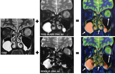 |
Clinical utility of turbo gradient and spin echo BLADE-DWI (TGSE-BLADE-DWI) for orbital tumors compared with readout-segmented echo-planar DWI
Qing Fu1,2, Xiang-chuang Kong1,2, Ding-xi Liu1,2, Yi-hao Guo3, Kun Zhou4, Zi-qiao Lei1,2, and Chuan-sheng Zheng1,2
1Department of Radiology, Union Hospital, Tongji Medical College, Huazhong University of Science and Technology, Wuhan, China, 2Hubei Province Key Laboratory of Molecular Imaging, Wuhan 430022, China, Wuhan, China, 3MR Collaboration, Siemens Healthcare Ltd., Guangzhou, China., Guangzhou, China, 4Siemens Shenzhen Magnetic Resonance Ltd., Shenzhen, China., Shenzhen, China
2D Turbo gradient and spin echo BLADE diffusion-weighted imaging (TGSE-BLADE-DWI) has been shown to improve image quality by reducing geometric distortions and susceptibility artifacts for middle ear cholesteatomas and optic neuritis; however, its use in depicting orbital tumors is still unknown. The results of this study demonstrated that TGSE-BLADE-DWI could provide significantly better image quality and comparable diagnostic performance for characterizing orbital tumors.
|
|||
3934.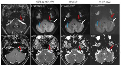 |
The efficacy 2D turbo gradient- and spin-echo diffusion-weighted imaging for cerebellopontine angle tumors
Qing Fu1,2, Xiang-chuang Kong1,2, Ding-xi Liu1,2, Yi-hao Guo3, Kun Zhou4, Zi-qiao Lei1,2, and Chuan-sheng Zheng1,2
1Department of Radiology, Union Hospital, Tongji Medical College, Huazhong University of Science and Technology, Wuhan, China, 2Hubei Province Key Laboratory of Molecular Imaging, Wuhan 430022, China, Wuhan, China, 3MR Collaboration, Siemens Healthcare Ltd., Guangzhou, China., Guangzhou, China, 4Siemens Shenzhen Magnetic Resonance Ltd., Shenzhen, China., Shenzhen, China
Geometric distortions and ghosting artifacts are found at bone-air interfaces using conventional diffusion-weighted imaging (DWI), which is a challenge for imaging cerebellopontine angle (CPA) tumors. Our study validated that geometric distortions and ghosting artifacts were not present on 2D turbo gradient- and spin-echo-BLADE-DWI scans, making this technique useful for visualizing CPA anatomic structures and diagnosing CPA tumors.
|
|||
3935.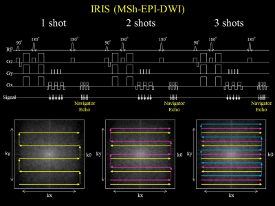 |
Improved visualization of optic nerve DWI using IRIS: comparison with conventional methods
Yutaka Hamatani1, Kayoko Abe2, Masami Yoneyama3, Jaladhar Neelavalli4, Yasuhiro Goto1, Isao Shiina1, Kazuo Kodaira1, Takumi Ogawa1, Mamoru Takeyama1, Isao Tanaka1, and Shuji Sakai2
1Department of Radioligical Services, Tokyo Women's Medical University Hospital, Tokyo, Japan, 2Department of Diagnostic imaging & Nuclear Medicine, Tokyo Women's Medical University Hospital, Tokyo, Japan, 3Philips Japan, Tokyo, Japan, 4Philips Healthcare, Bangalore, India
IRIS (Image Reconstruction using Image-space Sampling Function) is a DWI sequence for Multi shot (Mhs) echo planar imaging (EPI) with phase correction. The purpose of this study is to investigate the usefulness of IRIS for optic nerve, in which SSh EPI-DWI tends to show poor image quality due to magnetic susceptibility artifacts. As a result of comparing image quality among Single shot (SSh) EPI-DWI, SSh turbo spin echo (TSE)-DWI, MSh TSE-DWI, and IRIS, IRIS was the best sequence to visualize the optic nerve. IRIS suppressed magnetic susceptibility artifacts most clearly, and can provide excellent contrast with high robustness.
|
|||
3936.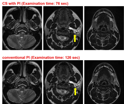 |
Compressed Sensing vs. Conventional Parallel Imaging: Utility of Head and Neck MRI for Image Quality and Inspection Efficacy
Hirotaka Ikeda1, Yoshiharu Ohno1, Kaori Yamamoto2, Kazuhiro Murayama3, Masato Ikedo2, Masao Yui2, Satomu Hanamatsu1, Akiyoshi Iwase4, Takashi Fukuba4, and Hiroshi Toyama1
1Radiology, Fujita Health University School of Medicine, Toyoake, Japan, 2Canon Medical System Corporation, Otawara, Japan, 3Joint Research Laboratory of Advanced Medical Imaging, Fujita Health University School of Medicine, Toyoake, Japan, 4Fujita Health University Hospital, Toyoake, Japan
There have been no major reports for assessing the utility of Compressed Sensing (CS) with Parallel Imaging (PI) as compared with routinely applied conventional PI for head and neck MR imaging. We hypothesized that a newly developed CS with PI could shorten examination time and improve the image quality for head and neck MR imaging as compared with conventional PI. The purpose of this study was to directly compare the capability for examination time shortening and image quality improvement of head and neck 3T MR imaging between CS with PI and conventional PI.
|
|||
3937.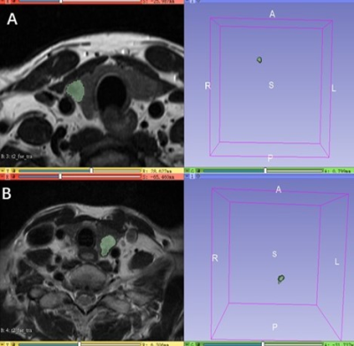 |
Can texture analysis of T2WI be used to predict extrathyroidal extension in papillary thyroid carcinoma?
Heng Zhang1, Shudong Hu1, Weiqiang Dou2, and Weiyin Vivian Liu2
1Department of Radiology, Affiliated Hospital, Jiangnan University, Wuxi, China, 2GE Healthcare, MR Research, Bejing, China
76 patients with pathologically confirmed papillary thyroid carcinoma (PTC) underwent preoperative thyroid T2-weighted (T2WI) MRI examination in this retrospective study. Using texture analysis for acquired T2WI imaging, entropy was found significantly higher in PTC patients with extrathyroidal extension (ETE) than without ETE, and thus considered an independent index for predicting PTC patients with ETE. With this finding, we therefore consider that preoperative T2WI-based texture features may be valuable for identifying ETE status in PTC patients and may help customize treatment strategies.
|
|||
3938.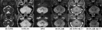 |
The clinical value of magnetic resonance imaging in patients with sudden hearing loss for pathogenic diagnosis and prognostic evaluation
Yanjun Wang1, Shenghong Ju1, Jilei Zhang2, and Yuancheng Wang1
1Radiology, Zhongda Hospital, School of Medicine, Southeast University, Nanjing, China, 2Philips Healthcare, Greater China, Shanghai, China
This study aimed to assess the clinical importance of 3D-FLAIR MR imaging in sudden sensorineural hearing loss (SSNHL) for head and neck radiologists and otolaryngologists. A novel MR protocol based on 3D-FLAIR and detailed clinical examinations were conducted among 60 patients. The results showed that the 3D-FLAIR MR protocol could identify the proper causes of SSNHL and reflect the severity of hearing loss, and the combination of clinical and MR features is beneficial for the prognostic evaluation of unilateral SSNHL, mainly for SSNHL with no recovery and complete recovery.
|
|||
3939.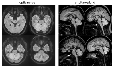 |
Development and optimization of diffusion-weighted imaging protocols for the optic nerve and pituitary gland
Zhiqiang Li1, Sharmeen Maze1, and John P Karis1
1Neuroradiology, Barrow Neurological Institute, Phoenix, AZ, United States
DWI helps reveal many neurological disorders. However, its application in the optic nerve and pituitary gland is hindered by strong distortion artifacts associated with ssEPI, the method of choice for clinical DWI. Recently, IRIS (an msEPI sequence) and SPLICE-PROPELLER (a distortion-free technique) have become available in the clinic. In this work, we develop and optimize DWI protocols for imaging the optic nerve and pituitary gland. Volunteer studies and patient scans demonstrate immunity to geometric distortions, and good overall image quality with clinically friendly scan times.
|
|||
3940.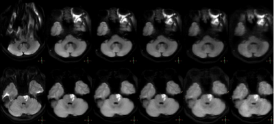 |
Application of Compressed Sensing Technology in the Fast Spin Echo Diffusion Weighted Imaging of the Skull Base
Haonan Zhang1, Qingwei Song1, Jiazheng Zhang2, Yishi Wang2, Renwang Pu1, Nan Zhang1, and Ailian Liu1
1Department of Radiology, the First Affiliated Hospital of Dalian Medical University, Dalian, China, 2PHILIPS——Philips Healthcare, beijing, China
Compared with echo planar imaging diffusion weighted iamging (EPI-DWI), turbo spin echo diffusion weighted imaging (TSE-DWI) can significantly reduce magnetic sensitivity artifacts in skull base imaging. However, the longer scan time limits its clinical promotion. The purpose of this study is to investigate the effect of the compression sensing acceleration factor on the image quality of TSE-DWI in the skull base area.
|
|||
3941.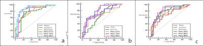 |
The Value of Apparent Diffusion Coefficient Gray Histogram in the Differential Diagnosis of Central Nervous System Lymphoma
Zhen MA1, Xin ZHAO1, Kaiyu Wang2, and Jinxia Guo3
1The Third Affiliated Hospital of Zhengzhou University, Zhengzhou City, China, 2GE Healthcare, MR Research China,, Beijing, China, 3GE Healthcare, MR Research China, Beijing, China
Retrospective analysis of patients with brain MRI examination and confirmed by pathology, respectively in three groups of MR ADC axial images, using Mazda software to delineate the region of interest at each level of the tumor, and carry out gray-scale global histogram analysis, and statistical analysis of the three groups of histogram parameter characteristics.The nine parameters, mean, variance, kurtosis, skewness, perc. 01%, perc. 10%, perc. 50%, perc. 90% and perc. 99% were obtained by gray histogram analysis (P < 0.05).
|
|||
3942.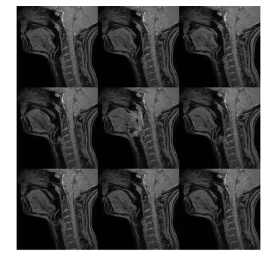 |
Semiautomated quantitative analysis of swallow function using dynamic MRI with image registration and intensity analysis
Timothy Bray1,2, Ruaridh Gollifer1,3, Simon Morley2, Susan Jawad2, Roganie Govender4, Stuart A Taylor1,2, Nicholas Hamilton4, and David Atkinson1
1Centre for Medical Imaging, University College London, London, United Kingdom, 2Department of Imaging, University College London Hospital, London, United Kingdom, 3Medical Physics, University College London Hospital, London, United Kingdom, 4Head and Neck Academic Centre, University College London, London, United Kingdom
Swallowing dysfunction (dysphagia) is a common problem and a major cause of morbidity in patients who have undergone radiotherapy for head and neck cancer. However, the pathophysiological basis of dysphagia in these patients is poorly understood and may differ between patients, creating a barrier to developing effective treatment strategies. Improved (and ideally quantitative) methods for characterising swallow function are therefore needed. We describe a method for imaging and analysis of pharyngeal and laryngeal motion during the swallow, based on joint image registration and modelling of intensity changes over a dynamic image series.
|
|||
3943.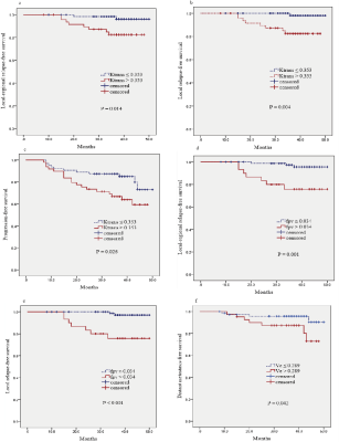 |
Prognostic value of quantitative dynamic contrast-enhanced magnetic resonance imaging and texture analysis for nasopharyngeal carcinoma
Yuhui Qin1,2, Xiaoping Yu2, Jing Hou2, and Fabao Gao1
1Department of Radiology, West China Hospital, Chengdu, China, 2Department of Radiology, Affiliated Cancer Hospital of Xiangya School of Medicine, Changsha, China
Although several studies have demonstrated that DCE-MRI is helpful in predicting the short-term therapeutic response for NPC, the relationship between the quantitative parameters and texture features (based on DCE-MRI and T2WI) and the prognosis of NPC is unclear. The present work is the first study to explore the performance of quantitative DCE-MRI and texture analysis in predicting the long-term outcome of NPC. Our data revealed that baseline quantitative DCE-MRI parameters and texture features might serve as predictors for the prognosis of NPC. These findings provide new insights to help optimize individual treatment regimen and improve the clinical outcome for patients.
|
|||
3944.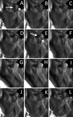 |
Real-time tyGA Imaging for diagnosis and treatment evaluation of articular disc displacements in the TMJ
Kilian Stumpf1, Mariam Seyfang2, Bernd Georg Lapatki2, and Volker Rasche1
1Department of Internal Medicine II, Ulm University Medical Center, Ulm, Germany, 2Department of Orthodontics, Ulm University Medical Center, Ulm, Germany
Magnetic resonance imaging (MRI) scans are the gold standard for confirming clinical diagnosis of temporomandibular disorders involving the condyle–articular disc complex, such as articular disc displacements (ADDs). Dynamic MRI has previously been applied for the visualization of motions of the temporomandibular joint. A combination of tiny golden angle (tyGA) profile ordering and a sliding window reconstruction allows retrospective reconstruction of images with nearly arbitrary temporal resolutions. In this contribution a real-time tyGA sequence with a frame rate of up to 125 ms was successfully applied for the diagnosis of ADD as well as occlusal splint therapy planning and evaluation.
|
|||
3945.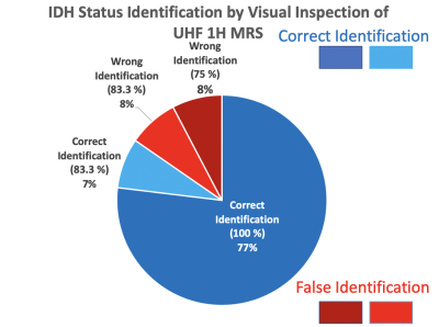 |
Diagnostic Consensus in the Interpretation of Ultra-High-Field MRS in Glioma Patients
Uzay E Emir1,2, Jannie Wijnen3, Olaf E Ansorge4, Evita Wiegers3, Anja van der Kolk5, Alexander Lin6, and Clark Chen7
1School of Health Sciences, Purdue University, West Lafayette, IN, United States, 2Weldon School of Biomedical Engineering, Purdue University, West Lafayette, IN, United States, 3Radiology, University Medical Centre Utrecht, Utrecht, Netherlands, 4University of Oxford, Oxford, United Kingdom, 5Antoni van Leeuwenhoek Hospital, Netherlands Cancer Center, Amsterdam, Netherlands, 6Brigham and Women’s Hospital / Dana Farber Cancer Institute, Harvard Medical School, Boston, MA, United States, 7Department of Neurosurgery, University of Minnesota, Minneapolis, MN, United States
We have recently initiated a 7T MRS glioma consortium, intending to bring together experts in the field to discuss pitfalls, promises, and potential research avenues of MRS in gliomas. During the "GlioMaRS-NET Workshop" in 2020, we tested the efficacy of UHF MRS for predicting the molecular characteristics by visual inspection by conducting a survey with a set of previously acquired UHF spectra from glioma patients. There was a high concordance in diagnostic interpretation of spectra for the identification of IDH-mutant gliomas versus IDH-wildtype gliomas.
|
|||
3946.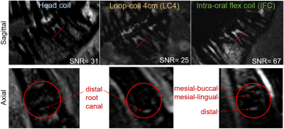 |
Intra-oral flexible coil for improved visibility of dental root canals in MRI
Agazi Samuel Tesfai1, Andreas Vollmer2, Ali Caglar Özen1,3, Wiebke Semper-Hogg2, Ute Ludwig1, and Michael Bock1
1Dept. of Radiology, Medical Physics, Medical Center – University of Freiburg, Faculty of Medicine, University of Freiburg, Freiburg, Germany, 2Department of Oral and Maxillofacial Surgery, Medical Center – University of Freiburg, Faculty of Medicine, University of Freiburg, Freiburg, Germany, 3German Consortium for Translational Cancer Research Partner Site Freiburg, German Cancer Research Center (DKFZ), Heidelberg, Germany
Accurate detection of dental root canals is vital to avoid complications in endodontic therapy; however, it is difficult to locate the root canals with their sub-millimeter diameter in dental CT. To display root canals with MRI, ex-vivo and in-vivo measurements were performed with a newly developed intra-oral flexible coil and were compared to conventional head and surface coil images. Ex-vivo, a minimum SNR gain of 6 could be achieved with the intra-oral coil setup, and in a volunteer a gain of 2.7 was seen with an improved delineation of the root canals.
|
The International Society for Magnetic Resonance in Medicine is accredited by the Accreditation Council for Continuing Medical Education to provide continuing medical education for physicians.