Digital Posters
Parkinson's Disease & Non-PD Dementia
ISMRM & SMRT Annual Meeting • 15-20 May 2021

Digital Posters
Parkinson's Disease & Non-PD Dementia
| Concurrent 6 | 15:00 - 16:00 |
3025.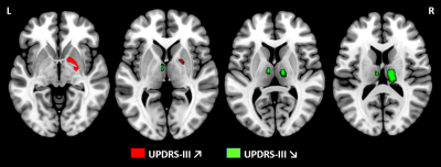 |
Subcortical grey matter changes associated with motor skills in Parkinson Disease: a longitudinal study
Céline Charroud1 and Luca Turella1
1Center for Mind/Brain Sciences - CIMeC, Mattarello, Italy This study investigated the subcortical structural modifications associated with motor symptoms of Parkinson Disease (PD) over time. Fifty subjects with PD performed two MRI sessions (baseline/48months). Motor symptoms were assessed using the Unified Parkinson’s Disease Rating Scale III. Our results showed an atrophy over time in all subcortical regions linked to the progression of PD. Furthermore, a reduced volume in thalamus and an increased volume in pallidum were associated with a decline in motor skills, consistent with the model of thalamo-cortical functional loops. Finally, we confirm that VBM and volumetry are complementary tools to assess brain degeneration in PD. |
|||
3026.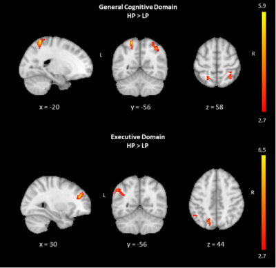 |
The perfusion deficits in general cognitive, executive, and visual dysfunction in Parkinson's disease measured by arterial spin labeling MRI
Dilek Betul Arslan1, Hakan Ibrahim Gurvit2, Ozan Genc1, Sevim Cengiz1, Ani Kicik3,4, Kardelen Eryurek4,5, Emel Erdogdu4,6, Zerrin Yildirim2, Zeynep Tufekcioglu2, Aziz Mufit Ulug1,7, Basar Bilgic2, Hasmet Hanagasi2, Tamer Demiralp4,8,
and Esin Ozturk-Isik1
1Biomedical Engineering, Biomedical Engineering Institution, Bogazici University, Istanbul, Turkey, 2Behavioral Neurology and Movement Disorders Unit, Department of Neurology, Istanbul Faculty of Medicine, Istanbul University, Istanbul, Turkey, 3Department of Physiology, Faculty of Medicine, Demiroglu Bilim University, Istanbul, Turkey, 4Neuroimaging Unit, Hulusi Behcet Life Sciences Research Center, Istanbul University, Istanbul, Turkey, 5Department of Neuroscience, Aziz Sancar Institute of Experimental Medicine, Istanbul University, Istanbul, Turkey, 6Department of Psychology, Faculty of Arts and Sciences, Isik University, Istanbul, Turkey, 7CorTechs Labs, San Diego, CA, United States, 8Department of Physiology, Istanbul Faculty of Medicine, Istanbul University, Istanbul, Turkey
The main aim of this study was to detect perfusion deficits at general executive, cognitive and visual domains of Parkinson's disease (PD) patients with mild cognitive impairment and normal cognition compared to healthy controls based on cerebral blood flow (CBF) maps obtained with arterial spin labeling MRI (ASL-MRI). CBF maps were calculated by fitting a general kinetic curve model for each pixel of ASL-MR images. A hypoperfusion pattern was observed in PD in line with general cognitive and executive dysfunction.
|
|||
3027.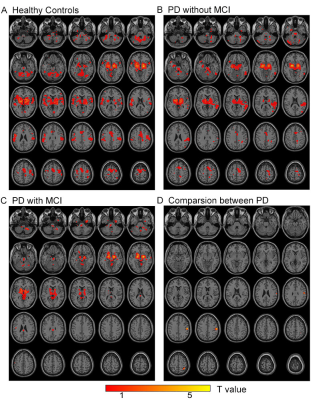 |
Disrupted Functional Connectivity of the Nucleus basalis of Meynert in Parkinson's Disease patients with Mild Cognitive Impairment
Chao Zhang1, Weiqiang Dou2, and Kai Xu1
1Department of Radiology, Xuzhou Medical University, Xuzhou, China, 2GE Healthcare,MR Research China, Beijing, China
Different functional connectivities of Basal nucleus of Meynert (BNM-FC) and the other brain regions among patients with Parkinson’s Disease (PD) accompanied and not accompanied with mild cognition impairment (PD-MCIs and PD-NCs, respectively) have been investigated in this study. A model based on Support-Vector-Machine with BNM-FC and neuropsychological results showed significantly decreased BNM-FC in the right superior parietal lobe of PD-MCIs than PD-NCs, allowing an accurate differentiation between PD-MCIs and PD-NCls. These results advanced our knowledge of the cholinergic system underpinning cognitive function in PD individuals.
|
|||
3028.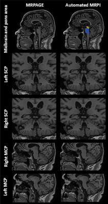 |
Automated Magnetic Resonance Parkinsonism Index: Test-Retest Reliability and 10 Year Changes in Aging and Parkinson’s Disease
Yao-Chia Shih1,2, Bénédicte Maréchal3,4,5, Ricardo Corredor Jerez3,4,5, Septian Hartono2,6, Hui-Hua Li2,7, Isabel Hui Min Chew1, Eng King Tan2,6, and Ling Ling Chan1,2
1Department of Diagnostic Radiology, Singapore General Hospital, Singapore, Singapore, 2Duke-NUS Medical School, Singapore, Singapore, 3Advanced Clinical Imaging Technology, Siemens Healthcare AG, Lausanne, Switzerland, 4Department of Radiology, Lausanne University Hospital and University of Lausanne, Lausanne, Switzerland, 5LTS5, École Polytechnique Fédérale de Lausanne, Lausanne, Switzerland, 6Department of Neurology, National Neuroscience Institute (Outram-campus), Singapore, Singapore, 7Health Services Research Unit, Singapore General Hospital, Singapore, Singapore
Progressive supranuclear palsy phenotypes are increasingly recognized and the Magnetic Resonance Parkinsonism Index (MRPI) useful in supporting clinical diagnosis. We examined the test-retest reliability of a landmark-based automated algorithm to derive MRPI from >2000 brain scans across different cohorts with neurodegeneration and MRI systems. We also investigated temporal changes in the MRPI derived from longitudinal scans 6–10 years apart in 53 subjects from case-control Parkinson’s disease cohorts. We found the automated MRPI fast (averaging 25s ± 2s per case) and reliable (average coefficient of variance <20%), without significant change in MRPI over time in aging and Parkinson’s disease.
|
|||
3029.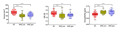 |
Magnetic resonance characteristics of iron and neuromelanin in early Parkinson’s disease: correlation with vesicular monoamine transporter 2
xueling Liu1, Pu-Yeh Wu2, and Yuxin Li1
1Department of Radiology, Huashan hospital affiliated to Fudan University, Shanghai, China, 2GE Healthcare, Beijing, China, Shang Hai, China
In this study, we investigated alterations of iron and NM in EPD, and correlations between them and VMAT2 binding in nigrostriatal system. Our results suggested that changes of neuromelanin and susceptibility on QSM images could quantitatively reflect the pathology of EPD and could be used as imaging biomarkers for diagnosing of EPD. The SUR measured by 18F-DTBZ images could reflect the lateralization in EPD patients. NM volume could reflect the striatal dopaminergic function, and both CR value and SUR could reflect the severity of motor impairment in EPD, which are expected to be used for monitoring the progression of EPD.
|
|||
3030.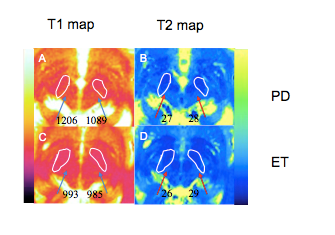 |
Value of magnetic resonance fingerprinting for differentiating tremor-dominant Parkinson’s disease from essential tremors
Yan Bai1,2, Rushi Chen1,2, Wei Wei1,2, Rui Zhang1,2, Zhun Huang2,3, Xianchang Zhang4, Mathias Nittka5, Gregor Koerzdoerfer5, and Meiyun Wang1,2
1Department of Radiology, Zhengzhou University People’s Hospital & Henan Provincial People’s Hospital, Academy of Medical Sciences., Zhengzhou, China, 2Henan Key Laboratory for Medical Imaging of Neurological Diseases, Zhengzhou, Henan, China, ZhengZhou, China, 3Department of Radiology, Henan University People’s Hospital & Henan Provincial People’s Hospital, School of Basic Medicine., Zhengzhou, China, 4MR Collaboration, Siemens Healthcare Ltd., Beijing, China, BeiJing, China, 5MR Pre-development, Siemens Healthcare GmbH, Erlangen, Germany, Erlangen, Germany
Conventional magnetic resonance imaging cannot reliably differentiate Parkinson’s disease (PD) from essential tremors (ET). Magnetic resonance fingerprinting (MRF) can simultaneously acquire T1 and T2 relaxometry. This study utilized MRF to obtain T1 and T2 values in substantia nigra (SN) of patients with tremor-dominant PD and ET. The T1 values of SN were significantly higher in patients with tremor-dominant PD than those with ET, whereas the T2 values showed no significant differences between groups. The findings suggest that MRF T1 mapping of the SN can potentially differentiate tremor-dominant PD from ET
|
|||
3031. |
STN/GP-nets: Fully automatic deep-learning based segmentation for DBS applications using ultra-high 7 Tesla MRI
Oren Solomon1, Tara Palnitkar1,2, Rémi Patriat1, Henry Braun1, Joshua Aman2, Michael C Park2,3, Guillermo Sapiro4, Jerrold Vitek2, and Noam Harel1,3
1Center for Magnetic Resonance Research, Department of Radiology, University of Minnesota, Minneapolis, MN, United States, 2Department of Neurology, University of Minnesota, Minneapolis, MN, United States, 3Department of Neurosurgery, University of Minnesota, Minneapolis, MN, United States, 4Department of Electrical and Computer Engineering, Department of Biomedical Engineering, Department of Computer Science, Department of Mathematics, Duke University, Durham, NC, United States
Deep brain stimulation (DBS) surgery has been shown to improve the quality of life for patients with various motor dysfunctions. The success of DBS is directly related to the proper placement of the electrodes, which requires accurate detection and identification of the relevant target structures. We present a deep-learning based automatic, robust and accurate segmentation technique from 7 Tesla MRI acquisitions of subcortical structures for DBS surgery planning and post-operative electrode localization. DBS targets and related structures include the subthalamic nucleus, substantia nigra, red nucleus and the internal and external compartments of the globus pallidus.
|
|||
3032.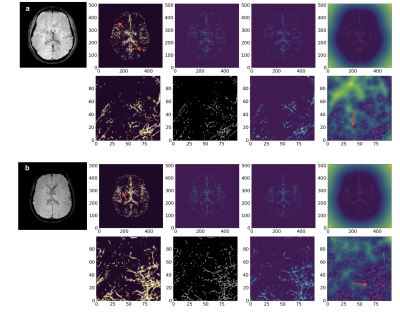 |
Evaluation and Quantification of SWI MRA of Cerebral Vasculature in Parkinson’s Disease Patients
Dmytro Pylypenko1, Yuhui Xiong1, Lanxin Ji1, Le He1, Yu Ma2, and Hua Guo1
1Center for Biomedical Imaging Research, Department of Biomedical Engineering, Tsinghua University, Beijing, China, 2Department of Neurosurgery, Tsinghua University Yuquan Hospital, Beijing, China
This study aimed to perform an evaluation of the vascular geometry in PD patients vs. healthy control group to quantify the structural changes in the cerebral vasculature by analyzing SWI MRA data. Findings show, that the PD’s vasculature network was found to have a higher tortuosity, fractality, and branching, yet lower volume and total length as well as smaller average diameter. It is also important to state that aging has a great impact on vascular impairment in PD patients.
|
|||
3033.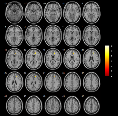 |
Cerebral hyper-perfusion associated with mild cognitive impairment in de novo Parkinson’s disease
Yong Zhang1, Chang-Peng Wang2, Jian Wang2, Li-Rong Jin2, and Bing Wu3
1GE Healthcare, Shanghai, China, 2Zhongshan Hospital, Shanghai, China, 3GE Healthcare, Beijing, China
We aimed to investigate the patterns of cerebral perfusion changes in early drug-naive PD patients with mild cognitive impairment (PD-MCI). Voxel-wise cerebral blood flow (CBF) were compared among PD-MCI patients, PD patients with normal cognition (PD-NC) and healthy controls. Increased CBF was detected in the right anterior cingulate cortex in the PD-MCI group as compared to the controls, as well as higher perfusion in the right middle frontal gyrus and superior frontal gyrus relative to PD-NC. Cerebral hyper-perfusion in frontal lobe might be associated with cognitive decline in early de novo PD.
|
|||
3034.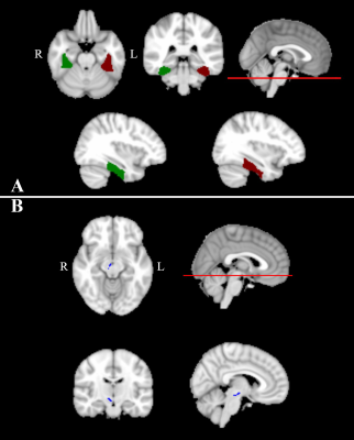 |
Simultaneous multi-parameter mapping to characterize Parkinson’s Disease using strategically acquired gradient echo imaging
Yu Shen1, Xianchang Zhang2, Yan Bai1, Yaping Wu1, Ewart Mark Haacke3, Bo Wu4, Yongsheng Chen4, Rui Zhang1, Rushi Chen1, Wei Wei1, and Meiyun Wang1
1Department of Medical Imaging, Henan Provincial People’s Hospital & Zhengzhou University, Zhengzhou, China, 2MR Collaboration, Siemens Healthcare Ltd. Beijing China, Beijing, China, 3Wayne State University, Detroit, MI, United States, 4Shanghai Zhuyan Medical Technology Company, Shanghai, China
Scanning patients with Parkinson’s disease (PD) is challenging because of tremors. Strategically acquired gradient echo (STAGE) imaging can acquire seven quantitative images relatively quickly. This method could provide comprehensive evaluations of cerebral alterations in PD patients while reducing motion artifacts. This study compared T1, T2*, R2*, and susceptibility-weighted imaging mapping (SWIM) acquired with STAGE imaging between 15 PD patients and 15 healthy controls (HC). Significant differences were found for the T1 and T2* values in several brain areas between PD and HC, suggesting that STAGE imaging is a convenient and powerful tool to investigate PD.
|
|||
3035.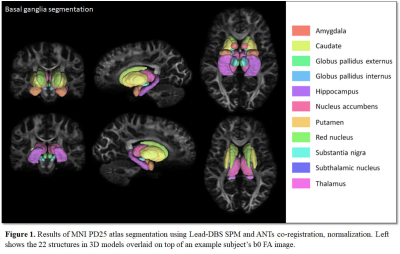 |
Assessing the differences in diffusion measures of basal ganglia and basal ganglia circuitry between controls and Parkinson’s disease patients
Jae-Hyuk Shim1 and Hyeon-Man Baek1
1Gachon University, Incheon, Korea, Republic of
The MNI PD25 subcortical atlas, which consists of key structures involved in Parkinson’s disease, was automatically segmented on each 3T control and Parkinson’s disease diffusion data obtained from the Parkinson’s disease Progression Marker Initiative (PPMI) database. Diffusion measures such as FA, AD, RD, MD and QA were obtained from each segmented ROI and interconnectivity for comparison between controls and Parkinson’s disease patients to observe for significant biomarkers that occur in basal ganglia due to Parkinson’s disease.
|
|||
3036.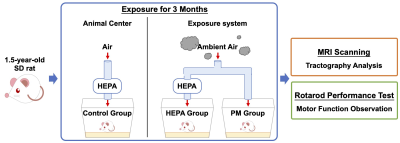 |
Exposure to Traffic-Related Particulate Matter Impact Motor Function and White Mater Integrity in Elder Rodent Model
Ting-Chieh Chen1, Yu-Chun Lo2, Yi-Chen Lin1, Ssu-Ju Li1, Ting-Chun Lin1, Ching-Wen Chang1, Yao-Wen Liang1, Hsiao-Chi Chuang3, and You-Yin Chen1,2
1Biomedical Engineering, National Yang-Ming University, Taipei, Taiwan, 2Ph.D. Program for Neural Regenerative Medicine, Taipei Medical University, Taipei, Taiwan, 3School of Respiratory Therapy, College of Medicine, Taipei Medical University, Taipei, Taiwan
Exposure to air pollution was demonstrated to be correlated with the neurodegenerative disease showing the cognitive deficits, but the impacts on motor function has yet to be investigated. The motor behavioral test and diffusion magnetic resonance imaging (MRI) tractography analysis were applied to the traffic-related particulate matter (PM)-exposed models in this study. We found the decline in motor function and the destruction of the microstructure after exposed to traffic-related PM for 3 months, in which the structural alterations of motor control circuit were similar to the characteristic of the Parkinson's disease (PD).
|
|||
3037.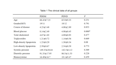 |
Excessive Brain Iron Deposition Detected by QSM in Extrapyramidal System in Parkinson's with Type 2 Diabetes Melitus Patients
Wanyao Li1, Yanwei Miao1, Bingbing Gao1, Yangyingqiu Liu1, Ailian Liu1, and Qingwei Song1
1First Affiliated Hospital of Dalian Medical University, Dalian, China
Type 2 diabetes melitus(T2DM) is associated with worse motor imbalance and cognitive decline in Parkinson’s disease.Abnormal cerebral iron deposition occur in extrapyramidal system in PD patients. However, whether the microscopic iron deposition will increase further in Parkinson’s patients with T2DM remains unknown. In this study, the iron deposition in extrapyramidal system measured QSM was compared between Parkinson's patients with T2DM (PDDM) and Parkinson's patients without T2DM (PDND).The increased magnetic sensitivity values (MSV) were found in most brain gray matter nucleus,especially for the left globus pallidus(GP) and bilateral substantia nigra(SN),in PDDM group rather than in PDND group.
|
|||
3038.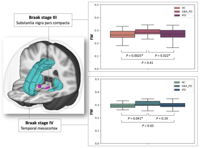 |
Brain white and gray matter alterations in early-stage Parkinson’s disease with GBA1 gene mutations evaluated using free water imaging
Christina Andica1, Koji Kamagata1, Masahiro Abe1, Wataru Uchida1,2, Yuya Saito1, Hayato Nozaki1,2, Kaito Takabayashi1, Masaaki Hori3, and Shigeki Aoki1
1Department of Radiology, Juntendo University Graduate School of Medicine, Tokyo, Japan, 2Department of Radiological Sciences, Graduate School of Human Health Sciences, Tokyo Metropolitan University, Tokyo, Japan, 3Department of Radiology, Toho University Omori Medical Center, Tokyo, Japan
Neuropathological mechanisms of Parkinson’s disease with GBA1 mutations (GBA-PD) are still unclear. We evaluated the white and gray matter of patients with early-stage GBA-PD using free water (FW) imaging. Patients with GBA-PD showed higher FW-corrected fractional anisotropy and lower FW-corrected mean diffusivity in the anterior thalamic radiation, forceps minor, and uncinate fasciculus, reflecting compensatory reorganization of neural circuits. Meanwhile, neuroinflammation (indexed by higher FW) was detected in the substantia nigra pars compacta (SNpc; Braak stage III), temporal mesocortex (Braak stage IV), forceps major, and cingulum cingulate gyrus. Furthermore, FW in the SNpc was positively correlated with the motor assessment scale.
|
|||
3039.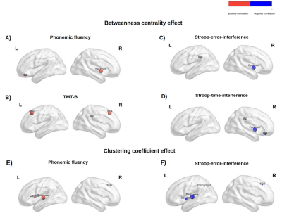 |
Evidence on the association between executive functions and tractography-derived nodal properties in Parkinson's disease
Giacomo Tomezzoli1,2, Lisa Novello2, Francesca Saviola2, Stefano Tambalo2, Beatrice Federica Luciani2, Enrica Pierotti2, Céline Charroud 2, Alessandro Gober2, Francesca Giacomoni2, Pamela Narduzzi2, Claudia Meli2, Marika Falla2, Alessandra Dodich2,
Luca Turella2, Costanza Papagno2, and Jorge Jovicich2
1DiPSCo, Department of Psychology and Cognitive Science, University of Trento, Rovereto, Italy, 2CIMeC, Center for Mind/Brain Sciences, University of Trento, Rovereto, Italy
Executive functions (EFs) play a crucial role for intact cognitive functioning, and their decline in Parkinson’s Disease (PD) patients is associated with decreased quality of life. We investigated the relationship between tractography-derived nodal properties in 8 brain regions involved in cognitive sub-components of executive control and EFs performance. We found preliminary evidence that graph-theory-derived nodal properties reveal associations with executive scores. As part of an ongoing longitudinal project, this initial evidence suggests that nodal metrics might be useful for investigating relationships between structural-connectivity properties and cognitive profiles in Parkinson’s disease.
|
|||
3040.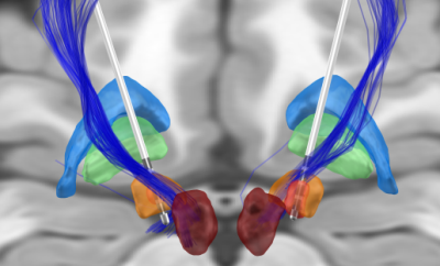 |
Patient-specific hyperdirect pathway activation in DBS for Parkinson’s Disease
Alba Segura Amil1,2 and T. A. Khoa Nguyen1,2
1Department of Neurosurgery, Inselspital, University Hospital Bern, Bern, Switzerland, 2ARTORG Center for Biomedical Engineering Research, University of Bern, Bern, Switzerland
Deep brain stimulation in the subthalamic nucleus is an effective treatment in advanced Parkinson’s disease. The aim of the current study was to determine an activation threshold of the hyperdirect pathway that results in the control of motor symptoms while avoiding the appearance of side effects. Patient-specific whole brain tractograpy, DBS leads reconstruction, and generation of volumes of tissue activated were performed in 9 subjects. The effect threshold was at 20% of hyperdirect pathway activation, while the side effect threshold was at 2% of corticospinal tract activation.
|
|||
3041.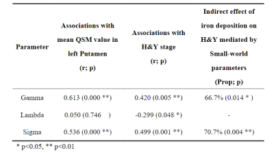 |
Brain structure network mediating the association between iron deposition and progression of Parkinson Disease
Bingxue Cheng1, Chenfei Ye2, and Ting Ma1,2,3
1Department of Information and Electronics, Harbin Institute of Technology at Shenzhen, GuangDong Province, China, 2Peng Cheng laboratory, Guangdong Province, China, 3National Clinical Research Center for Geriatric Disorders, Beijing, China
Our study integrated a high angular resolution diffusion imaging (HARDI) and Quantitative susceptibility mapping (QSM) to clarify whether the disease-specific patterns of the associated between iron deposits and the progress of PD are affected by the structural network. In brain sturctural network of PD, we observed that the increased small-worldness and decreased rich-club coefficients significantly mediate the association between the mean QSM value in left putamen and H&Y scale by using the causal mediation analysis.
|
|||
3042.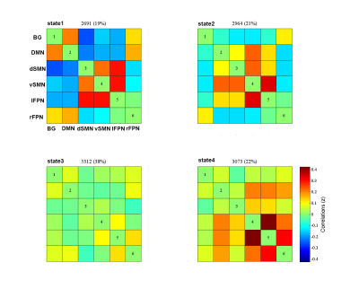 |
Abnormal intrinsic brain functional network dynamics associated with tremor in Parkinson’s disease
Junlan Zhu1, Guanxun Cheng1, Qiaoling Zeng1, Chao Lai1, Jiao Li1, and Shuwen Dong1
1Department of Medical Imaging, Peking University Shenzhen Hospital, Shenzhen, China
By applying group information guided independent component analysis to fMRI data, the dFNCs of each subject were estimated using a sliding window method and k-means clustering. Four dynamic functional states were identified. Interestingly, our study found that dwell time in State I was positively correlated with resting tremor scors in TD-PDs dFNC strength between BG and vSMN in State3 had significant positive correlation with MDS-UPDRS-III score in PD patients. Our study indicates that tremor in Parkinson’s disease is characterized by altered dFNC strength and temporal properties in dynamic connectivity, which provides a new insight into the pathological of Parkinsonian tremor.
|
|||
| 3043. | Motor Cerebro-Cerebellar Networks Breakdown Among Different Subtypes of Parkinson’s Disease
Silvia Basaia1, Federica Agosta1,2,3, Alessandro Francia1, Camilla Cividini1,3, Tanja Stojkovic4, Iva Stankovic4, Rosita Di Micco1, Luigi Albano1, Elisabetta Sarasso1, Noemi Piramide1,3, Vladana Markovic4, Elka Stefanova4, Vladimir S. Kostic4,
and Massimo Filippi1,2,3,5,6
1Neuroimaging Research Unit, Division of Neuroscience, IRCCS San Raffaele Scientific Institute, Milan, Italy, 2Neurology Unit, IRCCS San Raffaele Scientific Institute, Milan, Italy, 3Vita-Salute San Raffaele University, Milan, Italy, 4Clinic of Neurology, Faculty of Medicine, University of Belgrade, Belgrade, Italy, 5Neurorehabilitation Unit, IRCCS San Raffaele Scientific Institute, Milan, Italy, 6Neurophysiology Service, IRCCS San Raffaele Scientific Institute, Milan, Italy
We investigated the functional neural organization of the motor cerebro-cerebellar system in Parkinson’s disease (PD) patients with tremor-dominant (TD) or postural instability and gait disorder (PIGD) variant. A seed-cerebellar region (lobule VI of cerebellum) was defined based on motor task-based functional MRI in healthy controls. Functional connectivity was found to be disrupted in both PD subgroups between cerebellum, thalamus and default-mode regions. We identified different localization of functional over‐connectivity, PD-PIGD within inferior frontal gyrus and insula, while PD-TD in orbitofrontal gyrus. This study might provide novel insight into the underlying pathophysiological mechanism in PD subtypes.
|
|||
3044.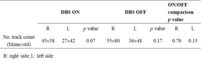 |
Diffusion Tensor Imaging and Tractography in Deep Brain Stimulation Postoperative Patients with Parkinson's Disease
Yan Li1, Yu Liu1, Naying He1, Chencheng Zhang2, Yijie Lai2, Hongyang Li2, Qing Li3, Caixia Fu4, Fuhua Yan1, and Ewart Mark Haacke1,5
1Department of Radiology, Ruijin Hospital, Shanghai Jiaotong University School of Medicine, Shanghai, China, 2Department of Neurosurgery, Center for Functional Neurosurgery, Ruijin Hospital, Shanghai Jiaotong University School of Medicine, Shanghai, China, 3MR Collaborations, Siemens Healthcare Ltd., Shanghai, China, 4MR Application Development, Siemens Shenzhen Magnetic Resonance Ltd., Shenzhen, China, 5Department of Radiology, Wayne State University, Detroit, MI, United States
Preliminary evidence supports the safety of diffusion weighted tractography in postoperative patients with deep brain stimulation. Here we further test the feasibility in both DBS-on and DBS-off status. Both DBS ON and OFF diffusion weighted imaging are safe and the tractography results are significantly comparable. Furthermore, we found that the nigrostriatal pathway's deterministic tractography is feasible. Our results highlight that DBS ON DTI is particularly fitting for those patients who cannot control their disease symptoms without stimulation but need MRI.
|
The International Society for Magnetic Resonance in Medicine is accredited by the Accreditation Council for Continuing Medical Education to provide continuing medical education for physicians.