Digital Posters
Neurofluids & Tissue Water
ISMRM & SMRT Annual Meeting • 15-20 May 2021

| Concurrent 6 | 17:00 - 18:00 |
2378.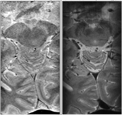 |
Water content for anatomical imaging with layer resolution at 7T
Ana-Maria Oros-Peusquens1, Jonas Kielmann1, and N. Jon Shah1,2,3,4
1INM-4, Research Centre Juelich, Juelich, Germany, 2Faculty of Medicine, JARA, RWTH Aachen University, Aachen, Germany, 3INM-11, JARA, Research Centre Juelich, Juelich, Germany, 4Department of Neurology, RWTH Aachen University, Aachen, Germany
An increasing number of structural studies attempt identification of distinct structural regions based on their layer signature. Interestingly, water content quantitative contrast is little used in these attempts, despite being a fundamental contrast in MRI, based on the proton equilibrium magnetisation, and closely reflecting cell density on which modern cytoarchitectonic studies are based. In this contribution, we demonstrate the usefulness of water content contrast at 7T and propose it for layer-related studies and high-resolution brain anatomy.
|
|||
2379.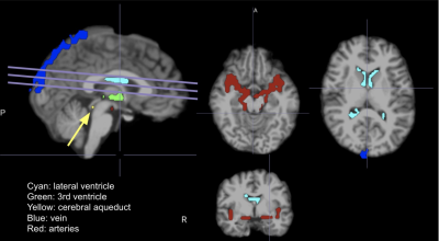 |
Vascular origins of low-frequency oscillations in cerebrospinal fluid resting-state fMRI signal: Interpretation using photoplethysmography
Ahmadreza Attarpour1, James Ward2, and J. Jean Chen1,2
1Medical Biophysics, University of Toronto, Toronto, ON, Canada, 2Rotman Research Institute, Baycrest, Toronto, ON, Canada
Due to its cardiogenic constituents, the cerebrospinal fluid (CSF) contribution to resting-state functional MRI (rs-fMRI) has been used for denoising. Recently, it has become increasingly used for characterizing CSF flow and glymphatic flow. However, the association between CSF and vascular oscillations in rs-fMRI remains to be fully understood. In this study, we quantify the relationship between rs-fMRI fluctuation in different CSF compartments and photoplethysmography (PPG) recordings, focusing on the frequency band around 0.1 Hz. We also quantify their relationship between the rs-fMRI signals (in CSF, tissue and vasculature) and other vascular measures derived from PPG traces.
|
|||
2380.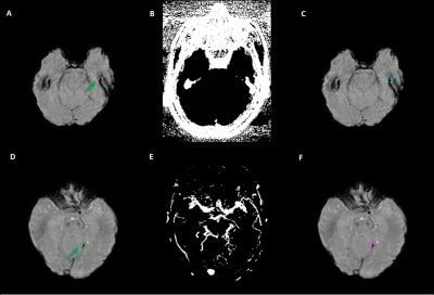 |
Improving Automatic Cerebral Microbleed Detection Using Algorithmic Methods in Multi-Echo STAGE Data
Miller Fawaz1, Sara Gharabaghi1, Mojtaba Jokar1, Ying Wang1,2, Chao Chai3, and E. Mark Haacke1,2
1Magnetic Resonance Innovations, Inc., Bingham Farms, MI, United States, 2Wayne State University, Detroit, MI, United States, 3Tianjin First Central Hospital, Tianjin, China
Automatic cerebral microbleed detection is attainable with our two step model for many disease states. We attributed previously shown lower performance in STAGE data to veins and edges, including some in the basal ganglia. We improved our existing pipeline for this detection by adding a false positive correction step to our pipeline using previously tested and new data. The results were improved overall, including on previously tested STAGE data, new STAGE data and our previously tested single echo data (multiple diseases). This makes our pipeline a viable and versatile real time automatic microbleed detection procedure.
|
|||
2381.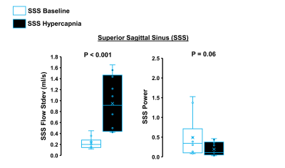 |
Real Time 4D flow MRI assessment of Low Frequency Oscillations in Large Intracranial Vessels at Rest and During Hypercapnia
Kathleen B Miller1, Leonardo A Rivera-Rivera1, Oliver Wieben1, Kevin M Johnson1, Sterling C Johnson1, and Jill N Barnes1
1University of Wisconsin-Madison, Madison, WI, United States
Low frequency oscillations (LFOs) of cerebral vessels may be important indicators of aging and disease related changes to cerebral autoregulation and glymphatic clearance. Assessing LFOs during a vasodilatory stimulus, such as hypercapnia, may reveal age-associated changes not apparent at rest. This study utilized 3D radial sampling and local low-rank reconstruction of 4D flow MRI scans to achieve real time temporal resolution to assess LFOs. Preliminary results suggest that there were no hypercapnia or age-associated changes in LFOs in the large intracranial arteries; however, there were both age and hypercapnia effects on LFOs in the venous circulation.
|
|||
2382.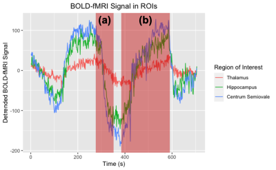 |
Breaking up Cerebrovascular Reactivity BOLD-fMRI to Investigate Dilation and Constriction Features
Kayley Marchena-Romero1,2, Xiang Ji2, Andrew Centen2, Joel Ramirez2, Andrew Lim2, Sandra E Black2, and Bradley J MacIntosh1,2
1Medical Biophysics, University of Toronto, Toronto, ON, Canada, 2Physical Sciences, Sunnybrook Research Institute, Toronto, ON, Canada
Cerebrovascular reactivity (CVR) is assessed by dynamic MRI scanning, such as BOLD-fMRI during a hypercapnia gas challenge. Conventional CVR metrics do not account for individual vasoactive challenges in series. We study the relationship between vasoconstriction after the 1st hypercapnia challenge and CVR during the 2nd challenge, using a non-parametric method, Sen’s Slope, to estimate BOLD change. We show that vasoconstriction is directly proportional to the amplitude of CVR across a range of adult ages, as assessed by linear regression analysis. These results support our hypothesis that temporal features of a cerebrovascular challenge can provide additional insight on cerebrovascular physiology.
|
|||
2383.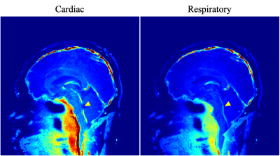 |
Investigation of Cardiac- and Respiratory-driven CSF Motions using Asynchronous Phase Contrast with Frequency Analysis
Satoshi Yatsushiro1, Tomohiko Horie2, Mitsunori Matsumae3, and Kagayaki Kuroda1
1Department of Human and Information Science, Tokai University, Hiratsuka, Japan, 2Department of Radiological Technology, Tokai University Hospital, Isehara, Japan, 3Department of Neurosurgery, Tokai University, Isehara, Japan
To investigate cardiac- and respiratory-driven CSF motions in the intracranial space, asynchronous phase contrast (Async-PC) imaging with Stockwell transform (ST) and power mapping was conducted for 3 healthy volunteers under free breathing. ST can separate the cardiac- and respiratory-driven CSF motions as spectrogram. Power mapping visualizes the power of the spectrum voxel-by-voxel. Async-PC with ST presented the time-frequency property of the cardiac- and respiratory-driven CSF motions, while that with the power mapping technique visualized the spatial distribution of both the motion contributions. Combination of those techniques may provide new findings related with CSF dynamics.
|
|||
2384.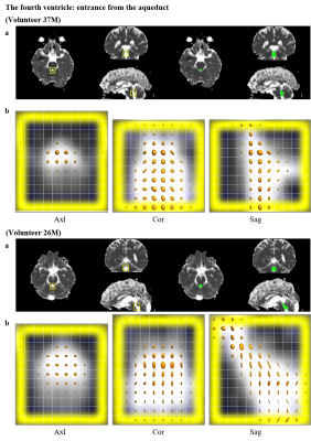 |
Low b-value DTI for Measuring Pseudo-random Flow of CSF: Region of Interest Analysis on Normal Volunteers
Yoshitaka Bito1,2, Hisaaki Ochi1,2, Kuniaki Harada2, and Kohsuke Kudo2
1Healthcare Business Unit, Hitachi, Ltd., Tokyo, Japan, 2Department of Diagnostic Imaging, Hokkaido University Graduate School of Medicine, Sapporo, Japan
Low b-value DTI (Low-b DTI) has been recently proposed for investigating the CSF physiology, and was reported to reflect the covariance of velocity distribution of the pseudo-random flow. The purpose of this study was to analyze diffusion properties of Low-b DTI (i.e. diffusion tensor, mean diffusivity, and fractional anisotropy) for normal volunteers. The diffusion properties show statistically high and anisotropic values at some ROIs such as around the foramen of Monro, the aqueduct, the prepontine cistern and the Sylvian fissure. It demonstrates that Low-b DTI can be used for evaluating the pseudo-random flow of CSF.
|
|||
2385.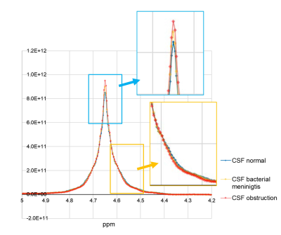 |
Feasibility of water peak MRS without water suppression for the evaluation of cerebrospinal fluid
Toshiaki Taoka1,2, Rintaro Ito1,2, Rei Nakamichi2, Takashi Abe2, Toshiki Nakane2, Hisashi Kawai2, Mayuko Sakai3, and Shinji Naganawa2
1Department of Innovative Biomedical Visualization (iBMV), Nagoya University, Nagoya, Japan, 2Radiology, Nagoya University, Nagoya, Japan, 3Canon Medical Systems Corporation, Otawara, Japan
We conducted water peak MRS without water suppression, in which the water peak itself is evaluated, to determine if the water peak is altered by the existence of solutes and to evaluate the feasibility of water peak MRS for detecting pathologic changes in CSF. The water peaks were modified by the solute concentrations of NaCl, Glu, and Alb. Differences in water peaks were observed among the CSF phantom simulating normal CSF, CSF with bacterial meningitis, and CSF with obstruction of the subarachnoid space.
|
|||
2386.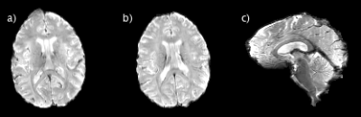 |
Characterization of Cerebrospinal Fluid Using ultra-high field MRI
Tiago Martins1, Tales Santini1, Minjie Wu1, Kristine Wilckens1, Davneet Minhas1, James W. Ibinson1, Howard J. Aizenstein1, and Tamer S. Ibrahim1
1University of Pittsburgh, Pittsburgh, PA, United States
In this work, we investigated the oscillations on the flow cerebrospinal fluid in the human brain using ultra-high field magnetic resonance imaging with a homogeneous head coil. Acquisition and analysis of images from 5 human volunteers results in identification of different frequency bands around 0.3, 0.8, 1.2, 2.3 and 3.4Hz. The analysis of the frequency spectrum and spatial localization of the signal yield results that could be correlated with physiological processes and CSF clearances.
|
|||
2387.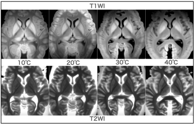 |
Temperature dependence of T1 and T2 values in formalin-fixed brains
Masatoshi Kojima1,2, Yohsuke Makino1,3, and Hirotarou Iwase1,3
1Department of legal of medicine, graduate school of medicine, Chiba university, Chiba, Japan, 2Department of radiology, Chiba medical center, Chiba, Japan, 3Department of forensic medicine, graduate school of medicine, Tokyo university, Tokyo, Japan
The purpose of this study was to investigate the variation of cerebral temperature and T1 and T2 values by varying the temperature of the formalin-fixed brain, assuming various temperatures during postmortem MRI. The T1 and T2 values were calculated by varying the temperature of the formalin-fixed brain in a thermostatic bath from 5°C to 40°C. THe T2 value prolonged with increasing temperature, and the T1 value was highest at 20°C and decreased at lower and higher temperatures. The results of this study will be useful for setting conditions for postmortem MRI imaging and image interpretation.
|
|||
2388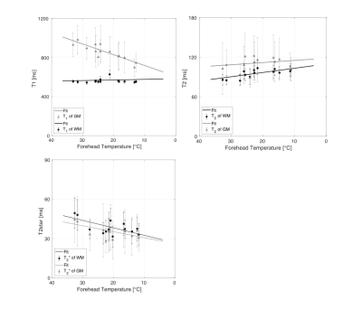 |
Real-time temperature correction of the relaxation parameters in in situ post-mortem neuro MRI Video Permission Withheld
Celine Berger1,2, Melanie Bauer1,2, Eva Scheurer1,2, and Claudia Lenz1,2
1Institute of Forensic Medicine, Department of Biomedical Engineering, Basel, Switzerland, 2Institute of Forensic Medicine, Health Department Basel-Stadt, Basel, Switzerland
The temperature sensitivity of the relaxation parameters represents a major issue in post-mortem MRI due to the passive cooling of the deceased. This study proposes a real-time non-invasive temperature correction method of MR neuroimaging parameters based on an MRI compatible forehead temperature probe. The observed significant linear relations between the forehead temperature and the relaxation times indicate that this real-time non-invasive temperature correction method is suitable for in situ post mortem MR neuroimaging.
|
|||
2389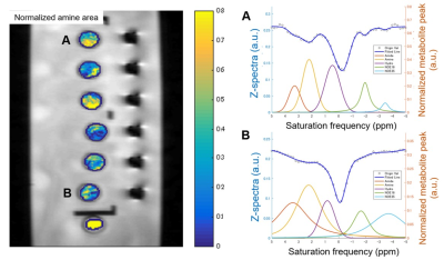 |
Biochemical Composition of the Cerebrospinal Fluid: Probing by MRI Video Permission Withheld
Khin Khin Tha1,2, Yuta Urushibata3, Hiroyuki Hamaguchi2, and Hideki Hyodoh4
1Global Center for Biomedical Science and Engineering, Hokkaido University Faculty School of Medicine, Sapporo, Japan, 2Department of Biomarker Imaging Science, Hokkaido University Graduate School of Biomedical Science and Engineering, Sapporo, Japan, 3Siemens Healthcare K.K., Tokyo, Japan, 4Department of Forensic Medicine, Hokkaido University Faculty of Medicine, Sapporo, Japan
This prospective study aimed to evaluate if CEST MRI can detect the biochemical composition of CSF. Fifty-two cadaveric CSF samples were tested for any correlation between the compounds detected by CEST MRI and the CSF biochemical analysis reports. The normalized area for intermediate exchanging amines showed a moderate positive correlation with protein concentration (r= 0.436, P= 0.001), a weak positive correlation with specific gravity (r=0.369, P=0.007), and a weak negative correlation with pH (r= -0.290, P= 0.037). The normalized area for intermediate exchanging amines may be sensitive to detect a change in CSF proteins.
|
|||
2390.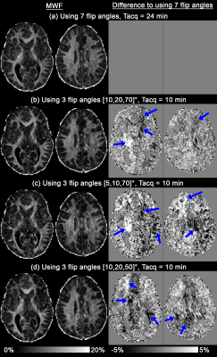 |
Optimisation of multi-compartment relaxometry myelin water imaging (MCR-MWI)
Kwok-Shing Chan1 and José P. Marques1
1Donders Institute for Brain, Cognition and Behaviour, Radboud University, Nijmegen, Netherlands
In this study, we optimise the variable flip angle acquisition protocol for GRE-based myelin water imaging using MCR model. We show numerically and via in vivo imaging that MCR-MWI can be performed with as few as 3 flip angles and still be able to achieve similar measurement performance as with 7 flip angles. We also conduct an analysis to investigate the usefulness of an image denoising method to improve MWI. Preliminary results suggest that MCR-MWI is very robust to noise and higher resolution data is needed to have a better insight.
|
|||
2391.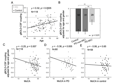 |
Decoupling of global brain activity and cerebrospinal fluid (CSF) flow was evident in Parkinson’s cognitive decline
Feng Han1, Gregory L Brown2,3, Yalin Zhu1, Aaron Belkin-Rosen1, Mechelle M Lewis3,4, Guangwei Du3, Yameng Gu1, Paul J Eslinger3,5, Richard B Mailman3,4, Xuemei Huang3,4,5,6,7,8, and Xiao Liu1,8
1Department of Biomedical Engineering, The Pennsylvania State University, State College, PA, United States, 2Department of Engineering Science and Mechanics, The Pennsylvania State University, State College, PA, United States, 3Department of Neurology, Pennsylvania State University Milton S. Hershey Medical Center, Hershey, PA, United States, 4Department of Pharmacology, Pennsylvania State University Milton S. Hershey Medical Center, Hershey, PA, United States, 5Department of Radiology, Pennsylvania State University Milton S. Hershey Medical Center, Hershey, PA, United States, 6Department of Neurosurgery, Pennsylvania State University Milton S. Hershey Medical Center, Hershey, PA, United States, 7Department of Kinesiology, Pennsylvania State University Milton S. Hershey Medical Center, Hershey, PA, United States, 8Institute for Computational and Data Sciences, The Pennsylvania State University, State College, PA, United States
Deposition and spreading of misfolded proteins have been linked to cognitive dysfunction in Parkinson’s disease (PD). The glymphatic system responsible for removing brain wastes thus may play a role in cognitive impairment in PD. This hypothesis is however difficult to test in clinical populations due to the lack of non-invasive measurements of glymphatic function. Low-frequency (< 0.1 Hz) resting-state fMRI (rsfMRI) signal was recently linked to glymphatic function primarily based on its sleep-dependent coupling with CSF flows. This study found early evidence that the coupling of global rsfMRI and CSF signals is indeed related to cognitive decline in PD.
|
|||
2392.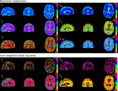 |
Optimised framework for myelin water imaging: data post-processing and Bayesian regression
Ivan Maximov1,2, Oliver Geier3, Elias Kellner4, Helle Pfeiffer3, Valerij G Kiselev4, and Marco Reisert4
1Western Norway University of Applied Sciences, Bergen, Norway, 2NORMENT, University of Oslo, Oslo, Norway, 3Oslo University Hospital, Oslo, Norway, 4University Medical Center Freiburg, Freiburg, Germany
Myelin water imaging (MWI) is a useful tool to probe and provide a quantitative measure of myelin content in the human brain in vivo. The most common MRI technique for MWI is based on multi-echo T2 measurements allowing one to estimate different T2 contributions into the signal decay. However, the conventional non-negative least squares algorithm is computationally very challenging and vulnerable to image artefacts. In the present work we developed an optimised framework for MWI enabling improved pipeline and fast metric estimations using Bayesian regression.
|
|||
2393.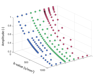 |
Evaluation of different b-value sampling strategies in cerebral IVIM: application to interstitial fluid
Gerhard Drenthen1, Jacobus Jansen1, Paulien Voorter1, Joost de Jong1, and Walter Backes1
1Maastricht University Medical Center, Maastricht, Netherlands
Recently, it was shown that besides the parenchymal diffusion and microvascular perfusion an additional, intermediate, component can be observed in the IVIM signal. The fraction of this intermediate diffusion ($$$f_{int}$$$) is suggested to be related to interstitial fluid in the perivascular spaces (PVS). In this study we examine several b-value sampling strategies for measuring the ($$$f_{int}$$$) using simulated IVIM data. When a large intermediate diffusion component is present in the IVIM signal (eg. white matter hyperintensities), b-value sampling strategies specifically aimed to quantify this component can provide better estimates of $$$f_{int}$$$ compared to linear or logarithmic spaced b-values.
|
|||
2394.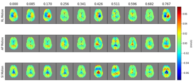 |
Retrospective Cardiac Gating of Simultaneous Coherent/Incoherent Motion Imaging (SCIMI) in the Brain
Isabelle Heukensfeldt Jansen1, Luca Marinelli1, J Kevin DeMarco2, Robert Y Shih2,3, Vincent B Ho2,3, and Thomas TK Foo1
1GE Global Research Center, Niskayuna, NY, United States, 2Walter Reed National Military Medical Center, Bethesda, MD, United States, 3Uniformed Services University of the Health Sciences, Bethesda, MD, United States
We use retrospective cardiac gating with phase-sensitive reconstruction of diffusion tensor data to create a 4D profile of motion in brain parenchyma with velocity encoding value (VENC) of 0.18 mm/s. We imaged the brain of a healthy volunteer in regions surrounding the ventricles with synchronized recording of a PPG signal. We created a velocity profile for the volume spanning the full cardiac cycle by binning individual images according to the local cardiac phase. Results show coherent periodic motion with distinct systolic/diastolic phases. This motion is thought to comprise of both tissue movement and interstitial fluid flow (ISF) in the region.
|
|||
2395.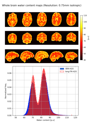 |
Isotropic water content mapping employing super-resolution reconstruction with acquisition in three orthogonal orientations
Dennis Thomas1,2, Ana-Maria Oros-Peusquens1, Dirk Poot3, and N. Jon Shah1,4,5,6
1Institute of Neuroscience and Medicine-4, Forschungszentrum Jülich, Jülich, Germany, 2RWTH Aachen University, Aachen, Germany, 3Department of Radiology and Nuclear medicine, Erasmus Medical Center, Rotterdam, Netherlands, 4Department of Neurology, RWTH Aachen University, Aachen, Germany, 5Institute of Neuroscience and Medicine 11, INM-11, JARA, Forschungszentrum Jülich, Jülich, Germany, 6JARA - BRAIN - Translational Medicine, Aachen, Germany
Tissue water content is highly regulated in the healthy brain, and even small changes are indicative of pathology. It also constitutes an important source of anatomic MRI contrast. However, this contrast remains insufficiently explored, partly due to lengthy measurement times and relatively low resolution. Super-resolution reconstruction techniques offer a trade-off between resolution, scan time and SNR.The goal of this work was to develop a technique to achieve high resolution, whole-brain water content maps by employing super-resolution reconstruction techniques. Results from the developed technique were evaluated with a carrageenan phantom and whole-brain water content maps acquired from a healthy volunteer.
|
|||
2396.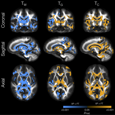 |
White and grey matter microstructural alterations and increased free-water content 13 years after very preterm birth
Claire Kelly1,2, Thijs Dhollander2, Ian Harding3,4, Wasim Khan3, Richard Beare2, Jeanie Cheong1,5,6, Lex Doyle1,5,6,7, Marc Seal2,7, Deanne Thompson1,2,7, and Peter Anderson1,8
1Victorian Infant Brain Studies (VIBeS), Murdoch Children's Research Institute, Melbourne, Australia, 2Developmental Imaging, Murdoch Children's Research Institute, Melbourne, Australia, 3Department of Neuroscience, Central Clinical School, Monash University, Melbourne, Australia, 4Monash Biomedical Imaging, Monash University, Melbourne, Australia, 5Newborn Research, The Royal Women's Hospital, Melbourne, Australia, 6Department of Obstetrics and Gynaecology, The University of Melbourne, Melbourne, Australia, 7Department of Paediatrics, The University of Melbourne, Melbourne, Australia, 8Turner Institute for Brain and Mental Health and School of Psychological Sciences, Monash University, Melbourne, Australia
We investigated the microstructural composition of the brain tissue in 130 adolescents born very preterm (VP) compared with 45 full-term (FT)-born controls. This involved a novel voxel-based analysis of white matter-like, grey matter-like, and fluid-like (free-water) diffusion tissue signal fractions derived by Single-Shell 3-Tissue Constrained Spherical Deconvolution. VP adolescents showed widespread, diverse microstructural alterations and increased free-water content across the brain parenchyma compared with FT controls, which were associated with perinatal risk factors and adverse neurodevelopmental outcomes. This study expands knowledge of the neurobiological mechanisms by which VP birth adversely affects brain development in the long-term.
|
The International Society for Magnetic Resonance in Medicine is accredited by the Accreditation Council for Continuing Medical Education to provide continuing medical education for physicians.