Digital Posters
Preclinical Models of CNS Disease
ISMRM & SMRT Annual Meeting • 15-20 May 2021

Digital Posters
Preclinical Models of CNS Disease
| Concurrent 6 | 19:00 - 20:00 |
2556.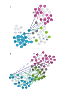 |
Longitudinal brain network changes in the GAERS rat model of absence epilepsy
Leo Hebbelmann1, Lydia Wachsmuth1, Henriette Lambers1, Cornelius Faber1, Annika Lüttjohann2, and Thomas Budde2
1Translational Research Imaging Center Clinic for Radiology, University of Münster, Münster, Germany, 2Physiology I, University of Münster, Münster, Germany
Resting-state fMRI under Isoflurane was performed to characterize brain networks in 4 and 8 month old Genetic Absence Epilepsy Rats from Strasbourg (GAERS) and in non-epileptic controls (NEC). Graph theoretical analysis identified major differences in intra-thalamic connectivity with age and compared to NEC, indicating that thalamus is strongly involved and probably modulated by frequently occurring seizures. In contrast, in NEC, brain networks did not change considerably in the age range studied.
|
|||
2557.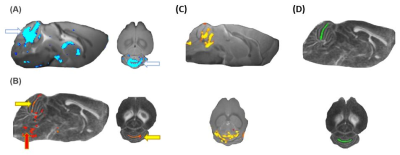 |
Anatomical and diffusion tensor MRI reveal microstructural effects of tau pathology in the inter-cerebellar fibres of the hTau.P301S mouse model
Ernest Eng1, Raimo Salo1, Heikki Tanila1, Mikko Kettunen1, and Olli Gröhn1
1A.I. Virtanen Institute for Molecular Sciences, Finland, Kuopio, Finland
The hTau.P301S-Tg mouse is an excellent model to study human tauopathy but knowledge about how tau affects the microstructure remains limited. We therefore performed anatomical and diffusion MRI to uncover structural regions likely affected by tauopathy and to characterise its progression. No initial morphometrical differences were found at 2.5 months of age. However, at 5 months, hTau.P301S mice exhibited local volumetric cerebellar changes and significantly lower FA and AD values, but higher RD values in inter-cerebellar fibres. Our data indicates that tauopathy results in structural and microstructural changes in the inter-cerebellar fibres, and possibly associates with the model’s motor declines.
|
|||
2558.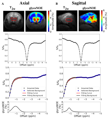 |
Preliminary study of a Lafora Disease mouse model using glycoNOE MRI
Chongxue Bie1,2,3, Yang Zhou4, Peter C. M. van Zijl1,2, Jiadi Xu1,2, Ramon C. Sun5, Matthew S. Gentry66, and Nirbhay N. Yadav1,2
1The Russell H. Morgan Department of Radiology, The Johns Hopkins University School of Medicine, Baltimore, MD, United States, 2F.M. Kirby Research Center for Functional Brain Imaging, Kennedy Krieger Institute, Baltimore, MD, United States, 3Department of Information Science and Technology, Northwest University, Xi'an, China, 4Institute of Biomedical and Health Engineering, Shenzhen Institutes of Advanced Technology, Chinese Academy of Sciences, Shenzhen, China, 5Department of Neuroscience, University of Kentucky, Lexington, KY, United States, 6Department of Molecular and Cellular Biochemistry, University of Kentucky, Lexington, KY, United States
Lafora disease (LD) is a glycogen storage disease marked by an intracellular accumulation of starch-like polyglucosan “Lafora bodies” (LBs) in brain and other tissues. There are no non-invasive approaches to quickly image brain glycogen and biopsies are difficult. We evaluate the feasibility of glycoNOE MRI to detect the accumulation of LBs in a laforin-deficient (Epm2a-/-) LD mouse model for this disease. Results suggest that the distribution of LBs in the brain can be mapped using glycoNOE MRI showing potential for studying this disease and its treatment non-invasively in vivo.
|
|||
2559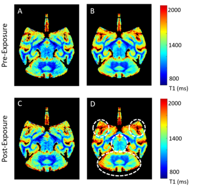 |
Quantitative Neuroimaging Study for a Non-human Primate Brain Infected with Intramuscular Ebola Virus Video Permission Withheld
Byeong-Yeul Lee1, Jeffrey M. Solomon2, Marcelo Castro1, Dong-Yun Kim3, Joseph Laux1, Becky Reeder1, Richard S. Bennett1, Dima Hammoud4,5, and Ji Hyun Lee1
1Integrated Research Facility at Fort Detrick, Integrated Research Facility at Fort Detrick, National Institute of Allergy and Infectious Diseases, National Institutes of Health, Frederick, MD, United States, 2Clinical Monitoring Research Program Directorate, Frederick National Laboratory for Cancer Research sponsored by the National Cancer Institute, Frederick, MD, United States, 3Office of Biostatistics Research, National Heart, Lung and Blood Institute, Bethesda, MD, United States, 4Radiology and Imaging Sciences, Clinical Center, National Institutes of Health, Bethesda, MD, United States, 5Center for Infectious Disease Imaging, National Institutes of Health, Bethesda, MD, United States
The goal of this study was to perform quantitative neuroimaging of rhesus monkey brains following exposure with Ebola virus (EBOV) via the intramuscular route. Using a high-resolution T1 relaxometry technique, we observed a significant increase in T1 values in the late stage (days 5-7 post inoculation). The most affected regions included the prefrontal-basal ganglia-cerebella pathway. These results are suggestive of CNS involvement in EBOV and provide new insights into the underlying pathophysiology in the brain.
|
|||
2560.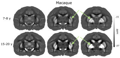 |
Brain aging in cynomolgus macaques and common marmosets explored by mapping the magnetic susceptibility and R2*
Rakshit Dadarwal1,2, Judith Mylius1, and Susann Boretius1,2,3
1Functional Imaging Laboratory, German Primate Center, Göttingen, Germany, 2Georg August Universität Göttingen, Göttingen, Germany, 3Leibniz Science Campus Primate Cognition, Göttingen, Germany
QSM and R2* both are promising MRI methods to detect subtle variations in tissue composition. The present study demonstrates the potential of QSM and R2* to characterize healthy brain aging in macaques and marmosets.
|
|||
2561.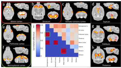 |
Brain connectivity impairments revealed by DTI and resting-state fMRI in a mouse model of Huntington’s disease
Jean-Baptiste Pérot1, Marina Célestine1, Miriam Riquelme-Pérez1, Carole Escartin1, Marc Dhenain1, Emmanuel Brouillet1, and Julien Flament1
1Université Paris-Saclay, Commissariat à l’Energie Atomique et aux Energies Alternatives (CEA), Centre National de la Recherche Scientifique (CNRS), Molecular Imaging Research Center (MIRCen), Laboratoire des Maladies Neurodégénératives, Fontenay-aux-Roses, France
Huntington’s Disease (HD) is a neurodegenerative disorder caused by the expansion of CAG repeats on the exon 1 of the HTT gene. Atrophy of the striatum is currently the main biomarker of the disease’s progression, but there is a need to find earlier and more functional biomarkers. Here, we evaluated Diffusion Tensor Imaging (DTI) and resting-state fMRI (rs-fMRI) as biomarkers in heterozygous zQ175 mice, a model mimicking the presymptomatic phase of HD. Our protocol allowed detection of vulnerable brain networks that would be of great interest for better understanding of the pathogenesis in clinical HD.
|
|||
2562.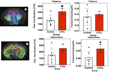 |
High Resolution DTI Tractographic Analysis in a Mouse Model of Pruritis
Talaignair N Venkatraman1, Ouyang Chen2, Allen W Song3, Ru-Rong Ji2, and Christopher D Lascola1
1Radiology, Duke University Medical Center, Durham, NC, United States, 2Neurobiology, Duke University Medical Center, Durham, NC, United States, 3BIAC, Duke University Medical Center, Durham, NC, United States
A high resolution tractographic ROI analysis was carried out on ex vivo brain in mice with cutaneous T-cell lymphoma (CTCL), a mouse model of chronic pruritis. Our results show statistically significant differences between CTCL and control mice in DTI markers of fiber tract integrity and fiber number primarily involving the thalamus and hippocampus.
|
|||
2563.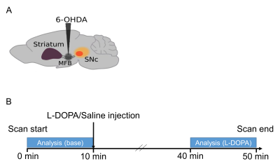 |
Alteration of basal ganglia thalamo-cortical loop in hemiparkinsonian mouse model
Tomokazu Tsurugizawa1, Yuki Nakamura2,3,4, Yukari Nakamura2,3,4, Assunta Pelosi2,3,4, Boucif Djemai5, Clement Debacker6, Jean-Antoine Girault2,3,4, and Denis Herve2,4,7
1Human Informatics and Interaction Research Institute, National Institute of Advanced Industrial Science and Technology (AIST), Tsukuba, Japan, 2Inserm UMR-S 1270, Paris, France, 3Sciences and Technology Faculty, Sorbonne Universite, Paris, France, 4Institut du Fer à Moulin, Paris, France, 5NeuroSpin/CEA-Saclay, Gif-sur-Yvette, France, 6Inserm, UMR1266, Paris, France, 7Sorbonne Universite, Paris, France
Despite a few studies, our knowledge of functional connectivity in the basal ganglia-thalamo-cortical loop remains incomplete in Parkinson’s disease mouse model. Here, we investigated alterations of functional connectivity and white matter structure in a hemi-parkinsonian mouse model. We found that the fractional anisotropy significantly decreased in the lesioned side in the subthalamic nucleus, such as medio-dorsal nucleus and centromedian nucleus of thalamus. The functional connectivity in the ipsilateral thalamic nuclei was also decreased in hemiparkinsonian mice. These results indicate that ipsilateral thalamic nuclei are key regions for functional and structural alterations in basal ganglia-thalamo-cortical loop in hemiparkinsonian mice.
|
|||
2564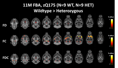 |
In vivo Fixel-Based Analysis of diffusion MRI in manifested Huntington’s disease in the zQ175 HD model Video Permission Withheld
Nicholas Vidas-Guscic1, Johan Van Audekerke1, Ben Jeurissen2, Jasmien Orije1, Tamara Vasilkovska1, Dorian Pustina3, Haiying Tang3, Roger Cachope3, Longbin Liu3, Mette Skinbjerg3, Celia Dominguez3, Ignacio Munoz-Sanjuan3, Annemie Van der Linden1,
and Marleen Verhoye1
1Bio-Imaging Lab, university of Antwerp, Antwerp, Belgium, 2Vision Lab, university of Antwerp, Antwerp, Belgium, 3CHDI foundation, Princeton, NJ, United States
Huntington’s disease (HD) is a debilitating neurodegenerative disease that affects the motor and cognitive abilities of patients. Diffusion-weighted imaging is often used in clinical evaluation of neurodegenerative disorders and recently a fixel-based analysis method was developed to analyse diffusion data. We implemented a multi-shell diffusion weighted acquisition and analysis in the zQ175 HD model. The results of the whole-brain and region-based diffusion tensor, diffusion kurtosis and fixel-based analysis indicate the presence of micro- and macrostructural differences in the zQ175 HD model in overall white matter fibers that can be attributed to significant changes in many fiber populations throughout the brain.
|
|||
2565.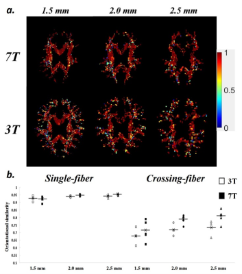 |
Reproducibility evaluation on resolving complex fiber orientations using diffusion spectrum imaging at 3T and 7T
Nan-Hao Chen1,2, Kuan-Hung Cho2, Yi-Ping Chao3,4, Sheng-Min Huang2, Norihiro Sadato5, Li-Wei Kuo2,6, and Masaki Fukunaga5
1Biomedical engineering and Environmental sciences, National Tsing Hua University, Hsinchu, Taiwan, 2Institute of Biomedical Engineering and Nanomedicine, National Health Research Institutes, Miaoli, Taiwan, 3Department of Computer Science and Information Engineering, Chang Gung University, Taoyuan, Taiwan, 4Graduate Institute of Biomedical Engineering, Chang Gung University, Taoyuan, Taiwan, 5Division of Cerebral Integration, Department of System Neuroscience, National Institute for Physiological Sciences, Okazaki, Japan, 6Institute of Medical Device and Imaging, National Taiwan University College of Medicine, Taipei, Taiwan
Diffusion spectrum imaging (DSI) is a model-free diffusion MRI technique capable of resolving complex fiber orientations. The capability of DSI at different field strengths and spatial resolutions remains unexplored. This study aimed to investigate the reproducibility of DSI quantified by orientational similarity and deviation angle at 3T and 7T. Our results show that DSI reproducibility at 3T and 7T for single-fiber group are comparable, whereas DSI at 7T could provide relatively better reproducibility than that at 3T for crossing-fiber group. The comparison of spatial resolutions for crossing-fiber group suggests that partial volume effect may be less dominant than signal-to-noise ratio.
|
|||
 |
2566.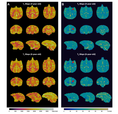 |
High-Resolution, Whole-Brain T1 and T2 Mapping of Monkey Using 3D Magnetic Resonance Fingerprinting at 9.4 T
Yuning Gu1, Lulu Wang2, Hongyi Yang2, Yun Wu2, Yong Chen3, Kai Zhong2, and Xin Yu1,3,4
1Biomedical Engineering, Case Western Reserve University, Cleveland, OH, United States, 2High Magnetic Field Laboratory, Chinese Academy of Sciences, Hefei, China, 3Radiology, Case Western Reserve University, Cleveland, OH, United States, 4Physiology and Biophysics, Case Western Reserve University, Cleveland, OH, United States
A 3D MRF method was developed for T1 and T2 mapping of the entire monkey brain with high spatial resolution (0.35x0.35x1 mm3). A conformal head coil was used for improved SNR, which enabled a 24-fold acceleration in data acquisition. B1 inhomogeneity was corrected in dictionary matching. Comparison of the T1 and T2 maps showed a ~20% reduction of T2 in the globus pallidus of 6-year-old monkeys compared to 4-year-old monkeys.
|
||
2567.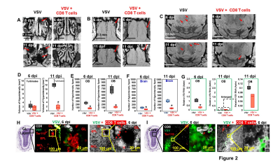 |
Early Adoptive Transfer of T Cells Decreases Brain Bleeding during Vesicular Stomatitis Virus Infection: A MRI Study
Li Liu1, Stephen Dodd1, Ryan Hunt1, Nikorn Pothayee1, Nadia Nadia Bouraoud1, Dragan Maric1, E Ashley Moseman1, Dorian B McGavern1, and Alan P Koretsky1
1National Institute of Neurological Disorders and Stroke, National Institute of Health, Bethesda, MD, United States
MRI was used to follow bleeding and T cell infiltration in mouse model of nasal infection of the brain with VSV. Microbleeds were identified as an early pathological and neuroimaging marker using high-resolution T2*-weighted MRI. Adoptive transfer of virus specific CD8 T cells helped clear VSV, decreased microbleeds but did not stop all microbleeds. Labeling T cells with MPIOs enabled MRI cell tracking and showed the earliest T cell infiltration in the brain. CD8 T cell infiltration and vessel rupture happened 1-day post-infection at glomerular layer, while T cells could be detected at the center of bulb before vessel breakdown.
|
|||
2568.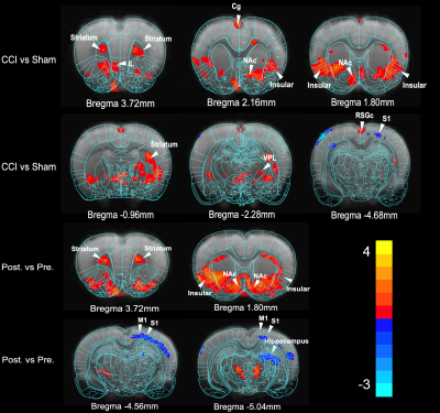 |
Regional brain MRI features reveal the histological alterations in chronic pain: an MRI-based cell imaging study
Lei Wei1, Ming Ding1, Xiao Xiao1, and He Wang1,2
1Institute of Science and Technology for Brain-Inspired Intelligence, Fudan University, shanghai, China, 2Human Phenome Institute, Fudan University, Shanghai, China, Shanghai, China
In our study, we used the ultra-high field magnetic imaging to show the brain functional and structure has changed in the rats of chronic pain model. then we also find the expression levels of the protein marker p-Erk has changed in some brain region, including insula, NAc, S1 and so on, and insula has been shown more significant activation. This results are same to the MRI functional data analysis. Finally, we hope to combine MRI and cell imaging technique could to verify the importance of insula in non-human chronic pain.
|
|||
2569.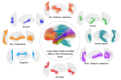 |
A fiber clustering-based atlas of the chimpanzee deep brain structural connectivity using diffusion MRI
Maëlig Chauvel1, Ivy Uszynski1, William Hopkins2, Jean-François Mangin1, and Cyril Poupon1
1Université Paris-Saclay, CEA, CNRS, BAOBAB, Neurospin, Gif-sur-Yvette, France, 2Keeling Center for Comparative Medicine and Research, The University of Texas MD Anderson Cancer Center, Bastrop, TX, United States
A way to better appreciate the ancient or evolved Homo sapiens brain characteristics relies on comparative investigations with homologous species. Chimpanzees remain our closest living hominid relatives, making it a pertinent model for brain studies. Few investigations address the structural connectivity of the chimpanzee brain, whereas its proximity with humans could be key to understanding the human brain. Thanks to the unique imaging data collection provided by the National Yerkes Primate Research Center and the access to elaborated diffusion MRI tools to investigate brain connectivity, we propose a novel long bundle atlas of the white matter of the chimpanzee brain.
|
|||
2570. |
Neurite Orientation Dispersion and Density Imaging in a Pre-clinical Model of Repetitive Mild Traumatic Brain Injury
Patrick McCunn1, Xiaoyun Xu2, Alex Li2, Arthur Brown2, and Robert Bartha2
1Patrick McCunn, SickKids Research Institute, Toronto, ON, Canada, 2Robarts Research Institute, London, ON, Canada
Identification of immediate microstructural changes following repetitive mild traumatic brain injury (mTBI) could shed light on the pathophysiology of second impact syndrome. The purpose of this study was to apply Neurite Orientation Dispersion and Density Imaging (NODDI) to a preclinical model of repetitive mTBI. In the corpus callosum, neurite density index (NDI) showed a significant increase within two hours following both the first and second injury, while orientation dispersion index (ODI) showed an increase after the first injury only. These results suggest an early microstructural response to repetitive mTBI which may follow a dose-dependent like response.
|
|||
2571.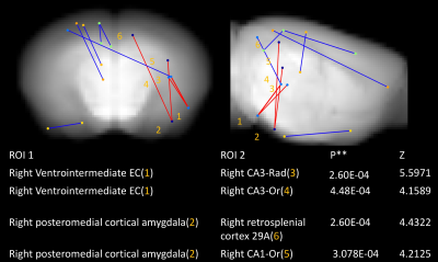 |
Increased functional connectivity but intact memory performance in tauopathy mouse at pre-tangle stage
Ling-Yun Fan1, Hsu-Lei Lee1, Robert Sullivan1, Elizabeth Coulson1,2, Juan Carlos Polanco1,2, and Kai-Hsiang Chuang1,3
1Queensland Brain Institute, University of Queensland, St Lucia, Australia, 2Clem Jones Centre for Ageing Dementia Research, University of Queensland, St Lucia, Australia, 3Centre for Advanced Imaging, University of Queensland, St Lucia, Australia
Although pre-tangle stage of tauopathy is regarded as an important pathogenic factor in neurodegenerative disease, their relationship with brain functional connectivity (FC) is still ambiguous. We investigated a tau mouse (rTg4510) model at 2.5 month-old age by resting-state functional MRI, spatial memory task and histopathology. We found increased FC between hippocampus and medial temporal area in rTg4510. Moreover, increased FC is negatively related to learning performance even though the memory is intact. On pathology, there is little phosphorylated tau over temporal areas. Our results suggest FC alternation may be an early sign of neural dysfunction at pre-tangle stage.
|
|||
2572.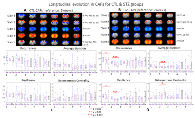 |
Spatio-temporal alterations in resting-state co-activation patterns in a rat model of sporadic Alzheimer’s disease
Yujian Diao1,2,3, Rolf Gruetter3, and Ileana O. Jelescu1,2
1CIBM Center for Biomedical Imaging, Lausanne, Switzerland, 2Animal Imaging and Technology, Ecole Polytechnique Fédérale de Lausanne, Lausanne, Switzerland, 3Laboratory of Functional and Metabolic Imaging, Ecole Polytechnique Fédérale de Lausanne, Lausanne, Switzerland
Impaired brain glucose consumption is a possible trigger of Alzheimer’s disease (AD). Previous work revealed affected brain structure and function by insulin resistance in terms of altered static functional connectivity (FC) in an intracerebroventricular-streptozotocin (icv-STZ) rat model of AD. Here, we used the co-activation patterns (CAP) method, a dynamic FC approach, to assess differences between icv-STZ rats and healthy controls. STZ rats displayed early higher predominance of states involving brain regions shown as hyperconnected by the static FC analysis. Longitudinally, specific brain states declined in the STZ rats only.
|
|||
2573.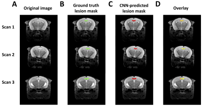 |
Automatic Segmentation of Brain Lesions in the Cuprizone Mouse Model of Multiple Sclerosis
Yuki Asada1, Luke Xie2, Skander Jemaa3, Kai H. Barck2, Tracy Yuen4, Richard A.D. Carano3, and Gregory Z. Ferl1
1Pharmacokinetics & Pharmacodynamics, Genentech, Inc., South San Francisco, CA, United States, 2Biomedical Imaging, Genentech, Inc., South San Francisco, CA, United States, 3PHC Data Science Imaging, Genentech, Inc., South San Francisco, CA, United States, 4Neuroscience, Genentech, Inc., South San Francisco, CA, United States
Here, we trained and evaluated a fully convolutional neural network for 3D images to automatically segment brain lesions in MRI images of a cuprizone mouse model of multiple sclerosis. To improve performance, several pre-processing and data augmentation methods were tested and compared. The impact of lesion size on network performance was evaluated and we applied the trained segmentation model to images from non-lesion control mice to assess the capacity of the network to detect the presence or absence of lesions. We conclude that the trained network can 1) detect the presence of a lesion and 2) accurately segment the volume.
|
|||
2574.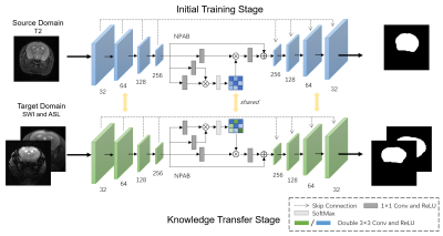 |
Ultrafast Skull Stripping of Mouse Brain Multi-Modality MR Images Using Deep Learning with Knowledge Transfer
Ziqi Yu1, Yuting Zhai1, Wenjing Xu1, Xiang Chen1, and Xiao-Yong Zhang1
1Institute of Science and Technology for Brain-Inspired Intelligence, Fudan University, Shanghai, China
Skull stripping of the mouse brain on MR images is a crucial step for rodent neuroimaging preprocessing. The traditional methods for this task are time-consuming. To solve the problem, we present a deep learning model, U-Net with Nonlocal Position-aware (NPA) block using domain knowledge transfer. The results demonstrated that our end-to-end method achieves high dice scores in several MR modalities with ultrafast processing speed which is two orders of magnitude faster than atlas-based methods. To conclude, our automatic skull stripping approach may provide an alternative to previous complex preprocessing pipelines for high-throughput rodent neuroimaging applications.
|
|||
|
2575.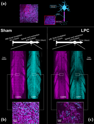 |
Inhomogeneous Magnetization Transfer (ihMT) MRI and Coherent Anti-stoke Raman Scattering (CARS) microscopy applied on LPC demyelinating model
Andreea Hertanu1,2, Cem Karakus3,4, Lucas Soustelle1,2, Victor N. D. Carvalho1,2,5, Gopal Varma6, David C. Alsop6, Bilal El Waly3,4, Olivier M. Girard1,2, Franck Debarbieux3,4, and Guillaume Duhamel1,2
1Aix Marseille Univ, CNRS, CRMBM, Marseille, France, 2APHM, Hôpital Universitaire Timone, CEMEREM, Marseille, France, 3Aix Marseille Univ, CNRS, INT, Marseille, France, 4Aix Marseille Univ, CNRS, CERIMED, Marseille, France, 5Aix Marseille Univ, CNRS, ICR, Marseille, France, 6Division of MR Research, Radiology, Beth Israel Deaconess Medical Center, Harvard Medical School, Boston, MA, United States
Intoxication of oligodendrocytes with lysophosphatidylcholine (LPC) is a useful model for the study of focal demyelination and more recently, neuronal damage. We propose an ihMT-CARS analysis on a rodent model of LPC-induced demyelination and a quantitative (T1, T2 and T1D, the dipolar relaxation time) characterization of the demyelinated area.
|
The International Society for Magnetic Resonance in Medicine is accredited by the Accreditation Council for Continuing Medical Education to provide continuing medical education for physicians.