Digital Posters
Healthy Aging vs. Dementia
ISMRM & SMRT Annual Meeting • 15-20 May 2021

| Concurrent 6 | 13:00 - 14:00 |
1904.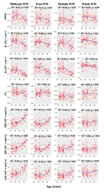 |
Maturation and degeneration of the human brainstem across the adult lifespan
Luis E. Cortina1, Richard G. Spencer1, and Mustapha Bouhrara1
1NIA, Baltimore, MD, United States
Brainstem tissue microstructural properties change across the adult lifespan. However, investigations elucidating the biological processes that govern brainstem maturation and degeneration are lacking. Here, we implement a multi-parameter approach to characterize the sex- and age-differences in a large cognitively unimpaired adult cohort. Our findings demonstrate quadratic associations between MR metrics and age as well as sexual dimorphism in certain MR metrics. Finally, our results indicate that myelination follows a temporally symmetric time course across the adult lifespan, while axons appear to degenerate significantly more rapidly than they mature.
|
|||
1905.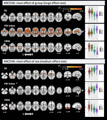 |
Sexual dimorphism in Alzheimer’s disease evaluated by free-water DTI and voxel-based morphometry
Maurizio Bergamino1, Elizabeth G Keeling1,2, Ryan R Walsh3, and Ashley M Stokes1
1Neuroimaging Research, Barrow Neurological Institute, Phoenix, AZ, United States, 2School of Life Sciences, Arizona State University, Tempe, AZ, United States, 3Muhammad Ali Parkinson Center at Barrow Neurological Institute, Phoenix, AZ, United States
The objective of this study was to investigate differences in white matter (WM) integrity and grey matter (GM) volumes between males and females in healthy aging and Alzheimer’s Disease using free-water diffusion tensor imaging (FW-DTI) and voxel-based morphometry (VBM) analyses, respectively.
|
|||
1906.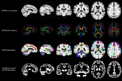 |
MIITRA atlas: Development and evaluation of high-resolution gray matter labels
Mohammad Rakeen Niaz1, Yingjuan Wu1, Abdur Raquib Ridwan1, Xiaoxiao Qi1, David A. Bennett2, and Konstantinos Arfanakis1,2
1Biomedical Engineering, Illinois Institute of Technology, Chicago, IL, United States, 2Rush Alzheimer's Disease Center, Rush University Medical Center, Chicago, IL, United States
The Multichannel Illinois Institute of Technology & Rush university Aging (MIITRA) atlas contains high resolution (0.5mm) T1-weighted (T1w) and diffusion tensor imaging (DTI) templates, constructed using high quality MRI data on a large (N=400), diverse, community cohort of non-demented older adults. The purpose of this work was twofold: a) to construct high resolution gray matter labels for the MIITRA atlas based on manually edited gray matter labels of the older adults included in the atlas, and b) to evaluate the performance of the new labels in labeling the gray matter of a separate group of older adults.
|
|||
1907.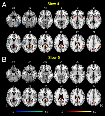 |
Frequency-dependent changes in the spontaneous neural activity are associated with cognitive impairment in patients with presbycusis
fei gao1, fuxin ren1, weibo chen2, and muwei li3
1Shandong Medical Imaging Research Institute, Shandong University, jinan, China, 2Philips Healthcare, shanghai, China, 3Vanderbilt University Institute of Imaging Science, Nashville, TN, United States
Previous studies have linked Presbycusis (PC) to cognitive impairment and incident Alzheimer’s disease. However, the neural mechanisms of cognitive impairment in patients with PC remain unclear. Although resting-state functional MRI studies have explored low-frequency oscillation (LFO) connectivity or amplitude of PC-related neural activity, it remains unclear whether the abnormalities occur within all frequency bands or within specific frequency bands. Therefore, we applied ALFF to examine changes in LFO amplitudes in PC patients at different frequency bands (slow-4 and slow-5). Then, we used brain regions showing altered ALFF as seeds to explore FC between these regions and all other brain voxels.
|
|||
1908.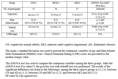 |
Myelin Water Fraction Imaging in Patients with Alzheimer’s Disease and Mild Cognitive Impairment
Geon-Ho Jahng1,2, Ji Yoon Lee3, Seung-Hyun Lim1, Hak Young Rhee4, Soonchan Park1,2, Jongho Lee5, and Chang-Woo Ryu1,2
1Radiology, Kyung Hee University Hospital at Gangdong, Seoul, Korea, Republic of, 2Medicine, Kyung Hee University, Seoul, Korea, Republic of, 3Biomedical Engineering, Kyung Hee University, Yongin-si, Korea, Republic of, 4Neurology, Kyung Hee University Hospital at Gangdong, Seoul, Korea, Republic of, 5Electrical and Computer Engineering, Seoul National University, Seoul, Korea, Republic of
The objective of this study was to investigate myelin loss in participants with AD, MCI, and cognitively normal (CN) elderly using the whole brain MWF map. The ViSTa-GRASE sequence provides the good MWF map in a reasonable scan time to evaluate myelin loss in AD patients. MWF was significantly reduced in AD compared with CN, indicating the presence of widespread demyelination in AD. In addition, MWF was significantly correlated with memory decline, indicating that MWF could serve as a potential imaging biomarker for AD for evaluating demyelination and predicting treatment outcomes.
|
|||
1909.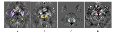 |
Cerebral Iron Deposition in Gray Nucleus Affect Cognitive Status in AD Patients: A Preliminary Quantitative Susceptibility Mapping Study
Yangyingqiu Liu1, Yanwei Miao1, Ailian Liu1, Lizhi Xie2, and Bing Wu2
1Department of Radiology, First Affiliated Hospital of Dalian Medical University, Dalian, China, 2GE healthcare, Beijing, China
Iron deposition of the gray matter nucleus is quantitatively assessed by magnetic sensitivity value (MSV)using quantitative susceptibility mapping (QSM), and its’correlation to cognitive scores is also analyzed in patients with Alzheimer's disease (AD).The results show that increased MSV value of gray matter nucleus in AD patients may affect cognitive scores.
|
|||
1910.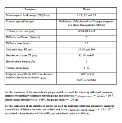 |
Microvascular MRI Signal Changes in the Presence of Amyloid-Beta Plaques or Microbleeds: A Simulation Study
Geon-Ho Jahng1,2, Chang Hyun Yoo3, Seokha Jin4, DongKyu Lee4, and HyungJoon Cho4
1Radiology, Kyung Hee University Hospital at Gangdong, Seoul, Korea, Republic of, 2Medicine, Kyung Hee University, Seoul, Korea, Republic of, 3Physics, Kyung Hee University, Seoul, Korea, Republic of, 4Biomedical Engineering, Ulsan National Institute of Science and Technology, Ulsan, Korea, Republic of
We simulated the changes in microvascular (MV) MRI signals with and without existing amyloid-beta plaques or microbleeds in an imaging voxel. The Monte Carlo simulations with the finite perturber method were used to calculate MV indices of mean vessel diameter (mVD), vessel size index (VSI), mean vessel density (Q), blood volume fraction (BVf), and microvessel-weighted imaging (MvWI). The simulation was performed with three different voxel environmental conditions: only the MV structures, MV structures with amyloid-beta plaques, and MV structures with microbleeds.
|
|||
1911.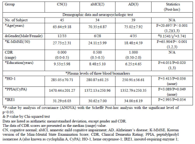 |
Association between Brain Tissue Loss and Blood Biomarkers of HO-1, PPIA, and IRE1 in Patients with Alzheimer’s Disease
Geon-Ho Jahng1,2, Ji Yoon Lee3, Hak Young Rhee4, Wonchae Choe5, and Chang-Woo Ryu1,2
1Radiology, Kyung Hee University Hospital at Gangdong, Seoul, Korea, Republic of, 2Medicine, Kyung Hee University, Seoul, Korea, Republic of, 3Biomedical Engineering, Kyung Hee University, Yongin-si, Korea, Republic of, 4Neurology, Kyung Hee University Hospital at Gangdong, Seoul, Korea, Republic of, 5Biochemistry and Molecular Biology, Kyung Hee University, Seoul, Korea, Republic of
We evaluated the relationship between imaging biomarker by GMV changes and blood biomarkers by plasma levels of HO-1, PPIA, and IRE1 in the groups of cognitively normal (CN), amnestic mild cognitive impairment (MCI), and AD participants. Our study demonstrates that subjects with AD have lower circulating levels of HO-1 than subjects with MCI and the plasma HO-1 levels were positively associated with the global GMV. Considering the potential roles of each enzyme in pathogenesis of AD, the plasma circulating levels of these enzymes possibly reflect the pathological changes of the brain in AD and would be candidates for blood-based biomarkers.
|
|||
1912.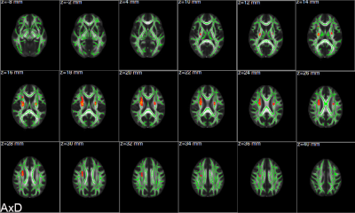 |
Alterations and associations between magnetic susceptibility of the basal ganglia and diffusion properties in Alzheimer's disease
Xiuxiu Liu1, Lei Du1,2, Wenwen Gao1, Bing Liu1,2, Yue Chen1, Yige Wang1,2, Guolin Ma1, and Weiyin Liu3
1Department of Radiology, China-Japan Friendship Hospital, Beijing, China, 2Graduate School of Peking Union Medical College, Beijing, China, 3GE Healthcare, Beijing, China Whether excessive iron deposition in basal ganglia of Alzheimer’s patients can affect the diffusion function of white matter is still unknown. There were 30 Alzheimer’s patients and 19 healthy controls in this study. We used diffusion tensor imaging and quantitative susceptibility mapping to discover the alterations of diffusion function and magnetic susceptibility of brain and associations between diffusion indexes, magnetic susceptibility, and cognitive function. This study revealed that diffusion indexes and magnetic susceptibility changed which were related to cognitive impairment. In addition, increasing magnetic susceptibility due to iron deposition may adversely affect the diffusion function of brain. |
|||
1913.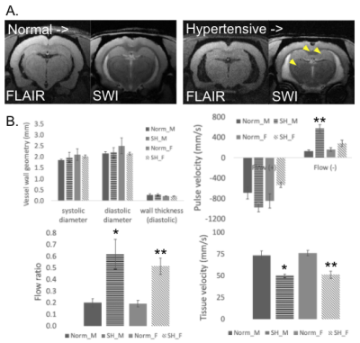 |
Relating Non-invasive Imaging Features of Vascular Aging in the Rodent Brain and Aorta
Erik Taylor1
1Radiology, University of New Mexico, Albuquerque, NM, United States
Diseases with age as the primary risk factor increase in prevalence as life expectancy increases. Aging is implicated in cardiovascular (CV) disease and neurodegeneration. As the stiffness of the proximal aorta increases, damaging pulses of pressure have been postulated to propagate into the brain. The purpose of this study was to relate vascular and brain pathology through non-invasive measures in normal and hypertensive rodents. Brain pathology was detected by multiple MRI contrast mechanisms, while specific vascular parameters were derived from ultrasound in the aorta. Non-invasive measures relating brain and vascular aging could aid in the future development of therapeutic strategies.
|
|||
1914.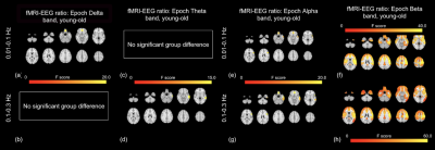 |
Associations between Resting-state EEG and fMRI signals in Brain Aging
Xiaole Zhong1 and J. Jean Chen1,2
1Rotman Research Institute, Baycrest Health Sciences, Toronto, ON, Canada, 2Department of Medical Biophysics, University of Toronto, Toronto, ON, Canada
Frequency and amplitude features for EEG and resting-state functional magnetic resonance imaging (rs-fMRI) data are individually used in human brain aging research. However, the association between the age effects on these two modalities is still unknown. In this study, we examined age effects on EEG and rs-fMRI from both frequency and amplitude perspectives. We found that while both EEG and fMRI are affected by age, there is no association between these respective age effects.
|
|||
1915.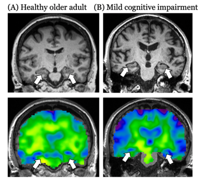 |
Hippocampal viscoelasticity is associated with risk of mild cognitive impairment
Lucy V Hiscox1, Emma M Tinney1, Peyton L Delgorio1, Matthew DJ McGarry2, Alyssa Lanzi3, James M Ellison4, Matthew L Cohen3, Chris R Martens5, and Curtis L Johnson1
1Department of Biomedical Engineering, University of Delaware, Newark, DE, United States, 2Dartmouth College, Hanover, NH, United States, 3Department of Communication Sciences & Disorders, University of Delaware, Newark, DE, United States, 4Swank Center for Memory Care and Geriatric Consultation, ChristianaCare, Wilmington, DE, United States, 5Kinesiology and Applied Physiology, University of Delaware, Newark, DE, United States
The health and integrity of the hippocampus is implicated in the development of mild cognitive impairment (MCI) and Alzheimer’s disease (AD). Neuroimaging techniques evaluate the characteristics of hippocampal health in vivo, including its size, diffusion properties, and blood flow. In this study, we show that the mechanical properties of the hippocampus, obtained through magnetic resonance elastography (MRE), are associated with an increased risk of MCI, when other techniques are not. Establishing risk based on practically relevant quantitative changes in neuroimaging variables can help identify participants likely to develop cognitive impairment and assist in establishing the effectiveness of treatment interventions.
|
|||
1916.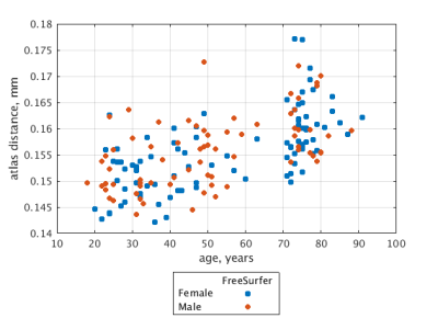 |
Brain rejuvenation improves accuracy of automatic segmentation of the aged brain
Roman Fleysher1, Mohammad Mansouri1, and Michael L Lipton1
1Radiology, Albert Einstein College of Medicine, Bronx, NY, United States
Despite best efforts, performance of automatic brain segmentation is inferior in older adults. We hypothesize that worsening of segmentation quality with age is related to brain atrophy and changes to image contrast. To disentangle the two possibilities, we simulated brain atrophy and confirmed worse segmentation performance. We then propose and demonstrate “brain rejuvenation” as a method to improve segmentation of the aged brain.
|
|||
1917.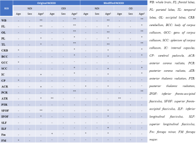 |
Neurite dispersion and density across the adult lifespan investigated using a modified NODDI approach
Maryam H. Alsameen1, Wenshu Qian1, Matthew Kiely1, Curtis Triebswetter1, Zhaoyuan Gong1, and Mustapha Bouhrara1
1National Institute on Aging, NIH, Baltimore, MD, United States The NODDI approach has been shown to overestimate the CSF and neurite density (NDI) fractions in white matter. We propose a new modification to the NODDI algorithm to address these issues. Our approach requires minimal extension of the total acquisition time. While the neurite orientation dispersion (ODI) values were consistent between the two approaches, the modified NODDI approach provides lower, more physiologically plausible, NDI values as compared to those derived using the original NODDI approach. Further, NDI and ODI exhibit, overall, quadratic associations with age. These associations were more pronounced and significant from results derived using the modified NODDI approach.
|
|||
1918.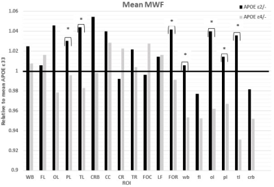 |
APOE genotype influences cerebral myelination in normative aging
Curtis G. Triebswetter1, Nikkita Khattar1, Matthew Kiely1, Maryam H. Alsameen1, Zhaoyuan Gong1, Susan M. Resnick2, and Mustapha Bouhrara1
1Laboratory of Clinical Investigation, National Institute on Aging, Baltimore, MD, United States, 2Laboratory of Behavioral Neuroscience, National Institute on Aging, Baltimore, MD, United States
Little is known about the role of APOE isoforms in cerebral myelination. Although several quantitative MRI studies have demonstrated the influence of APOE on cerebral tissue integrity, its specific implication in brain myelination remains to be established. In this work, we investigated associations between APOE isoforms ε2 or ε4 and myelination, probed using specific and nonspecific measures of myelin content, in multiple brain regions in a cohort of cognitively unimpaired participants spanning a wide age range. We show significant correlations between APOE isoforms ε2 or ε4 and myelin content in several brain structures.
|
|||
1919.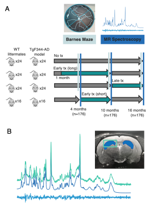 |
A neuroimaging study of the effects of early versus late anti-inflammatory treatment in the TgF344-AD rat model of Alzheimer’s disease
Caitlin F Fowler1,2, Dan Madularu3, Gabriel A Devenyi4,5, John Breitner5,6, and Jamie Near1,4,5
1Biological and Biomedical Engineering, McGill University, Montreal, QC, Canada, 2Cerebral Imaging Centre, Douglas Hospital Research Centre, Verdun, QC, Canada, 3Center for Translational Neuroimaging, Northeastern University, Boston, MA, United States, 4Cerebral Imaging Centre, Douglas Hospital Research Institute, Verdun, QC, Canada, 5Psychiatry, McGill University, Montreal, QC, Canada, 6Division of Human Neurosciences, Douglas Hospital Research Centre, Verdun, QC, Canada
Alzheimer’s disease (AD) is a progressive neurodegenerative disorder with no effective treatments or known biomarkers for definitive diagnosis, substantiating the need for early detection and intervention. This project employs Magnetic Resonance Spectroscopy to quantify neurochemical changes in the TgF344-AD rat model of AD in response to early versus late administration of a common non-steroidal anti-inflammatory drug, specifically addressing the critical question of treatment timing. Preliminary results suggest the TgF344-AD rat recapitulates most neurochemical features of human AD and that early treatment is more effective than late treatment at mitigating disease-related neurochemical changes.
|
|||
1920.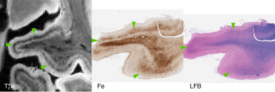 |
T2*-weighted ex vivo whole-hemisphere 7 T MRI localizes novel focal iron-rich pathology in frontotemporal lobar degeneration
M. Dylan Tisdall1, Daniel Ohm2, Rebecca Lobrovich2, Sandhitsu R Das1, Gabor Mizsei1, Karthik Prabhakaran2, Ranjit Ittyerah1, Sydney Lim1, Corey T McMillan2, James Gee1, John Q Trojanowski3, Edward B Lee3, David Wolk2, John A
Detre1,2, Paul Yushkevich1, Murray Grossman2, and David Irwin2
1Radiology, Perelman School of Medicine, University of Pennsylvania, Philadelphia, PA, United States, 2Neurology, Perelman School of Medicine, University of Pennsylvania, Philadelphia, PA, United States, 3Pathology and Laboratory Medicine, Perelman School of Medicine, University of Pennsylvania, Philadelphia, PA, United States
We describe novel, focal iron-rich pathology in frontotemporal lobar degeneration (FTLD), detected via combined ex vivo 7 T whole-hemisphere MRI and MRI-guided histopathology. While previous work has shown iron-positive plaques in AD, and focal iron-positive pathology in the motor cortex of ALS patients, we find novel focal pathologic features across a diverse range of FTLD patients, including both FTLD-tau and FTLD-TDP. These focal features occurred in regions often missed with standardized diagnostic sampling, demonstrating the sensitivity of an MRI-guided approach to histopathology. Additionally, these focal iron deposits are candidates for future in vivo detection, providing new sources of imaging contrast.
|
|||
1921.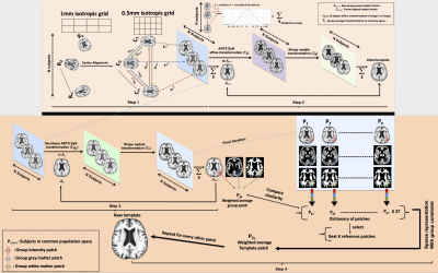 |
Construction of an unbiased high resolution and detail-preserving structural T1-weighted template for use in studies on older adults
Abdur Raquib Ridwan1, Yingjuan Wu1, Mohammad Rakeen Niaz1, David A. Bennett2, and Konstantinos Arfanakis1,2
1BIOMEDICAL ENGINEERING, ILLINOIS INSTITUTE OF TECHNOLOGY, Chicago, IL, United States, 2Rush Alzheimer’s Disease Center, RUSH UNIVERSITY MEDICAL CENTER, Chicago, IL, United States
Construction of an optimal T1-weighted (T1w) template for older adults often requires selection of a target anatomy for nonlinear-registrations, however a poorly chosen template may introduce bias due to large structural discrepancies between template and subjects used for template construction. Such a bias may introduce misregistration and cause loss of important anatomical details. In addition, large structural brain differences across older adults may introduce registration errors despite using state-of-the-art registration algorithms. The purpose of this work was to construct a minimum deformation T1w template of the older adult brain using a sparse, patch-based, detail-preserving template construction method that minimizes biases.
|
|||
1922.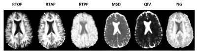 |
Assessing White Matter Microstructural Changes Associated with Aging & Dementia using Mean Apparent Propagator (MAP) MRI
Jason F. Moody1, Douglas C. Dean III1,2,3, Steven R. Kecskemeti 3, Sterling C. Johnson4,5, Barbara B. Bendlin4, and Andrew L. Alexander1,3,6
1Department of Medical Physics, University of Wisconsin Madison, Madison, WI, United States, 2Department of Pediatrics, University of Wisconsin-Madison, Madison, WI, United States, 3Waisman Center, University of Wisconsin Madison, Madison, WI, United States, 4Wisconsin Alzheimer’s Disease Research Center, University of Wisconsin Madison, Madison, WI, United States, 5Geriatric Research Education and Clinical Center, Middleton Memorial VA Hospital, University of Wisconsin-Madison, Madison, WI, United States, 6Department of Psychiatry, University of Wisconsin-Madison, Madison, WI, United States
We implement mean apparent propagator (MAP) MRI to investigate distinct white matter (WM) microstructural changes associated with aging and AD dementia. Age trajectories of MAP MRI parameters extracted from the cingulum, corpus callosum, and superior longitudinal fasciculus reveal evidence for structurally affected axons in aging populations. Return to origin probability (RTOP) and non-Gaussianity (NG) age trajectories are significantly flatter in AD dementia subjects compared to healthy controls, indicating that these measures could serve as markers for WM deterioration characteristic of dementia. Our findings provide an early quantitative framework for identifying specific WM microstructural deficiencies associated with aging and dementia.
|
|||
1923.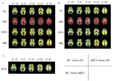 |
The DKI value in detecting microstructural white matter alterations in Alzheimer's disease and amnestic mild cognitive impairment: A TBSS study
Tongtong Li1, Yu Zhang1, Xiuwei Fu2, Xianchang Zhang3, Yuan Luo4, and Hongyan Ni5
1First Central Clinical College, Tianjin Medical University, Tianjin, China, 2Tianjin Medical University General Hospital, Tianjin, China, 3MR Collaboration, Siemens Healthcare Ltd, Beijing, China, 4Department of Radiology, China-Japan Friendship Hospital, Beijing, China, 5Department of Radiology, Tianjin First Central Hospital, Tianjin, China
Previous studies that used diffusion kurtosis imaging to study Alzheimer's disease (AD) mostly analyzed data based on regions of interest method, which may be biased by subjectivity. This study used Tract-based Spatial Statistics (TBSS) method to analyze the DKI data and found that kurtosis fractional anisotropy (KFA) values outperformed other DKI-derived parameters in sensitive detection of microstructural white matter damage for early diagnosis of amnestic mild cognitive impairment (aMCI) and AD.
|
The International Society for Magnetic Resonance in Medicine is accredited by the Accreditation Council for Continuing Medical Education to provide continuing medical education for physicians.