Digital Posters
New Advances in Diffusion for the Brain
ISMRM & SMRT Annual Meeting • 15-20 May 2021

Digital Posters
New Advances in Diffusion for the Brain
| Concurrent 5 | 19:00 - 20:00 |
1710.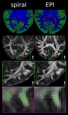 |
Spiral diffusion imaging at 800 µm resolution using a scanner with 300 mT/m gradients and gradient field monitoring
Luke Joel Edwards1, Kerrin J. Pine1, Shubhajit Paul1, Fakhereh Movahedian Attar1, Michael Herbst2, Mirsad Mahmutović3, Boris Keil3, Harald Möller4, Evgeniya Kirilina1,5, and Nikolaus Weiskopf1,6
1Department of Neurophysics, Max Planck Institute for Human Cognitive and Brain Sciences, Leipzig, Germany, 2Gengenbach, Germany, 3Institute of Medical Physics and Radiation Protection, TH Mittelhessen University of Applied Sciences, Giessen, Germany, 4NMR Group, Max Planck Institute for Human Cognitive and Brain Sciences, Leipzig, Germany, 5Center for Cognitive Neuroscience Berlin, Freie Universität Berlin, Berlin, Germany, 6Felix Bloch Institute for Solid State Physics, Leipzig University, Leipzig, Germany
Spiral diffusion weighted imaging (DWI) with field monitoring and iterative reconstruction offers potentially reduced echo time (TE) and higher effective resolution (less blurring) compared to EPI. Coupled with a scanner with ultra-strong gradients, it enabled an 800 µm DWI protocol for imaging fine structures of the brain in vivo. Compared to EPI, the shorter TEs provided distinctly different contrast in iron-rich areas (U-fibres and sub-cortical nuclei), which could enhance investigations of these regions. The protocol did, however, come with a reduction in SNR/(unit time) compared to EPI due to differences in readout time.
|
|||
1711.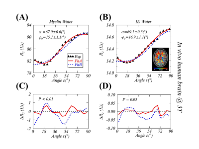 |
Anisotropic transverse relaxation in the human brain white matter induced by restricted rotational diffusion
Yuxi Pang1
1Dept. of Radiology, University of Michigan, Ann Arbor, MI, United States
Orientation-dependent transverse ($$$R_2$$$ and $$$R_2^*$$$) relaxation phenomena have been documented in the human brain white matter, yet the underlying relaxation mechanisms still remain not well understood. This work is to propose an alternative relaxation pathway through restricted molecular rotational diffusion, in terms of a generalized magic angle effect (gMAE) model, to better characterize recently reported anisotropic $$$R_2$$$ of myelin water and intra- and extracellular water in vivo at 3T. The proposed gMAE model is intrinsically connected with anisotropic translational diffusion from DTI, elucidating not only previously reported $$$R_2^*$$$ anisotropy ex vivo but also its temperature-dependence at 7T.
|
|||
1712.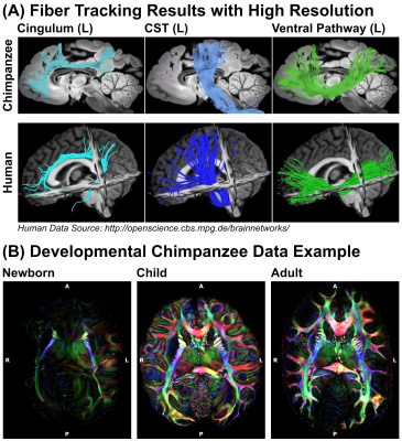 |
High-Resolution Post-Mortem Diffusion MRI Acquisitions for Connectivity Analyses in Chimpanzees
Cornelius Eichner1, Michael Paquette1, Guillermo Gallardo1, Christian Bock2, Jenny E. Jaffe3,4, Carsten Jäger1, Evgeniya Kirilina1,5, Ilona Lipp1, Toralf Mildner1, Torsten Schlumm1, Felizitas C Wermter2, Harald E. Möller1, Nikolaus Weiskopf1,
Catherine Crockford4,6, Roman Wittig4,6, Angela D Friederici1, and Alfred Anwander1
1Max Planck Institute for Human Cognitive and Brain Sciences, Leipzig, Germany, 2Alfred Wegener Institute Helmholtz Centre for Polar and Marine Research, Bremerhaven, Germany, 3Project Group Epidemiology of Highly Pathogenic Microorganisms, Robert Koch Institute, Berlin, Germany, 4Tai Chimpanzee Project, Centre Suisse de Recherches Scientifiques en Cote d'IVoire, Abidjan, Cote D'ivoire, 5Center for Cognitive Neuroscience Berlin, Freie Universität Berlin, Berlin, Germany, 6Max Planck Institute for Evolutionary Anthropology, Leipzig, Germany
Detailed neuroanatomical comparisons between humans and chimpanzees could greatly benefit evolutionary neuroscience. However, ethical considerations regarding primate research disallow acquisitions of chimpanzee MRI data in vivo for multiple years. Hence, the availability of diffusion MRI (dMRI) and tractography data from chimpanzees is limited to a few previously acquired datasets. Here we optimize diffusion acquisitions for an interdisciplinary approach to great ape neuroimaging, using post-mortem dMRI data from naturally deceased wild and captive animals. The optimization of data quality from two acquisition strategies allowed to us acquire chimpanzee diffusion MRI data of unpreceded quality and reopen a gateway for evolutionary neuroscience.
|
|||
1713.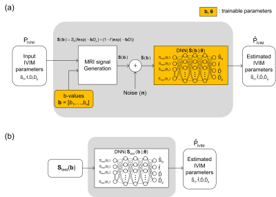 |
IVIM quantification and b-value optimization using deep neural network
Wonil Lee1, Byungjai Kim1, Jongyeon Lee1, and HyunWook Park1
1KAIST, Daejeon, Korea, Republic of
Many studies have been performed to show that IVIM could be used as a biomarker for various diseases(1-5). Since IVIM is formulated by a biexponential model, it is difficult to quantify the IVIM parameters. Researchers have tried to solve the inverse problems of the biexponential model using two approaches:improving fitting method and selecting optimized b-values(6-8). The trained DNN and the optimized b-values by the proposed method quantified IVIM parameters more accurately than combination of the conventional b-value optimization schemes with DNN fitting method. The optimized b-values by the proposed method showed superior performance even when combined with other fitting methods.
|
|||
 |
1714.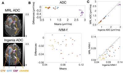 |
Technical performance of ADC and IVIM measurements in glioma and normal brain on a 1.5T MR-Linac
Liam S. P. Lawrence1, Rachel W. Chan2, Hanbo Chen3, Brian Keller3, James Stewart3, Mark Ruschin3, Brige Chugh3,4, Mikki Campbell3, Aimee Theriault3, Greg J. Stanisz1,2,5, Scott MacKenzie3, Sten Myrehaug3, Jay Detsky3, Pejman
J. Maralani6, Chia-Lin Tseng3, Greg J. Czarnota1,2,3, Arjun Sahgal3, and Angus Z. Lau1,2
1Medical Biophysics, University of Toronto, Toronto, ON, Canada, 2Physical Sciences, Sunnybrook Research Institute, Toronto, ON, Canada, 3Department of Radiation Oncology, Sunnybrook Health Sciences Centre, Toronto, ON, Canada, 4Department of Physics, Ryerson University, Toronto, ON, Canada, 5Department of Neurosurgery and Paediatric Neurosurgery, Medical University of Lublin, Lublin, Poland, 6Department of Medical Imaging, University of Toronto, Sunnybrook Health Sciences Centre, Toronto, ON, Canada
Technical performance evaluation of diffusion parameters on MR-Linacs (MRLs) is important for cancer applications. We evaluated the accuracy and repeatability of 1.5T MRL measurements of apparent diffusion coefficient (ADC) and intravoxel incoherent motion blood volume fraction (IVIM-f) in the brain via comparison with a diagnostic-quality scanner, in patients undergoing treatment. ADC measurements agree in normal and tumour tissue, but are biased in cerebrospinal fluid. IVIM-f measurements are likely negatively biased. Repeatability is comparable between scanners. The majority of high-grade glioma patients demonstrated significant ADC changes, but not IVIM-f changes.
|
||
1715.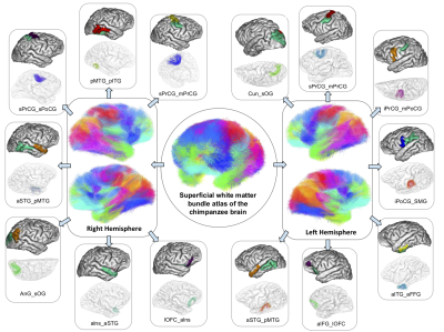 |
A new superficial white matter connectivity atlas of the chimpanzee brain
Maëlig Chauvel1, Ivy Uszynski1, William Hopkins2, Jean-François Mangin1, and Cyril Poupon1
1Université Paris-Saclay, CEA, CNRS, BAOBAB, Neurospin, Gif-sur-Yvette, France, 2Keeling Center for Comparative Medicine and Research, The University of Texas MD Anderson Cancer Center, Bastrop, TX, United States
Mapping the chimpanzee brain connectome is a key to the comprehension of the singularity of the human brain evolution. Contrary to the macaque species, few studies have been performed in vivo on chimpanzees due to its proximity with humans. While different atlases of the structural connectivity exist for humans, we established the first superficial atlas of the white matter connectivity in chimpanzees, using diffusion MRI-based tractography and advanced fiber clustering techniques.
|
|||
1716. |
Internal gradient distribution tensors of white matter tracts models
Jesus E. Fajardo1 and Gonzalo A. Álvarez1,2,3
1Centro Atómico Bariloche, CONICET, CNEA, 8400, San Carlos de Bariloche, Argentina, 2Instituto Balseiro, CNEA, Universidad Nacional de Cuyo, 8400, San Carlos de Bariloche, Argentina, 3Instituto de Nanociencia y Nanotecnología, CONICET, CNEA, San Carlos de Bariloche, Argentina
Detecting non-invasively tissue-microstructural changes associated with pathologies constitutes a promising diagnosis paradigm. Susceptibility-induced magnetic field gradients constitute a way to obtain microstructural information which is expected to complement Diffusion Weighted Imaging (DWI) methods, since these methods arise from distinct physical phenomena. We performed simulations that show the potential of extracting quantitative tissue-microstructure information of axons in the white matter based on internal-gradient-distribution tensors. We show promising results that can allow detecting alterations in axon diameters, axon density and demyelination using internal gradient distributions as a diagnostic tool.
|
|||
1717.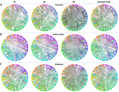 |
Resolution and b Value Dependent Structural Connectome for Ex Vivo Mouse Brain
Stephanie Allan Crater1 and Nian Wang2
1Duke University, Durham, NC, United States, 2Radiology and Imaging Sciences, Indiana University, Indianapolis, IN, United States
Tractography and structural connectome have been widely used in preclinical studies in recent years. However, assessment of the experimental parameters for the final outputs of diffusion MRI is still lack. In this study, we acquired an ex vivo mouse brain high spatial (50 µm isotropic) and angular resolution (384 diffusion encoding directions) diffusion MRI dataset in a preclinical 9.4T system. We analyzed the tractography and connectome under different experimental conditions, including b value, spatial resolution, and angular resolution.
|
|||
1718.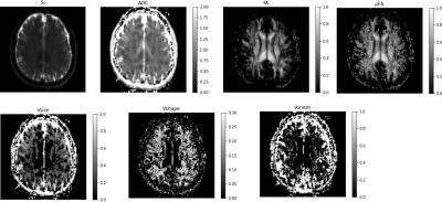 |
In Vivo Diffusion Tensor Distribution MRI of the Human Brain Using 300 mT/m Gradients
Kulam Najmudeen Magdoom1, Alexandru V. Avram1, Dario Gasbarra2, Qiuyun Fan3, Thomas Witzel3, Susie Y Huang3, and Peter J Basser1
1National Institute of Health, Bethesda, MD, United States, 2University of Helsinki, Helsinki, Finland, 3Massachusetts General Hospital and Harvard Medical School, Charlestown, MA, United States
Measuring and mapping the diffusion tensor distribution (DTD) via MRI holds the promise of revealing the tissue microstructure at sub-voxel resolution. In this study, we show the DTD results obtained on a human brain in-vivo using the new framework at MGH Connectome scanner with 300 mT/m gradients. The DTD within a voxel is assumed to be a normal tensor variate distribution constrained (CNTVD) within the manifold of 3 x 3 symmetric positive definite matrices. The estimated DTD is used to obtain a family of metrics which aim to disentangle size, shape and orientation heterogeneities present within a voxel.
|
|||
1719.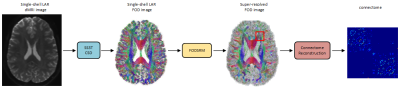 |
A Deep Learning Method for Connectome Reconstruction Using Clinical MRI Protocols
Rui Zeng1, Jinglei Lv2, He Wang3, Luping Zhou2, Michael Barnett2, Fernando Calamante2, and Chenyu Wang2
1School of Biomedical Engineering, The University of Sydney, Sydney, Australia, 2The University of Sydney, Sydney, Australia, 3Fudan University, Shanghai, China
In this study, a deep learning model called FODSRM was developed for fiber orientation distribution (FOD) super-resolution, which enhances single-shell low-angular-resolution FOD computed from clinic-quality dMRI data (e.g., 32 directions b=1000) to obtain the super-resolved high-angular resolution quality that would have been produced from advanced research scanners (e.g., multi-shell HARDI data). The results demonstrate that the super-resolved FOD data generated by the proposed method can generate high-definition structural connectome from clinical acquisition protocols, even when applied to data from a protocol not included in the trained dataset.
|
|||
1720.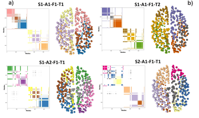 |
Estimation of individual brain signature and node-wise sensibility by a community-based DW-MRI connectome analysis
Juan Luis Villarreal Haro1,2, Gabriel Girard1,3,4, Jean Philippe Thiran1,4, and Alonso Ramírez-Manzanares2
1Signal Processing Lab (LTS5), École Polytechnique Fédérale de Lausanne, Lausanne, Switzerland, 2Computer Science Department, CIMAT, Centro de Investigación en Matemáticas, Guanajuato, Mexico, 3CIBM Center for BioMedical Imaging, Lausanne, Switzerland, 4Radiology Department, Centre Hospitalier Universitaire Vaudois and University of Lausanne, Lausanne, Switzerland
In this work, we study the effect of the reconstruction pipeline s on the reproducibility and sensitivity of the DW-MRI brain connectivity graphs. The brain database we analyze contains several scan repetitions that allow us to characterize the impact of different reconstruction pipeline parameters on the connectomes' topology. We use a novel methodology to detect robust graph communities and show the level of reproducibility of the brain structure/biomarkers. Moreover, we proposed a tool to identify the less-robust graph nodes that suffer the effects of the reconstruction pipelines' variability.
|
|||
1721.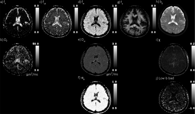 |
Multimodal Apparent Diffusion (MAD) Magnetic Resonance Imaging with comprehensive quantification of diffusion in the brain
Frederick C. Damen1, Alessandro Scotti1, Frederick W. Damen2, Nitu Saran1, Tibor Valyi-Nagy3, Mirko Vukelich1, and Kejia Cai1
1Radiology, University of Illinois at Chicago, Chicago, IL, United States, 2Biomedical Engineering, Purdue University, West Lafayette, IN, United States, 3Pathology, University Of Illinois at Chicago, Chicago, IL, United States
In physiological and pathological conditions in which multiple underlying tissue properties are expected to vary the diffusion weighted signal, the diagnostic value of conventional apparent diffusion coefficient (ADC) notably decreases. The proposed method extracts four separate distributions, i.e., modes, of diffusion weighted signal, which includes flow and unimpeded, hindered, and restricted diffusion, thereby increasing the precision of quantification of the hindered diffusion in grey and white matters.
|
|||
1722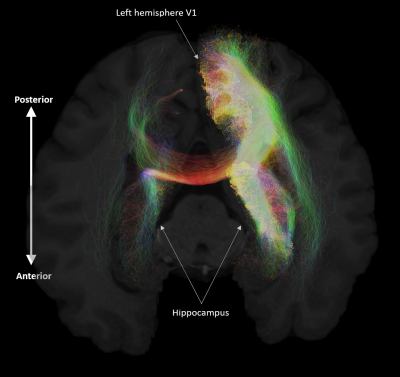 |
Anatomical connectivity of the anterior-posterior axis of the human hippocampus: new insights using quantitative fibre-tracking Video Permission Withheld
Marshall Axel Dalton1, Arkiev D'Souza2, Jinglei Lv1, and Fernando Calamante3
1School of Biomedical Engineering, Faculty of Engineering, University of Sydney, Sydney, Australia, Sydney, Australia, 2DVC Research, Brain and Mind Centre, University of Sydney, Sydney, Australia, Sydney, Australia, 3Sydney Imaging, University of Sydney, Sydney, Australia, Sydney, Australia
The hippocampus is a brain structure central to a broad range of cognitive functions including episodic memory but we know surprisingly little about how different parts of the human hippocampus anatomically connect with cortical regions to support key functions such as memory. We combined high-quality data from the Human Connectome Project with cutting-edge fibre-tracking methods to quantitatively characterise structural connectivity (SC) between the anterior/middle/posterior portions of the hippocampus and whole brain. We also mapped the distribution of endpoints within the hippocampus for streamlines connecting from cortical regions. Our results provide key contributions to ongoing efforts to characterise human hippocampal SC.
|
|||
1723.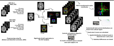 |
Semi-automated assessment of the principal diffusion direction in the corpus callosum: application across brain diseases
Maria Eugenia Caligiuri1, Andrea Quattrone2, Alessandro Mechelli2, and Aldo Quattrone1
1Neuroscience Research Center, University "Magna Graecia", Catanzaro, Italy, 2Institute of Neurology, University "Magna Graecia", Catanzaro, Italy
We propose a novel, semi-automated approach to assess corpus callosum integrity at the individual level, based on the spatial distribution of the principal diffusion direction orientation. We applied the method to the clinical challenge of differentiating normal pressure hydrocephalus (iNPH, N=23) from progressive supranuclear palsy (PSP, N=27) and Alzheimer’s Disease (AD, N=35), whose shared clinical and radiological features can make early differential diagnosis difficult. We found differential involvement of PDD distribution across neurodegenerative (AD, PSP) and non-degenerative diseases (such as iNPH), a finding that could shed light on the different mechanisms that underlie tissue damage.
|
|||
|
1724.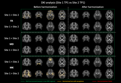 |
Harmonization of diffusion kurtosis imaging metrics with rotational invariant spherical harmonics (RISH) to remove cross-site biases
Alberto De Luca1,2, Suheyla Cetin Karayumak3, Alexander Leemans2, Yogesh Rathi3, Stephan Swinnen4,5, Jolien Gooijers4,5, Amanda Clauwaert4,5, Roald Bahr6, Stian Bahr Sandmo6, Nir Sochen7,8, David Kaufmann9, Marc Muehlmann10, Geert-Jan Biessels1,
Inga K Koerte3,11, and Ofer Pasternak3
1Department of Neurology, University Medical Center Utrecht, Utrecht, Netherlands, 2PROVIDI Lab, Image Sciences Institute, University Medical Center Utrecht, Utrecht, Netherlands, 3Brigham and Women's Hospital, Harvard Medical School, Boston, MA, United States, 4Movement Control and Neuroplasticity Research Group, KU Leuven, Leuven, Belgium, 5Brain Institute, KU Leuven, Leuven, Belgium, 6Oslo Sports Trauma Research Center, Norwegian School of Sport Sciences, Oslo, Norway, 7Department of Applied Mathematics, Tel Aviv University, Tel Aviv, Israel, 8Sagol School of Neuroscience, Tel Aviv University, Tel Aviv, Israel, 9Department of Radiology, Charite University Hospital, Berlin, Germany, 10Department of Radiology, LMU Munich, Munich, Germany, 11cBRAIN, Department of Child and Adolescent Psychiatry, LMU Munich, Munich, Germany
Diffusion Kurtosis Imaging (DKI) metrics computed from diffusion MRI (dMRI) are affected by different acquisition protocols and scanner properties which limits their implementation in multicentric studies. We investigated whether harmonizing multi-shell dMRI with the rotation invariant spherical harmonics (RISH) method allows to remove cross-site differences in DKI metrics while retaining longitudinal effects. 46 subjects underwent a longitudinal two-shell dMRI protocol in 3 imaging sites. Our results show that the RISH method removes inter-site differences both at whole-brain and voxel-level while maintaining the effect-size of longitudinal changes, and is thus promising for the implementation of multi-site DKI studies.
|
||
1725.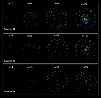 |
Subsampling Diffusion Gradients via Poisson Sphere Elimination
Ye Wu1, Sahar Ahmad1, Lei Ma1, Erkun Yang1, and Pew-Thian Yap1
1Biomedical Research Imaging Center, University of North Carolina, Chapel Hill, Chapel Hill, NC, United States
Subsampling from a set of diffusion gradient directions is useful for evaluation of image acquisition and reconstruction strategies. However, a challenge associated with gradient subsampling is the requirement to ensure uniform distribution of a predetermined number of subsampled directions. Here, we introduce a method for near-uniform subsampling for an arbitrary target number of gradient directions.
|
|||
1726.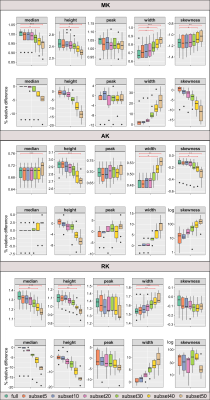 |
Subsampling an existing diffusion MRI multi-shell scheme: impact on histogram measures derived from DTI and DKI
Ana R Fouto1, Rita G Nunes1, Amparo Ruiz-Tagle1, Marc Golub1, Inês Esteves1, Athanasios Vourvopoulos1, Raquel Gil-Gouveia2, Andreia C Freitas1, Nuno A Silva3, and Patrícia Figueiredo1
1ISR-Lisboa/LARSyS and Department of Bioengineering, Instituto Superior Técnico, Universidade de Lisboa, Lisbon, Portugal, 2Neurology Department, Hospital da Luz, Lisbon, Portugal, 3Learning Health, Hospital da Luz, Lisbon, Portugal
Multi-shell diffusion MRI sampling schemes require long acquisition times and are hence potentially compromised by poorer subject compliance. There is therefore a need to consider strategies to enable shortening the exam to ensure clinical feasibility, while maintaining cross-exam comparability. We analyzed the impact of subsampling a multi-shell scheme on the estimation of diffusion maps. In particular, we evaluated its effect on histogram-derived metrics computed over skeletonized maps of DTI and DKI parameters. Several metrics showed a significant effect of subsampling. This should be carefully considered depending on the effect size we are trying to measure on a patient’s population.
|
|||
1727.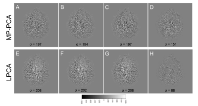 |
Evaluation of noise/signal leaking in PCA-based DWI denoising methods
Hu Cheng1
1Indiana University, Bloomington, IN, United States
We propose a novel but simple method to evaluate noise/signal leaking in PCA-based denoising for diffusion MRI. This method was applied on MP-PCA and LPCA on a human dataset acquired with the ABCD protocol. The results show that MP-PCA is more conservative in removing the noise than LPCA and therefore has little signal leaking. On the other hand, LPCA has less noise leaking as it removed more noise than MP-PCA. Our proposed method can be an effective tool in assessing denoising performance in practice.
|
|||
1728.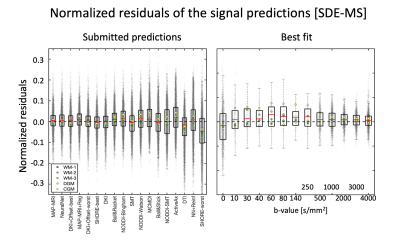 |
On the generalizability of diffusion MRI signal representations across acquisition parameters: chronicles of the MEMENTO challenge.
Alberto De Luca1,2, Andrada Ianus3,4, Alexander Leemans2, Marco Palombo5, Hui G Zhang5, Daniel C Alexander5, Markus Nilsson6, Geert-Jan Biessels1, Mauro Zucchelli7, Matteo Frigo7, Enes Albay7,8, Sara Sedlar7, Abib Alimi7,
Samuel Deslauriers-Gauthier7, Rachid Deriche7, Rutger Fick9, Maryam Afzali10, Tomasz Pieciak11,12, Fabian Bogusz11, Santiago Aja-Fernandez12, Evren Özarslan13,14, Derek K Jones10, Haoze Chen15, Mingwu Jin16, Zhijie Zhang15, Fengxiang Wang15,
Vishwesh Nath17, Prasanna Parvathaneni18, Jan Morez19, Jan Sijbers19, Ben Jeurissen19, Shreyas Fadnavis20, Stefan Endres21, Ariel Rokem22, Eleftherios Garyfallidis20, Irina Sanchez23, Vesna Prchkovska23, Paulo Rodrigues23, Bennett A Landman24,
and Kurt G Schilling24
1Department of Neurology, University Medical Center Utrecht, Utrecht, Netherlands, 2PROVIDI Lab, Image Sciences Institute, University Medical Center Utrecht, Utrecht, Netherlands, 3Champalimaud Research, Champalimaud Centre for the Unknown, Lisbon, Portugal, 4Centre for Medical Imaging Computing, University College London, London, United Kingdom, 5Centre for Medical Image Computing, University College London, London, United Kingdom, 6Clinical Science, Department of Radiology, Lund University, Lund, Sweden, 7Inria Sophia Antipolis – Méditerranée, Université Côte d'Azur, Sophia Antipolis, France, 8Istanbul Technical University, Instanbul, Turkey, 9Therapanacea, Paris, France, 10Cardiff University Brain Research, Imaging Centre (CUBRIC), School of Psychology, Cardiff University, Cardiff, United Kingdom, 11AGH University of Science and Technology, Krakow, Poland, 12LPI, ETSI Telecomunicacion, Universidad de Valladolid, Valladolid, Spain, 13Department of Biomedical Engineering, Linköping University, Linköping, Sweden, 14Center for Medical Image Science and Visualization, Linköping University, Linköping, Sweden, 15School of Instruments and Electronics, North University of China, Taiyuan, China, 16Department of Physics, University of Texas at Arlington, Arlington, TX, United States, 17NVIDIA Corporation, Bethesda, MD, United States, 18National Institute of Neurological Disorders and Stroke, National Institute of Health, Bethesda, MD, United States, 19Imec-Vision Lab, Department of Physics, University of Antwerp, Antwerp, Belgium, 20Intelligent Systems Engineering, Indiana University Bloomington, Bloomington, IN, United States, 21Leibniz Institute for Materials Engineering - IWT, University of Bremen, Bremen, Germany, 22Department of Psychology and the eScience Institute, University of Washington, Seattle, WA, United States, 23QMENTA Inc, Barcelona, Spain, 24Institute of Imaging Science, Vanderbilt University, Nashville, TN, United States
The acquisition and modelling of diffusion MRI (dMRI) data offer many opportunities to explore the organization of the brain. A variety of methods has been proposed to this end, but their generalizability to different diffusion encodings and stronger gradients remains unknown. In the MEMENTO challenge, we asked participants to predict dMRI signals collected with single, double and double oscillating diffusion encodings in combination with a variety of weighting parameters. We received eighty-three submissions ranging from signal representations to multicompartment models and deep learning-based predictions, which offer a unique perspective to investigate the status quo of the field.
|
|||
1729. |
Measurement of radiofrequency absorption and thermal diffusion coefficients of brain tissue
David H Gultekin1, Peter H Siegel2,3, John T Vaughan1, and John C Gore4
1Zuckerman Mind Brain Behavior Institute, Columbia University, New York, NY, United States, 2Jet Propulsion Laboratory, National Aeronautics and Space Administration, Pasadena, CA, United States, 3Department of Electrical Engineering, California Institute of Technology, Pasadena, CA, United States, 4Vanderbilt University Institute of Imaging Science, Nashville, TN, United States
A method is introduced to measure the absorption coefficient and the thermal diffusion coefficient of ex vivo brain tissue exposed to radiofrequency radiation emitted from a half wavelength dipole antenna and a rectangular waveguide using MRI and thermocouples. By analyzing the spatial and temporal variations of thermal gradients, it is possible to measure simultaneously both the absorption coefficient and the thermal diffusion coefficient in brain tissue in a single experiment.
|
The International Society for Magnetic Resonance in Medicine is accredited by the Accreditation Council for Continuing Medical Education to provide continuing medical education for physicians.