Digital Posters
CNS Disease Mechanisms by Quantitative MRI: Clinical
ISMRM & SMRT Annual Meeting • 15-20 May 2021

Digital Posters
CNS Disease Mechanisms by Quantitative MRI: Clinical
| Concurrent 6 | 19:00 - 20:00 |
2576.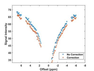 |
Determining Spinal Cord pH using Chemical Exchange Saturation Transfer (CEST) MRI
Alicia Cronin1,2, Patrick Liebig3, Sarah Detombe4, Neil Duggal4, and Robert Bartha1,2
1Medical Biophysics, University of Western Ontario, London, ON, Canada, 2Centre for Functional and Metabolic Mapping, Robarts Research Institute, London, ON, Canada, 3Siemens Healthineers, Erlangen, Germany, 4Clinical Neurological Sciences, University Hospital, London Health Sciences Centre, London, ON, Canada
Degenerative cervical myelopathy (DCM) is one of the most common forms of spinal cord dysfunction. Predicting functional recovery after surgery remains elusive. Pathophysiological mechanisms, like ischemia and hypoxia in the spinal cord, could impact recovery after surgery. Chemical Exchange Saturation Transfer (CEST) produces image contrast based on the rate of exchange of amine and amide protons. This exchange rate is dependent on tissue pH, creating a pH-weighted contrast. CEST imaging in the spinal cord incorporating respiratory correction could be used to examine tissue pathology caused by hypoxia in DCM and other spinal cord injuries.
|
|||
2577.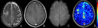 |
The Added Value of Inflow-Based Vascular-Space-Occupancy and Diffusion-Weighted Imaging in Preoperative Grading of Gliomas
Haimei Cao1, Xiang Xiao1, Jun Hua2,3, Guanglong Huang4, Xiaodan Li1, Wenle He1, Jie Qin1, and Yuankui Wu1
1Department of Medical Imaging, Nanfang Hospital, Southern Medical University, Guangzhou, China, 2Neurosection, Division of MRI Research, Department of Radiology, Johns Hopkins University School of Medicine, Baltimore, MD, United States, 3F.M. Kirby Research Center for Functional Brain Imaging, Kennedy Krieger Institute, Department of Radiology, Johns Hopkins University School of Medicine, Baltimore, MD, United States, 4Department of Neurosurgery, Nanfang Hospital, Southern Medical University, Guangzhou, China
Glioma grading is vital for planning therapeutic approaches and assessing prognosis and response to treatment. Advanced MR imaging provides physiological information of brain tumors in microcirculation, cell and molecular levels, thus improving the preoperative prediction of glioma grade. In this study, we studied the value of combined inflow-based vascular-space-occupancy (iVASO) MR imaging and diffusion-weighted imaging (DWI) in preoperative prediction of gliomas grade. The results showed that combined iVASO and DWI improved the diagnostic performance of glioma grading. This suggests that the combined application of iVASO and DWI might be used as part of the routine brain tumor imaging protocol.
|
|||
2578.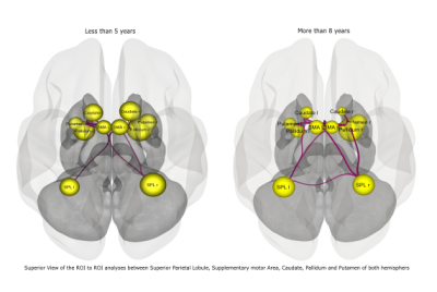 |
Alterations in brain connectivity as duration of disease increases in Parkinson's disease.
Priyanka Bhat1, S Senthil Kumaran2, Achal K Srivastava1, and Vinay Goyal3
1Neurology, AIIMS, Delhi, India, 2Nuclear Magnetic Resonance, AIIMS, Delhi, India, 3Neurology, Medanta, The Medicity, Gurgaon, India Parkinson’s disease (PD) is charcterized by progressive degenration of dopaminergic neurons in substantia nigra. This leads to motor symptoms like bradykinesia with rigidity and tremor. PD patients also experience difficulty in traversing through narrow spaces. Indicating towards altered motor execution and planning and decision making. This study was aimed thus to explore the progressively deteriorating brain connectivity in PD. Functional connectivity was observed to be altered with duration of disease. |
|||
2579.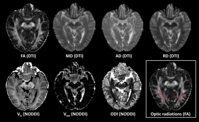 |
DTI and NODDI assessment of posterior optic pathway function in sellar and parasellar tumor patients
Eun-Jung Choi1, Koung Mi Kang2, Woojin Jung1, Jongho Lee1, Seung Hong Choi2, and Yong Hwy Kim3
1Department of Electrical and Computer Engineering, Seoul National University, Seoul, Korea, Republic of, 2Department of Radiology, Seoul National University Hospital, Seoul, Korea, Republic of, 3Department of Neurosurgery, Seoul National University Hospital, Seoul, Korea, Republic of
We investigated the impairment in the optic radiations affected by the anterior visual pathway compression using the diffusion tensor imaging (DTI), and neurite orientation dispersion and density imaging (NODDI). The results were correlated with the visual field impairment score (VFIS), which assesses the function of posterior visual pathway. The DTI and NODDI parameters in the optic radiations were significantly correlated with postoperative visual field improvement as well as preoperative visual field impairment in patients with the compression of optic chiasm. The study demonstrated the anterior visual pathway compression influences the visual field defect of the posterior visual pathway.
|
|||
2580.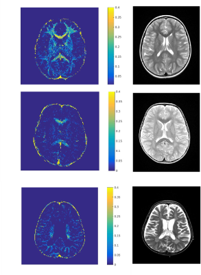 |
White Matter in Metachromatic Leukodystrophy as Assessed by Myelin Water Fraction and Diffusion Tensor Imaging
Laleh Eskandarian1,2, Safak Parlak3, Onur Afacan4,5, Ceren Günbey6, Nesibe Gevher Ertuğrul6, Banu Anlar6, and Kader Karli Oguz2,3
1Neuroscience Department, Bilkent University, Ankara, Turkey, 2National Magnetic Resonance Research Center (UMRAM), Bilkent University, Ankara, Turkey, 3Faculty of Medicine, Department of Radiology, Hacettepe University, Ankara, Turkey, 4Department of Radiology, Boston Children’s Hospital, Boston, MA, United States, 5Department of Radiology, Harvard Medical School, Boston, MA, United States, 6Department of Pediatrics, Hacettepe University, Ankara, Turkey
Metachromatic leukodystrophy (MLD) is a dysmyelinating autosomal recessive lysosomal storage disease. Conventional T2WI may not show the disease involvement accurately, especially in early phases of the disease where a bone marrow transplant can be a treatment option. Therefore we used Myelin Water Fraction (MWF) and Diffusion Tensor Imaging to investigate WM in patients with MLD. MWF and metrics derived from DTI, especially fractional anisotropy (FA), showed diffuse abnormality in WM and were significantly correlated. These imaging techniques can better assess involvement of the cerebral WM in patients with MLD.
|
|||
2581.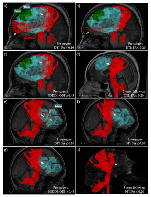 |
Exploring edematous nerve fibers by using Neurite Orientation Dispersion and Density Imaging
Shin Tai Chong1, Xinrui Liu2, Hung-Wen Kao3, Chien-Yuan Eddy Lin4, Sanford PC Hsu5, and Ching-Po Lin1
1Institute of Neuroscience, National Yang-Ming University, Taipei, Taiwan, 2Department of Neurosurgery, First Hospital of Jilin University, Jilin, China, 3Tri-Service General Hospital, National Defense Medical Center, Taipei, Taiwan, 4GE Healthcare, Taipei, Taiwan, 5Department of Neurosurgery, Neurological Institute, Taipei Veterans General Hospital, Taipei, Taiwan
We hypothesized that the Neurite Orientation Dispersion and Density Imaging (NODDI)-based tractography could improve the reconstruction of the fiber tracts that by tracking through regions of peritumoral edema. In visual comparison with diffusion tensor imaging (DTI), we found that NODDI-based tractography of the corticospinal tract (CST) showed higher sensitivity and specificity in the region of peritumoral edema. The NODDI-based tractography was validated in two patients with tumors resected for five years. With this technique, neurosurgeons may develop better surgical planning to maximize tumor resection and minimize functional loss.
|
|||
2582.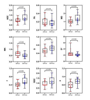 |
Assessment of IDH1 Mutation Status and MGMT Promoter Methylation Status of Gliomas Using DWI, IVIM and DKI
Yan Xie1, Shihui Li1, Nanxi Shen1, Weiyin Vivian Liu2, and Wenzhen Zhu1
1Department of Radiology, Tongji Hospital, Tongji Medical College, HUST, Wuhan, China, 2MR Research, GE Healthcare, Beijing, China
The purpose of this study was to evaluate the efficacy of conventional diffusion weighted imaging (DWI), intravoxel incoherent motion (IVIM) imaging and diffusion kurtosis imaging (DKI) in assessing IDH1 mutation status and MGMT promoter methylation status in gliomas. In lower-grade gliomas, diffusion MRI parameters were able to significantly distinguish the mutation status of IDH1 and the methylation status of MGMT promoter, which is helpful for predicting prognosis and sensitivity to alkylating chemotherapeutic agents of patients. However, in glioblastomas, there was no significant difference between mutant and wild-type IDH1 as well as methylation and unmethylation status of the MGMT promoter.
|
|||
2583.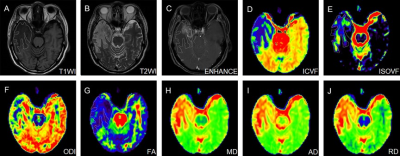 |
NODDI in detecting cognitive decline in patients with radiation-induced brain injury: comparison with DTI
Weike Zeng1 and Mengzhu Wang2
1Sun Yat-sen Memorial Hospital, Sun Yat-sen University, Guangzhou, China, 2MR Scientific Marketing, Siemens Healthcare, Guangzhou, China
This study aimed to demonstrate the feasibility of neurite orientation dispersion and density imaging (NODDI) for in exploring the pathological mechanism of cognitive impairment in RI compared to DTI. We evaluated the differences of all diffusion parameters between patients with and without cognitive decline confirmed by Montreal Cognitive Assessment (MoCA) scores, as well as the correlation between diffusion maps and cognitive decline. Results demonstrated that NODDI was more sensitive than DTI in detecting cognitive decline in patients with radiation-induced brain injury. Cognitive decline in radiation-induced brain injury was associated with the reduction of neurite density
|
|||
2584.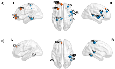 |
Post-surgery network reorganization in glioma patients: a longitudinal study of functional segregation and centrality
Beatrice Federica Luciani1, Francesca Saviola1, Luca Zigiotto2,3, Stefano Tambalo1, Domenico Zacà1, Lisa Novello1, Silvio Sarubbo2,3, and Jorge Jovicich1
1CIMeC Center for Mind/Brain Sciences, University of Trento, Rovereto (Trento), Italy, 2Department of Neuroscience, Division of Neurosurgery, S.Chiara Hospital, APSS, Trento, Italy, 3Structural and Functional Connectivity Lab, S.Chiara Hospital, APSS, Trento, Italy
In glioma patients, tumor lateralization and grade can critically affect the underlying brain functional connectivity. Little is known about how functional connectivity changes after surgical glioma resection. We employed graph theory analysis to investigate post-surgery longitudinal reorganization of functional networks at global (network-wide) and local (nodal) level. We found that left-lateralized and/or high-grade gliomas show reduced segregation and centrality properties over time, compared to right-lateralized and low-grade gliomas. Our results suggest the importance of pre-surgical mapping of hub networks like the default mode network, in addition to visual, motor and language networks.
|
|||
2585.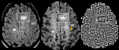 |
Fast 3D Wave-CAIPI Susceptibility Weighted Imaging and SPACE FLAIR for Comprehensive Evaluation of Demyelinating Lesions in Multiple Sclerosis
Augusto Lio M. Goncalves Filho1,2, Azadeh Tabari1,2, Chanon Ngamsombat3, Ilena George4, Stephen F. Cauley2, Wei Liu5, Daniel N. Splitthoff6, Wei-Ching Lo7, Pamela W. Schaefer1, Otto Rapalino1, Eric C. Klawiter4, John Conklin1,2, and Susie Y. Huang1,2
1Department of Radiology, Massachusetts General Hospital, Boston, MA, United States, 2Department of Radiology, Athinoula A. Martinos Center for Biomedical Imaging, Charlestown, MA, United States, 3Department of Radiology, Siriraj Hospital, Bangkok, Thailand, 4Department of Neurology, Massachusetts General Hospital, Boston, MA, United States, 5Siemens Shenzhen Magnetic Resonance Ltd., Shenzhen, China, 6Siemens Healthcare GmbH, Erlangen, Germany, 7Siemens Medical Solutions Inc., Boston, MA, United States
The central vein sign (CVS) and paramagnetic rims are imaging signs specific for demyelinating lesions in multiple sclerosis (MS) that can be detected using high-resolution susceptibility-weighted imaging (SWI) and FLAIR sequences. We developed an optimized protocol for fast, comprehensive evaluation of demyelinating lesions including highly accelerated Wave-CAIPI SWI and FLAIR. In 65 patients undergoing evaluation for demyelinating disease, we found that 72% of MS and CIS patients had at least one CVS, and 45% showed at least one paramagnetic rim. The application of highly accelerated Wave-SWI and Wave-FLAIR may improve the characterization of MS lesions in clinically feasible acquisition times.
|
|||
2586.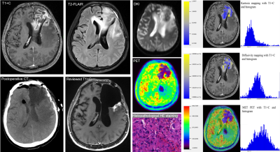 |
Glioblastoma Recurrence vs. Radiotherapy Injury: Combined Model of DKI and 11C-MET Using PET/MR May Increase Accuracy of Differentiation
Haodan Dang1, Ruimin Wang1, Jiajin Liu1, Huaping Fu1, Mu Lin2, Jiahe Tian1, Jinming Zhang1, and Baixuan Xu1
1Department of nuclear medicine, Chinese PLA General Hospital, Beijing, China, 22. MR Collaboration, Diagnostic Imaging, Siemens Healthcare, Shanghai, China
The purpose of this study was to evaluate the diagnostic potential of decision-tree model of diffusion kurtosis imaging (DKI) and 11C-methionine (11C-MET) PET imaging, for the differentiation of radiotherapy injury from glioblastoma recurrence using integrated PET/MR. Eighty-six glioblastoma cases with suspected lesions after radiotherapy were retrospectively enrolled. Compared to models of DKI-alone (AUC=0.85) and PET-alone (AUC=0.89), the combined model demonstrated the best diagnostic accuracy (AUC=0.95). The decision-tree model has the potential to further increase diagnostic accuracy for discrimination between radiotherapy injury and glioblastoma recurrence. 11C-MET PET/MR may thus contribute to the management of glioblastoma patients with suspected lesions after radiotherapy.
|
|||
2587.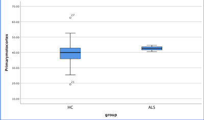 |
Usefulness of Quantitative Susceptibility MRI for the Detection of Iron in the Motor Cortex in Amyotrophic Lateral Sclerosis
qianwen li1, juan Wei2, and jie Lu1
1Xuanwu Hospital,Capital Medical University, Beijing, China, 2GE Healthcare, Beijing, China
Quantitative Susceptibility Mapping (QSM), a newly developed quantitative and accurate measurement method that can be used to detect iron-related motor cortex alterations in patients of Amyotrophic lateral sclerosis (ALS) that may be relevant to pathologic changes.
|
|||
2588.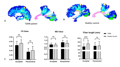 |
Disturbed interhemispheric functional and structural connectivity in Type 2 diabetes
Ying Cui1, Tian-yu Tang2, and Shenghong Ju1
1Department of Radiology, Zhongda hospital, Southeast University, Nanjing, China, 2Southeast University, Nanjing, China
This study combined the resting-state functional MRI (rs-fMRI) and diffusion tensor imaging (DTI) to explore whether the interhemispheric coordination is altered in type 2 diabetes (T2DM). Results showed that the occipital lobe was disrupted in both functional and anatomic connections and such alteration is strongly associated with endocrine parameters, especially for insulin resistance.
|
|||
2589.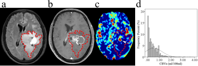 |
Noninvasive assessment of MGMT promoter methylation status in grade II-IV gliomas using inflow-based vascular-space-occupancy
Yuankui Wu1, Wenle He1,2, Wensheng Wang2, Jun Hua3,4, Xiang Xiao1, Xiaomin Liu1, Yikai Xu1, and Yingjie Mei5
1Department of Medical Imaging, Nanfang Hospital, Southern Medical University, Guangzhou, China, 2Department of Radiology, Guangdong 999 Brain Hospital, Guangzhou, China, 3Neurosection, Division of MRI Research, Department of Radiology, Johns Hopkins University School of Medicine, Baltimore, MD, United States, 4F.M. Kirby Research Center for Functional Brain Imaging, Kennedy Krieger Institute, Department of Radiology, Johns Hopkins University School of Medicine, Baltimore, MD, United States, 5Philips healthcare, Guangzhou, China
Accurate prediction of O(6)-methylguanine-DNA methyltransferase (MGMT) promoter methylation status preoperatively is important. DSC-MRI can predict MGMT promoter methylation status in glioblastomas but not in lower-grade gliomas. Inflow-based vascular-space-occupancy (iVASO), a novel perfusion technique without the need for exogenous contrast agents, emphasizes the perfusion blood volume in arteries and arterioles. In this study, the predictive ability for MGMT promoter methylation status of iVASO histogram features was investigated. The results showed that iVASO-based histogram features accurately predicted the methylation status of MGMT promoter in gliomas. This suggests that iVASO may be a promising noninvasive imaging tool in predicting MGMT promoter methylation.
|
|||
2590.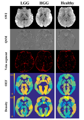 |
Susceptibility-Based Characterization of Venous Distribution and Oxygen Extraction Fraction in Brains with Glioma at 7T
Shihui Zhou1,2, Huilou Liang2,3, Yuchao Liang4, Siqi Cai1,2, Chunxiang Jiang1,2, Rong Xue2,3, Lei Wang4, and Lijuan Zhang*1
1Shenzhen Institutes of Advanced Technology, Chinese Academy of Sciences, Shenzhen, China, 2University of Chinese Academy of Sciences, Beijing, China, 3State Key Laboratory of Brain and Cognitive Science, Beijing MRI Center for Brain Research, Institute of Biophysics, Chinese Academy of Sciences, Beijing, China, 4Neurosurgery, Beijing Tiantan Hospital of Capital Medical University, Beijing, China
Vein density and oxygen extraction fraction were quantified in brains with glioma using a susceptibility-based approach at 7T. Brains with low grade glioma featured more extensive areas with significant difference in vein density and OEF between the homotopic ROIs that mainly involve fronto-parietal areas and basal ganglia, in contrast to brains with high grade glioma with the differential vein density and OEF distribution mainly involving bilateral thalami. The inter-homotopic measurements of vein density and OEF were significantly correlated, suggesting that the oxygen metabolism malfunction in brains with high grade glioma might be partially ascribed to the altered venous distribution.
|
|||
2591.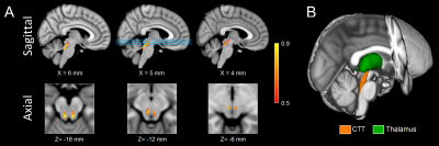 |
Tau-mediated microstructural changes in the central tegmental tract in APOE-ε4 positive mild cognitive impairment
Jason Langley1, Sana Hussain2, Daniel E Huddleston3, Ilana Bennett4, and Xiaoping P Hu1,2
1Center for Advanced Neuroimaging, University of California Riverside, Riverside, CA, United States, 2Department of Bioengineering, University of California Riverside, Riverside, CA, United States, 3Department of Neurology, Emory University, Atlanta, GA, United States, 4Department of Psychology, University of California Riverside, Riverside, CA, United States
In mild cognitive impairment, hyperphosphorylated tau proteins and tau inclusions first appear in the locus coeruleus and transentorhinal cortex and spread to other brain regions, including the thalamus. Locus coeruleus axons project to the thalamus via the central tegmental tract (CTT). Relative to APOE-ε4 negative subjects, a decrease in MD (p=0.038) and an increase in ficvf (p=0.007) was seen in the CTT of APOE-ε4 positive subjects. In the APOE-e4 positive group, CTT microstructural measures were positively correlated with thalamus tau-PET SUVR but no correlations between CTT microstructure and tau-PET SUVR were observed in the APOE-ε4 negative group.
|
|||
2592.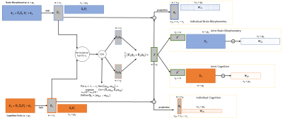 |
Joint and individual statistical analysis of brain MRI and cognition measures in Alzheimer's Disease
Raphiel Jamale Murden1, Deqiang Qiu2, and Benjamin B Risk2
1Biostatistics and Bioinformatics, Emory University, Atlanta, GA, United States, 2Emory University, Atlanta, GA, United States
Canonical Joint and Individual Variation Explained (CJIVE) provides a method for jointly analyzing multi-block datasets collected from the same individuals. We apply this method to measures of brain morhometry and cognition from a sample of older adults. We found latent patterns of joint variation across data types which were statistically associated with diagnoses of Alzheimer's Disease and mild cognitive impairment. We also found that the unique patterns of variation within each data type were not associated with diagnoses.
|
|||
2593.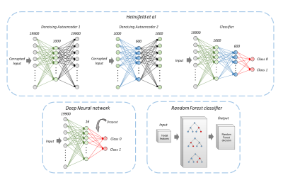 |
Functional connectivity-based prediction of Autism on site harmonized ABIDE dataset
Madhura Ingalhalikar1, Sumeet Shinde1, Arnav Karmarkar1, Archith Rajan1, Rangaprakash D2, and Gopikrishna Deshpande3
1Symbiosis Centre for medical image analysis, Symbiosis international university, Pune, India, 2Martinos Center for Biomedical Imaging, Massachusetts General Hospital, Boston, MA, United States, 3Department of Electrical and Computer Engineering, Auburn University, Auburn, AL, United States
Functional MRI connectivity based analysis that ranges between simple univariate methods to complex deep-learning pipelines has been employed to differentiate autistic patients from healthy controls on benchmark datasets such as ABIDE. However, the variability induced via multi-site acquisition of data may perturb the underlying prediction model with undesirable consequences. We illustrate that statistical elimination of scanner effects using COMBAT harmonization yields better results and also facilitates in gaining insights into the discriminative connectivity patterns that emerge post harmonization and which correlate with clinical markers.
|
|||
2594.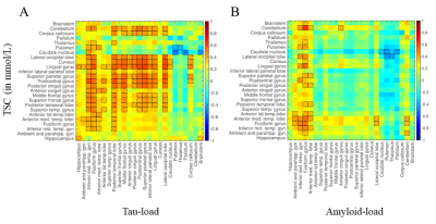 |
Ultra-High Field Sodium MRI in Alzheimer’s Disease Reveals Stage-dependent Metabolic Alterations Associated with Tau-pathology
Alexa Haeger1,2,3, Michel Bottlaender1,4, Julien Lagarde4,5,6, Renata Porciuncula Baptista1, Cécile Rabrait-Lerman1, Volker Luecken2,3, Jörg Bernhard Schulz2,3, Alexandre Vignaud1, Marie Sarazin4,5,6, Kathrin Reetz2,3, Sandro Romanzetti2,3, and Fawzi Boumezbeur1
1BAOBAB, CNRS, Paris-Saclay University, CEA-NeuroSpin, Gif-sur-Yvette, France, 2Department of Neurology, RWTH Aachen University, Aachen, Germany, 3JARA-BRAIN Institute of Molecular Neuroscience and Neuroimaging, Forschungszentrum Jülich GmbH, Julich, Germany, 4BioMaps, CNRS, Inserm, Paris-Saclay University, CEA-SHFJ, Orsay, France, 5Neurology of Memory and Language, GHU Paris Psychiatrie & Neurosciences, Sainte-Anne Hospital, Paris, France, 6Université de Paris, Paris, France
Deficits in brain cells’ homeostasis and metabolism are suspected to occur ahead of the atrophy observed throughout the brain of AD patients with potential interactions between Amyloid/Tau deposits and the Na/K-pump activity leading to increased cerebral sodium concentrations. Yet this increase remains to be confirmed. We present a multimodal imaging study combining structural 1H-MRI, quantitative 23Na MRI at 7T in association with Tau- and Amyloid-PET. We show that total sodium concentration is increased in multiple brain regions in AD compared to cognitively healthy controls, and that these changes are more strongly correlated with local Tau- than Amyloid-loads.
|
|||
2595.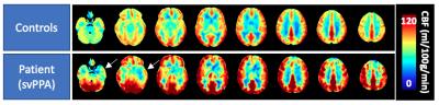 |
Feasibility of Arterial Spin Labeling for Detection of Longitudinal Changes in Perfusion in Elderly and Frontotemporal Dementia Patients
Tracy Ssali1,2, Lucas Narciso1,2, Matthais Günther3, Frank Prato1,2, Udunna Anazodo1,2, Elizabeth Finger4, and Keith St Lawrence1,2
1Lawson Health Research Institute, London, ON, Canada, 2Department of Medical Biophysics, Western University, London, ON, Canada, 3Fraunhofer Institute for Medical Image Computing MEVIS, Bremen, Germany, 4Department of Clinical Neurological Sciences, Western University, London, ON, Canada
Recent advances in the understanding of frontotemporal dementia (FTD) and subsequently, the development of novel disease modifying treatments, has stimulated the need for tools to assess treatment efficacy. While perfusion imaging by arterial spin labeling (ASL) is an attractive approach, its sensitivity to detect longitudinal changes in perfusion in dementia patients remains unknown. Longitudinal variability in perfusion was assessed on a voxel-by-voxel basis over a month. Power analysis revealed ASL has the sensitivity to longitudinal changes as low as 7-8%. These results highlight the potential of ASL for detecting longitudinal changes in FTD populations.
|
The International Society for Magnetic Resonance in Medicine is accredited by the Accreditation Council for Continuing Medical Education to provide continuing medical education for physicians.