Digital Posters
Quantitative Neuro: Technical Advances
ISMRM & SMRT Annual Meeting • 15-20 May 2021

Digital Posters
Quantitative Neuro: Technical Advances
| Concurrent 5 | 15:00 - 16:00 |
1272.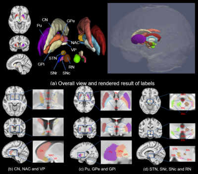 |
Precise localization of Deep brain nuclei in MNI-space guided by Hybrid Multi-modal MRI Brain Atlas
Chenyu He1, Xiaojun Guan2, Boliang Yu1, Hongjiang Wei3, and Yuyao Zhang1,4,5
1School of Information Science and Technology, ShanghaiTech University, Shanghai, China, 2Department of Radiology, The Second Affiliated Hospital, Zhejiang, University School of Medicine, Hangzhou, China, 3Institute for Medical Imaging Technology, School of Biomedical Engineering, Shanghai, Jiao Tong University, Shanghai, China, 4Shanghai Engineering Research Center of Intelligent Vision and Imaging, ShanghaiTech University, Shanghai, China, 5iHuman Institute, ShanghaiTech University, Shanghai, China
Our work successfully provides precious manual depiction of typical DBNs in the standard MNI space with the guidance of constructed MNI-spaced average multi-modal template (T1-weighted and quantitative susceptibility mapping (QSM)). 10 pairs of DBN and their sub-nuclei, are manually depicted including caudate nucleus (CN), putamen (Pu), ventral pallidum (VP), globus pallidus (GP) (internal and external divisions, GPi & GPe), nucleus accumbens (NAC), subthalamic nucleus (STN), substantia nigra (SN) (substantia nigra pars compacta (SNc) & substantia nigra pars reticulata (SNr)) and red nucleus (RN).
|
|||
 |
1273.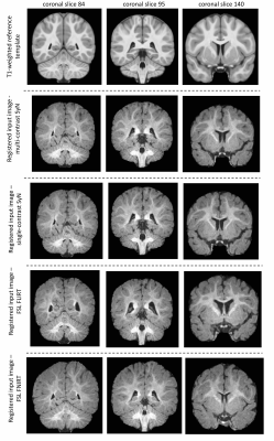 |
Multi-contrast Brain Image Registration for Low-contrast Paediatric Magnetic Resonance Imaging
Cassandra M.J. Wannan1, Shane Tonissen2, Bruce Tonge3, Efstratios Skafidas 1,2, Christos Pantelis1,2, and Warda T. Syeda1
1Melbourne Neuropsychiatry Centre, The University of Melbourne, Parkville, Australia, 2Department of Electrical and Electronic Engineering, The University of Melbourne, Parkville, Australia, 3Psychiatry Monash Health, Faculty of Medicine, Nursing and Health Sciences School of Clinical Sciences, Monash University, Melbourne, Australia
A multi-contrast brain imaging technique has been presented, that leverages information from T1- and T2-weighted images to register iso-intense paediatric MRI data. The proposed method is applied to T1-weighted images of 6-months old infants, and the performance is evaluated in comparison to commonly employed single-contrast registration techniques.
|
||
1274.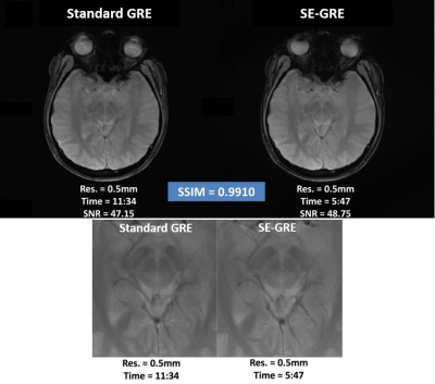 |
Comprehensive Human Brain Imaging Enhancement with the Single-frequency Excitation Wideband MRI (SE-WMRI) Technique
Jordan Wang1, Po-Wei Cheng1, Ming-Jang Chiu2, Tzi-Dar Chiueh3, and Jyh-Horng Chen3
1Graduate institute of biomedical electronics and bioinformatics, National Taiwan University, Taipei, Taiwan, 2National Taiwan University Hospital, Taipei, Taiwan, 3Department of electrical engineering, National Taiwan University, Taipei, Taiwan
The Single-frequency Excitation Wideband MRI (SE-WMRI) technique was implemented on a Siemens PRISMA 3T human MRI system to demonstrate its multifaceted benefits for human brain imaging, including acceleration, SNR improvement, and spatial resolution enhancement. The accelerated Wideband brain results showed high consistency with standard scans, and the resolution enhanced Wideband images revealed more detailed brain structures details and clearer blood vessel due to reduced partial volume effect. Since SE-WMRI can enhance MR efficiency without any hardware upgrade, it’s highly cost-effective for medical institutions in pursuit of higher MR imaging quality.
|
|||
1275.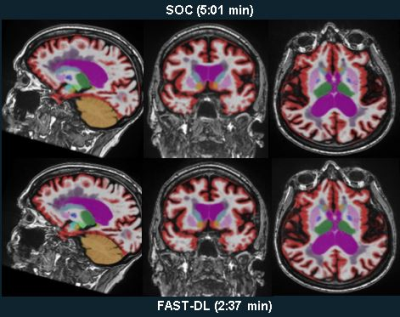 |
Deep Learning Enables 60% Accelerated Volumetric Brain MRI While Preserving Quantitative Performance – A Prospective, Multicenter Trial
Suzie Bash1, Long Wang2, Chris Airriess3, Sara Dupont2, Greg Zaharchuk4, Enhao Gong2, Tao Zhang2, Ajit Shankaranarayanan2, and Lawrence Tanenbaum5
1Neuroradiology, RadNet, Woodland Hills, CA, United States, 2Subtle Medical, Menlo Park, CA, United States, 3Cortechs.ai, San Diego, CA, United States, 4Stanford University, Stanford, CA, United States, 5RadNet, New York, NY, United States
In this prospective, multireader, multicenter study, we explore the impact of deep learning (DL) enhancement of 60% accelerated 3D T1 weighted brain MR image acquisitions. We found that the DL processed images demonstrated high volumetric quantification accuracy, matched clinical disease status predictability, and provided what readers perceived as superior image quality when compared with the longer standard-of-care exams, suggesting good generalizability, accuracy, and potential utility of DL enhancement in routine clinical settings. The results of this trial support the use of DL enhancement to shorten clinical MR brain examinations, even when additional quantitative tools such as volumetric analysis are applied.
|
|||
1276.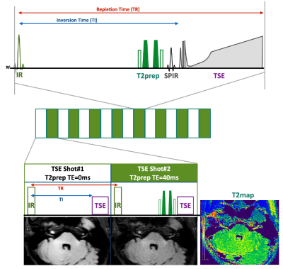 |
T2 Mapping of the Cranial Nerves with Multi-Interleaved X-prepared Turbo-spine Echo with Intuitive Relaxometry (MIXTURE) FLAIR
Hajime Yokota1, Takayuki Sakai2, Masami Yoneyama3, Yansong Zhao4, and Takashi Uno1
1Department of Diagnostic Radiology and Radiation Oncology, Chiba University, Chiba, Japan, 2Department of Radiology, Eastern Chiba Medical Center, Togane, Japan, 3Philips Japan, Tokyo, Japan, 4Philips Healthcare, Cleveland, OH, United States
Multi-Interleaved X-prepared tse with inTUitive Relaxometry (MIXTURE) is a 3D TSE sequence that can produce high-resolution morphology images and T2 map simultaneously. The purpose of this study was to evaluate MIXTURE FLAIR for T2 measurement of the cranial nerves. MIXTURE FLAIR produced reliable T2 values in the fantom study. The variance of T2 value measured by MIXTURE FLAIR was relatively small, and the T2 value was significantly smaller than multi-echo TSE and MIXTURE T2 in the volunteer study. MIXTURE FLAIR enabled the T2 value measurement of the cranial nerves, which was not previously feasible.
|
|||
1277.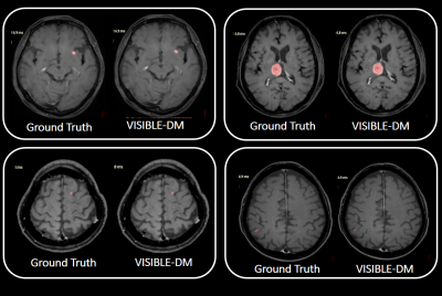 |
Interleaved Black- and Bright-Blood Acquisition for Automatic Brain Metastasis Detection using Deep Learning Convolutional Neural Network
Makoto Obara1, Yoshitomo Kikuchi2, Akio Hiwatashi2, Alexander Fischer 3, Yuta Akamine1, Tetsuo Ogino1, Masami Yoneyama1, Ronee Asad1, Yu Ueda1, Jihun Kwon1, and Marc Van Cauteren4
1Philips Japan, Tokyo, Japan, 2Departments of Clinical Radiology, Graduate School of Medical Sciences, Kyushu University, Fukuoka, Japan, 3Philips GmbH Innovative Technologies, Aachen, Germany, 4Philips Healthcare, Tokyo, Japan
Volume isotropic simultaneous interleaved bright- and black-blood examination (VISIBLE) is an established approach for brain metastasis screening. We investigated the potential of VISIBLE in deep learning model development. The results suggest VISIBLE’s usefulness for automatic metastasis detection.
|
|||
1278.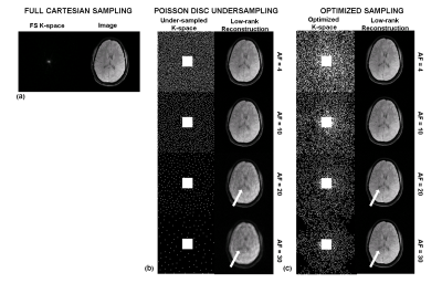 |
Performance of data driven learned sampling patterns for accelerating brain 3D-T1ρ MRI
Rajiv G Menon1, Marcelo V.W. Zibetti1, and Ravinder R. Regatte1
1New York University Langone Health, New York, NY, United States
3D-T1ρ has many useful biomedical applications but requires long data acquisition times. The goal of the study was to apply a fast data-driven optimization approach, bias- accelerated subset selection (BASS), to generate optimal sampling patterns (SPs) for compressed sensing (CS) reconstruction for brain 3D-T1ρ MRI. Five healthy volunteers were recruited and fully sampled (FS) Cartesian, 3D-T1ρ MRI was obtained. The performance of Poisson disc (PD) and optimized SP were compared using normalized root mean square error (NRMSE). The data-driven optimized SP provides upto 2 times (NRMSE=0.09 optimized SP vs 0.18 PD-SP) improvement in images at the highest AFs tested.
|
|||
1279.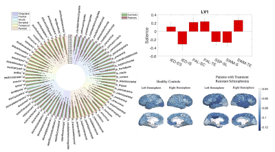 |
A Multiblock Partial Least Squares Correlation Framework for Covariate Adjustment and Interpretation of Latent Associations in Multimodal Data
Warda T. Syeda1, Bjørn H. Ebdrup1,2,3, Cassandra M.J. Wannan1, Micah Cearns1, Rigas Soldatos1, Antonia Merritt1, Mahesh Jayaram 1, Andrew Zalesky 1, Jayachandra M. Raghava2,4, Birgitte Fagerlund 2, Egill Rostrup 2, Birte Glenthøj 2,3, Leigh A. Johnston5,6,
Chad Bousman 1,7, Ian Everall8, Efstratios Skafidas 1,9, and Christos Pantelis1,9
1Melbourne Neuropsychiatry Centre, The University of Melbourne, Parkville, Australia, 2Center for Neuropsychiatric Schizophrenia Research and Center for Clinical Intervention and Neuropsychiatric Schizophrenia Research, Mental Health Centre Glostrup, Copenhagen University Hospital, Glostrup, Denmark, 3Faculty of Health and Medical Sciences, Department of Clinical Medicine, University of Copenhagen, Denmark, Denmark, 4Functional Imaging Unit, Department of Clinical Physiology, Nuclear Medicine and PET, Rigshospitalet, Glostrup, Denmark, 5Department of Biomedical Engineering, The University of Melbourne, Melbourne, Australia, 6Melbourne Brain Centre Imaging Unit, The University of Melbourne, Parkville, Australia, 7Departments of Medical Genetics, Psychiatry, and Physiology & Pharmacology, University of Calgary, Calgary, AB, Canada, 8Institute of Psychiatry, Psychology & Neuroscience, King's College London, London, United Kingdom, 9Electrical and Electronic Engineering Department, The University of Melbourne, Parkville, Australia
Partial least squares (PLS) methods enable identification of multimodal patterns of latent associations between neuroimaging, cognitive, functional and clinical measures. Here, we propose a multiblock PLS correlation (MB-PLS-C) technique to enable covariate representation in the latent space and an interpretation framework to assess results from PLS-C analyses in clinical contexts. We investigate latent structure-cognition patterns in a multivariate dataset of individuals with treatment-resistant schizophrenia and healthy controls using the proposed MB-PLS-C method, and compare with standard PLS-C with and without covariate adjustment through residualization.
|
|||
1280.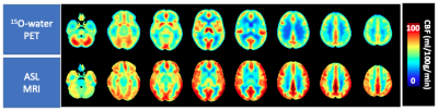 |
Concordance of Regional Hypoperfusion by ASL MRI and 15O-water PET in Frontotemporal Dementia: Is ASL an Efficacious Alternative?
Tracy Ssali1, Lucas Narciso1,2, Justin Hicks1,2, Matthais Günther3, Frank Prato1,2, Udunna Anazodo1,2, Elizabeth Finger4, and Keith St Lawrence1,2
1Lawson Health Research Institute, London, ON, Canada, 2Department of Medical Biophysics, Western University, London, ON, Canada, 3Fraunhofer Institute for Medical Image Computing MEVIS, Bremen, Germany, 4Department of Clinical Neurological Sciences, Western University, London, ON, Canada
The ability of arterial spin labeling (ASL) to detect perfusion abnormalities in clinical populations can be limited by poor signal to noise and transit-time artefacts. This is evident in studies involving frontotemporal dementia patients, where reports on the diagnostic value of ASL have been inconsistent. This study presents a head-to-head comparison of regional hypoperfusion detected by ASL and PET with radiolabeled water (15O-water), the gold standard for measuring CBF. T-maps depicting hypoperfusion were generated using absolute and relative CBF. There was good agreement between regional hypoperfusion identified by ASL and 15O-water, particularly for relative CBF maps which reduced inter-subject variability.
|
|||
1281.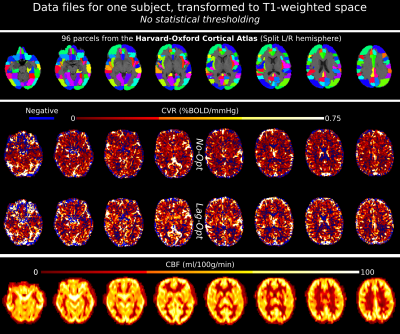 |
The relationship between Cerebrovascular Reactivity and baseline Cerebral Blood Flow: the effect of acquisition and analysis choices
Rachael C Stickland1, Kristina M Zvolanek1,2, Stefano Moia3,4, Apoorva Ayyagari1,2, César Caballero-Gaudes3, and Molly G Bright1,2
1Physical Therapy and Human Movement Sciences, Feinberg School of Medicine, Northwestern University, Chicago, IL, United States, 2Biomedical Engineering, McCormick School of Engineering, Northwestern University, Evanston, IL, United States, 3Basque Center on Cognition, Brain and Language, Donostia, Spain, 4University of the Basque Country EHU/UPV, Donostia, Spain
Blood Oxygen Level Dependent (BOLD) signals can be modulated by the baseline vascular and metabolic state. Understanding how baseline vascular physiology relates to dynamic neurovascular processes will lead to more accurate interpretations of BOLD Cerebrovascular Reactivity (CVR) measurements. We investigated the relationship between baseline Cerebral Blood Flow (bCBF) and BOLD-CVR, generally reporting positive correlations. Optimizing for vascular delays, and modelling with simple breathing task data, can improve CVR correlations with bCBF. Future work should investigate individual differences and include larger samples.
|
|||
1282.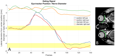 |
Image-based self-gating for motion artifact free imaging of the eye
Kilian Stumpf1, Hanna Frantz1, Patrick Metze1, Thomas Hüfken1, Tobias Speidel1, and Volker Rasche1
1Department of Internal Medicine II, Ulm University Medical Center, Ulm, Germany
MR acquisitions of the eyes are often complicated by eye motions leading to the occurrence of motion artifacts in the images. Using a tiny golden angle profile ordering scheme and a sliding window reconstruction the position of the optic nerve is identified and tracked via peak analysis of signal intensities along a 1-D-line containing the optic nerve. An image-based self-gating signal is derived from the calculated nerve locations. This approach not only leads to distinct reduction of motion artifacts but also enables the visualization of various motion phases occurring during the data acquisition without the use of external tracking devices.
|
|||
1283.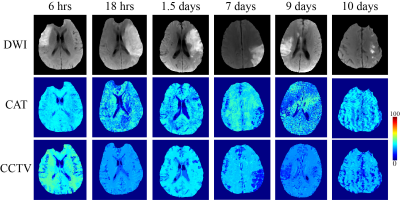 |
Combined cluster analysis of time evolution and tissue type with total variation denoising (CCTV) for QQ-based oxygen extract fraction mapping
Junghun Cho1, Pascal Spincemaille1, Thanh D Nguyen1, Ajay Gupta1, and Yi Wang1,2
1Radiology, Weill Cornell Medicine, New York, NY, United States, 2Biomedical Engineering, Cornell University, Ithaca, NY, United States
A Combined Cluster analysis of time evolution and tissue type with Total Variation denoising (CCTV) was developed to suppress noise propagation in oxygen extraction fraction (OEF) maps based on the QSM+qBOLD (QQ) model of multi-echo gradient echo data without vascular challenge. Compared to cluster analysis of time evolution (CAT), the developed CCTV provided more accurate OEF in simulation and greater contrast to noise ratio between lesion and its healthy contralateral side in ischemic stroke patients.
|
|||
1284 |
Impact of b-value on the estimation of white matter fiber orientation dependent R2* Video Permission Withheld
Melanie Bauer1,2, Celine Berger1,2, Claudia Lenz1,2, Eva Scheurer1,2, and Christoph Birkl3
1Institute of Forensic Medicine, Biomedical Engineering, University of Basel, Basel, Switzerland, 2Institute of Forensic Medicine, Health Department Basel-Stadt, Basel, Switzerland, 3Department of Neuroradiology, Medical University of Innsbruck, Innsbruck, Austria
To generate precise diffusion images in post mortem MRI, higher b-values are necessary than for in vivo MRI due to decreased diffusion. In this study, the influence of different b-values on the fiber orientation dependency of R2* was assessed in 12 post mortem and 4 in vivo cases. Our results show that R2* values are higher but their orientation dependency is lower post mortem than in vivo. However, the chosen b-values did not affect the estimation of the white matter fiber angle and, hence, the orientation dependency of R2* for both post mortem in situ and in vivo MRI.
|
|||
1285.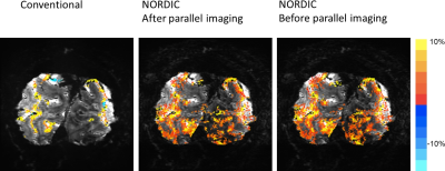 |
NORDIC denoising before image reconstruction.
Steen Moeller1, Cheryl Olman2, Luca vizioli1, Logan Dowdle1, Essa Yacoub1, Mehmet Akcakaya1,3, and Kamil Ugurbil1
1University of Minnesota, MINNEAPOLIS, MN, United States, 2Psychology, University of Minnesota, MINNEAPOLIS, MN, United States, 3ELECTRICAL AND COMPUTER ENGINEERING, University of Minnesota, Minneapolis, MN, United States
Investigating the utility of using the recently proposed NORDIC denoising prior to GRAPPA based unaliasing, for establishing the feasibility of integration with deep learning image reconstruction techniques.
|
|||
1286.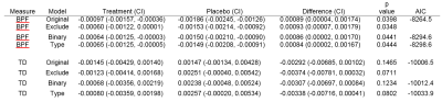 |
Influence of equipment changes on a longitudinal trial
Ken Sakaie1, Janel Fedler2, Jon Yankey2, Kunio Nakamura1, Josef Debbins3, Mark J. Lowe1, Paola Raska1, and Robert J. Fox1
1The Cleveland Clinic, Cleveland, OH, United States, 2University of Iowa, Iowa City, IA, United States, 3Barrow Neurological Institute, Phoenix, AZ, United States
There is an urgent need for imaging biomarkers to develop new therapies for progressive multiple sclerosis. Hardware upgrades can confound the outcomes of imaging in clinical trials of such therapies. We examine different analytic approaches to evaluating the impact of and correcting for the effects of hardware changes in a retrospective analysis of SPRINT-MS, a multi-center clinical trial. Brain parenchymal fraction (BPF), a measure of atrophy, and transverse diffusivity (TD), a measure of demyelination are examined.
|
|||
1287.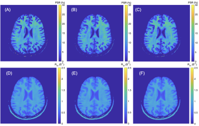 |
Rapid Whole-Brain Myelin Mapping via Selective Inversion Recovery and Compressed SENSE
Ping Wang1, Nicholas Sisco1, and Richard Dortch1
1Barrow Neurological Institute, Phoenix, AZ, United States
There is a significant need for specific biomarkers of myelin damage and repair. Quantitative magnetization transfer (qMT) using selective inversion recovery (SIR) is able to quantify the macromolecular-to-free proton pool size ratio (PSR), which has been shown to relate closely with myelin content and disability. For clinical applications, SIR is often hampered by long scan times. As a result, we employed compressed sensing and parallel imaging to significantly reduce scan times, with little effect on the precision and accuracy of PSR estimates in white matter.
|
|||
1288.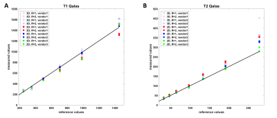 |
Reproducibility and Multi-vendor Accuracy Comparison of T1- and T2- Mapping Using 2D and 3D Synthetic MRI with GRAPPA, SENSE and Compressed Sense
Maarten Naeyaert1, Tim Vanderhasselt1, Marcel Warntjes2, and Hubert Raeymaekers1
1Radiology, Universitair Ziekenhuis Brussel, Brussels, Belgium, 2SyntheticMR AB, Linköping, Sweden
To evaluate intra- and inter-scan repeatability of quantitative scans, T1-, T2- and PD maps were acquired simultaneously using the 2D multi-delay-multi-echo sequence and the 3D-QALAS sequence, for 3T scanners of three different manufacturers. All scans were acquired with and without acceleration factor of 2, using GRAPPA, SENSE or compressed sensing, depending on the vendor. On one scanner measurements were repeated 10 times. The estimated values using synthetic MRI were compared to the reference values of the ISMRM-NIST phantom. The results for T1- and T2- mapping show a linear curve close to the reference values, with good reproducibility.
|
|||
1289.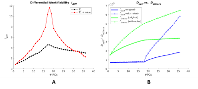 |
Improving T1-weighted MRI brain images by optimizing differential identifiability
Bradley Fitzgerald1, Kausar Abbas2, Thomas M. Talavage1,3, and Joaquin Goni2
1Electrical & Computer Engineering, Purdue University, West Lafayette, IN, United States, 2Industrial Engineering, Purdue University, West Lafayette, IN, United States, 3Biomedical Engineering, University of Cincinnati, Cincinnati, OH, United States
Analysis of scan-rescan subject identifiability of T1 anatomical brain MRI could provide insight into intersession differences. Here we examine a principal component analysis-based method of maximizing differential identifiability and its effect on T1 brain images with and without added noise. We demonstrate that differential identifiability can be maximized via dataset reconstruction with reduced principle components. This reconstruction results in increased similarity between repeated scans for a given subject as well as apparent reduced intersession noise in images. We conclude that further analysis of maximized differential identifiability could provide insight for future applications in reducing intersession MRI noise.
|
|||
1290. |
GAN-based analysis for investigation of disease specific image pattern in SWI data of patients suffering from multiple sclerosis
Alina Lopatina1,2, Stefan Ropele3, Renat Sibgatulin1, Jürgen R Reichenbach1,2,4, and Daniel Güllmar1
1Medical Physics Group / IDIR, Jena University Hospital, Jena, Germany, 2Michael-Stifel-Center for Data-Driven and Simulation Science, Jena, Germany, 3Department of Neurology, Medical University of Graz, Graz, Austria, 4Center of Medical Optics and Photonics Jena, Jena, Germany
We propose a method to transform susceptibility-weighted images of multiple sclerosis (MS) patients to images reflecting healthy volunteers based on generative adversarial networks (GANs). This method helps to identify MS by changing voxel information corresponding to the disease. The results showed that voxels around the central veins and ventricles are identified as MS-specific by the method. This finding may contribute to improvements in MS diagnosis and encourage future studies based on the presented findings.
|
|||
1291.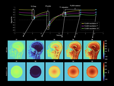 |
3D Quantitative MRI of the Brain: Effects of B1 Inhomogeneity in 3D-QALAS
Anders Tisell1,2, Peter Lundberg1,2, Marcel Jan Bertus Warntjes1, and Frederik Testud3
1CMIV, Linköping University, Linköping, Sweden, 2Medical Radiation Physics, Linköping University, Linköping, Sweden, 3Siemens Healthcare AB, Malmö, Sweden
We have implemented the 3D QALAS sequence for a 3 T MR-scanner and validated the accuracy and evaluated the effect of B1+ inhomogeneities on the resulting R1 and R2 estimations. When using a small flip angle of 4° the determined R1 and R2 maps showed a high accuracy; however, for 10° there was a significant bias and dependence on B1+ inhomogeneities.
|
The International Society for Magnetic Resonance in Medicine is accredited by the Accreditation Council for Continuing Medical Education to provide continuing medical education for physicians.