Digital Posters
Quantitative Neuro: Translational Studies
ISMRM & SMRT Annual Meeting • 15-20 May 2021

Digital Posters
Quantitative Neuro: Translational Studies
| Concurrent 5 | 15:00 - 16:00 |
1292.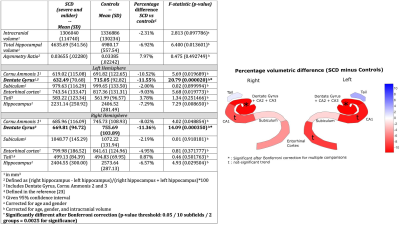 |
Hippocampal segmentation from 7T images showed reduced subfields volume in Sickle Cell Disease subjects
Tales Santini1, Minseok Koo1, Nadim Farhat1, Vinicius P. Campos2, Salem Alkhateeb1, Marcelo A. C. Vieira2, Meryl A Butters1, Caterina Rosano1, Howard J Aizenstein1, Joseph Mettenburg1, Enrico M. Novelli1, and Tamer S Ibrahim1
1University of Pittsburgh, Pittsburgh, PA, United States, 2University of Sao Paulo, Sao Carlos, Brazil
Sickle cell disease (SCD) is an inherited hemoglobinopathy that can cause organ dysfunction such as cerebral vasculopathy and neurological complications. We explored whether SCD may be also associated with abnormalities in hippocampal subregions. We analyzed 7T MRI images from individuals with SCD and matched controls. Individuals with SCD had a significantly smaller volume of the DG+CA2+CA3 hippocampal region. Other hippocampal subregions also showed a trend towards smaller volumes in the SCD group. Further studies are necessary to investigate the mechanisms that lead to structural changes in the hippocampus subfields and their relationship with cognitive performance in SCD.
|
|||
1293.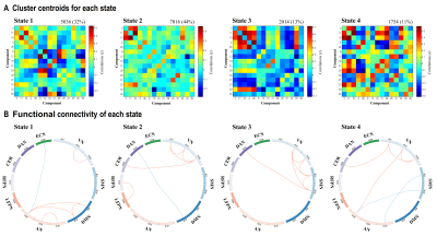 |
Altered intrinsic brain functional network dynamics in patients with end-stage renal disease undergoing maintenance hemodialysis
Baolin Wu1, Feifei Zhang1, Zhiyun Jia1,2, and Qiyong Gong1,3
1Huaxi MR Research Center (HMRRC), Department of Radiology, West China Hospital of Sichuan University, Chengdu, China, 2Department of Nuclear Medicine, West China Hospital of Sichuan University, Chengdu, China, 3Psychoradiology Research Unit of Chinese Academy of Medical Sciences (2018RU011), Chengdu, China
Although patients with end-stage renal disease (ESRD) have shown disrupted connectivity within and between resting-state functional networks, the patterns of change in dynamic functional network connectivity (FNC) remain unclear. The present work is the first study to investigate the dynamic functional connectivity in patients with ESRD. Patients with ESRD showed state-specific FNC disruptions and altered dynamic FNC properties. Furthermore, the total number of transitions was related to cognitive performance in those patients. These findings provided new insights into the pathophysiological mechanism of their cognitive deficits.
|
|||
1294.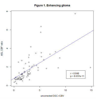 |
Diagnostic accuracy of ASL in comparison with DSC perfusion in the surveillance of different types of brain tumors
Anna Lavrova1, Wouter Teunissen2, Esther Warnert2, Martin van den Bent3, Vladimir Cheremisin1, and Marion Smits2
1Radiology, Saint Petersburg University, Saint Petersburg, Russian Federation, 2Radiology, Erasmus MC, Rotterdam, Netherlands, 3Neurology, Erasmus MC, Rotterdam, Netherlands
Dynamic susceptibility contrast-enhanced (DSC) perfusion is an established standard in the assessment of brain tumor perfusion. This study aims to assess the feasibility of using non-contrast arterial spin-labeling (ASL) instead of DSC.
|
|||
1295.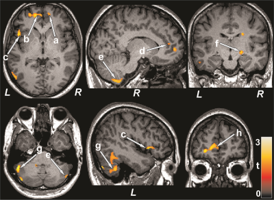 |
Regional Brain Perfusion Changes in Cognition and Mood Regulatory Sites in Patients with Type 2 Diabetes Mellitus
Bhaswati Roy1, Sarah Choi2, Matthew J. Freeby 3, and Rajesh Kumar1,4,5,6
1Anesthesiology, University of California Los Angeles, Los Angeles, CA, United States, 2School of Nursing, University of California Los Angeles, Los Angeles, CA, United States, 3Medicine, Endocrinology - Diabetes and Metabolism, University of California Los Angeles, Los Angeles, CA, United States, 4Bioengineering, University of California Los Angeles, Los Angeles, CA, United States, 5Radiological Sciences, University of California Los Angeles, Los Angeles, CA, United States, 6Brain Research Institute, University of California Los Angeles, Los Angeles, CA, United States
Patients with Type 2 diabetes mellitus (T2DM) show cognitive and mood changes, and brain tissue injury in those regulatory regions. However, underlying cause of tissue damage in cognition and mood regulatory sites, and their associations with these functional deficits in T2DM remain unclear. We evaluated cerebral blood flow (CBF) in T2DM patients over controls, and found changes in the frontal and prefrontal cortices, cerebellum, hippocampus, cingulate, insula, thalamus, and basal-forebrain, sites involved regulating mood and cognition. Significant correlations emerged between CBF and functional deficits in T2DM, including mood and cognition symptoms, implying the altered hemodynamic contributing to the functional deficits.
|
|||
1296.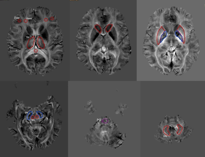 |
Application of quantitative susceptibility mapping in assessment of iron content in brain regions of normal children
Shilong Tang1 and Lisha Nie2
1Children's Hospital of Chongqing Medical University, Chongqing, China, 2GE Healthcare, MR Research China, Beijing, Beijing, China
In the central nervous system, iron is a cofactor for many metabolic processes and aminergic neurotransmitter synthesis, and iron plays an important role in brain development from the fetal to adolescent stages. There have been many reports about the application of quantitative magnetic susceptibility mapping (QSM) to detect iron content in the normal adult brain at home and abroad and few reports about the application of QSM to detect iron content in the normal brain of children.
|
|||
 |
1297.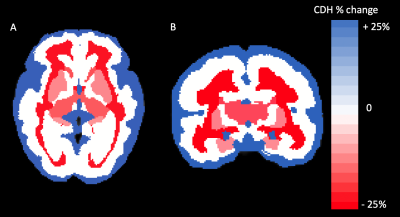 |
Regional Brain Growth in Fetuses with Congenital Diaphragmatic Hernia
Fedel Machado-Rivas1,2, Lina Acosta Buitrago3, Jungwhan J Choi1,2, Onur Afacan1,2, Clemente Velasco-Annis1, Simon K Warfield1,2, Ali Gholipour1,2, and Camilo Jaimes1,2
1Radiology, Boston Children's Hospital, Boston, MA, United States, 2Radiology, Harvard Medical School, Boston, MA, United States, 3Universidad del Rosario, Bogota, Colombia
Brain growth trajectories of fetal subjects with congenital diaphragmatic hernia (CDH) were compared to typically developing fetuses. T2-weighted SSFSE acquisitions were reconstructed to a super-resolution volume and atlas-based segmentations of brain structures were propagated. Gestational age, sex, segment volumes, and morphological hernia metrics (for CDH subjects) were analyzed in 71 fetuses with CDH and in 50 controls. We found lower brain volumes relative to normal fetuses in the developing white matter, hippocampus, diencephalon, and deep gray structures. Metrics associated with greater hernia severity were associated with lower volumes. Furhermore, our analysis shows that right-sided hernias were associated with lower volumes.
|
||
1298.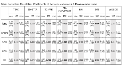 |
Inter-rater reliability of sciatic nerve evaluation with MR neurography: a comparison study of multiple sequences
Ryuna Kurosawa1, Hajime Yokota2, Takafumi Yoda1, Takayuki Sada1, Koji Matsumoto1, Takashi Namiki3, Masami Yoneyama3, Yoshitada Masuda1, and Takashi Uno2
1Department of Radiology, Chiba University Hospital, Chiba, Japan, 2Diagnostic Radiology and Radiation Oncology, Graduate School of Medicine, Chiba University, Chiba, Japan, 3Philips Japan, Tokyo, Japan
MR neurography (MRN) is a useful technique to evaluate damaged peripheral nerves showing increased signal intensity and enlarged nerve diameter. Although various MRN sequences have been developed, the inter-rater reliability of MRN have rarely been evaluated. Therefore, the purpose of this study was to evaluate inter-rater reliability of sciatic nerve evaluation with MRN sequences: T2WI, 3D-STIR, T2-FFE, 3D-iNerveVIEW, Diffusion-neurography, DTI, and pcDSDE. Measured diameters varied among the sequences. 3D-iNerveVIEW showed a high inter-rater reliability for signal measurements. In MRN, it is important to understand the characteristics of each sequence.
|
|||
|
1299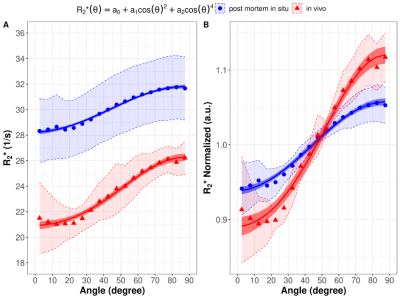 |
White Matter fiber orientation dependent R2*: comparison between post mortem in situ and in vivo Video Permission Withheld
Celine Berger1,2, Claudia Lenz1,2, Melanie Bauer1,2, Eva Scheurer1,2, and Christoph Birkl3
1Institute of Forensic Medicine, Department of Biomedical Engineering, Basel, Switzerland, 2Institute of Forensic Medicine, Health Department Basel-Stadt, Basel, Switzerland, 3Department of Neuroradiology, Medical University of Innsbruck, Austria
White matter R2* imaging is sensitive to the fiber orientation with respect to the main magnetic field. This study compared fiber orientation dependent R2* of white matter in post mortem (pm) in situ and in vivo based on an identical technical approach using diffusion tensor imaging to estimate the fiber angle. R2* increased with increasing fiber angle in both groups, whereby a decreased R2* orientation sensitivity was observed post mortem compared to in vivo. This may be mainly caused by the decreased pm brain temperature compared to in vivo conditions leading to reduced diffusion and by tissue decomposition after death.
|
||
|
1300.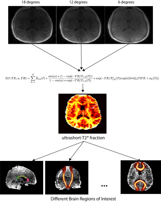 |
Reproducibility of brain ultrashort-T2* component measurements in healthy volunteers
Nikhil Deveshwar1,2 and Peder E. Z. Larson1,2
1Department of Radiology and Biomedical Imaging, University of California, San Francisco, San Francisco, CA, United States, 2UC Berkeley - UCSF Graduate Program in Bioengineering, Berkeley and San Francisco, CA, United States
This work presents a study detailing the reproducibility of ultrashort-T2* measurements using a novel UTE relaxometry method. Generated parameter maps show similar looking structures in terms of intensity and definition. Furthermore, the distributions of the ultrashort-T2* fractional component in various brain ROIs show similar distributions and have low coefficients of variance. These results suggest the proposed UTE relaxometry method is reproducible and can reliably measure parameters of the brain ultrashort-T2* component.
|
||
1301.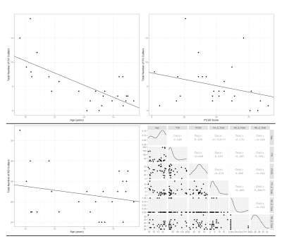 |
Correlating Concussion-Related Symptoms to the Personalized MRI Assessment of Brain Abnormalities in Children
Ethan Danielli1,2, David Stillo1,2, Rachelle Ho3,4, Carol DeMatteo3,5, Geoffrey B Hall4, Nicholas A Bock4, John F Connolly1,4,5,6, and Michael D Noseworthy1,2,5,7,8
1School of Biomedical Engineering, McMaster University, Hamilton, ON, Canada, 2Imaging Research Centre, St. Joseph's Healthcare Hamilton, Hamilton, ON, Canada, 3School of Rehabilitation Sciences, McMaster University, Hamilton, ON, Canada, 4Department of Psychology, Neuroscience & Behaviour, McMaster University, Hamilton, ON, Canada, 5ARiEAL Research Centre, McMaster University, Hamilton, ON, Canada, 6Department of Linguistics, McMaster University, Hamilton, ON, Canada, 7Department of Electrical and Computer Engineering, McMaster University, Hamilton, ON, Canada, 8Department of Radiology, McMaster University, Hamilton, ON, Canada
Although concussion diagnosis is often subjective, DTI can effectively quantify brain white matter damage. In this study, demographic and DTI metrics were compared between 26 paediatric concussion subjects against 49 healthy age and sex matched controls. FA was significantly reduced in injured brain regions and correlated with younger age and worsened symptoms. RD was significantly increased in injured brain regions and correlated with younger age and the interaction between time to scan and PCSS score. Based on these results, age affects concussion severity in younger children resulting in worse symptoms and a greater number of abnormal brain regions.
|
|||
1302.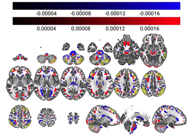 |
Cerebral perfusion network analysis to understand cognition in old age: a principal component analysis of ASL-MRI
Jodi Karlyn Watt1,2,3, Stefan Pszczolkowski1,2,3, Yue Xing1,2,3, Christopher Tench1,2,3, Dorothee Auer1,2,3, and Alzheimer's Disease Neuroimaging Initiative4
1Division of Clinical Neuroscience, University of Nottingham, Nottingham, United Kingdom, 2Sir Peter Mansfield Imaging Centre, University of Nottingham, Nottingham, United Kingdom, 3NIHR Nottingham Biomedical Research Centre, University of Nottingham, Nottingham, United Kingdom, 4Alzheimer's Disease Neuroimaging Initiative, Los Angeles, CA, United States
A functional biomarker of age-related cognitive decline is yet to be elucidated but would be desirable to better understand detrimental and protective processes in order to promote cognitive health for the elderly. Challenges arise from multiple sources of variance across individuals and within brain perfusion maps with an unknown link between local or network function and clinical tests of global cognition. Principal Component Analysis provides the opportunity to address some of these knowledge gaps, by defining this relationship and relating this to underlying anatomical regions.
|
|||
1303.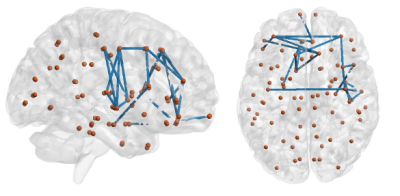 |
Altered structural connectivity and impairment of brain network-cognition relationship in obstructive sleep apnea (OSA)
Tasfiya Islam1, Mengting Liu1, Dae Lim Koo2, Ryan Cabeen1, Eunyeon Joo3, and Hosung Kim1
1USC Stevens Neuroimaging and Informatics Institute, Keck School of Medicine of University of Southern California, Los Angeles, CA, United States, 2Department of Neurology, Boramae Medical Center, Seoul National University College of Medicine, Seoul, Korea, Republic of, 33Department of Neurology, Samsung Medical Center, Sungkyunkwan University School of Medicine, Samsung Biomedical Research Institute, Seoul, Korea, Republic of
Obstructive sleep apnea (OSA) possibly affects individuals cognitively. To identify potential neurocognitive drawbacks for these patients, diffusion MRI and T1 neuroimaging data were collected from 148 OSA and healthy subjects. Structural brain networks were constructed using diffusion MR images. Network topological characteristics and localized connectivity were performed on diffusion related network along with brain and behavior relationships. It was found that the OSA individuals generally had greater modularity network measure, and impaired connectivity in several regions in the frontal lobe. Also, some of the brain connections and behavior relationships that were established in healthy subjects were disrupted for OSA patients.
|
|||
|
1304.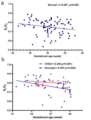 |
QSM detects early alterations of brain venous blood oxygenation in fetuses with complex congenital heart diseases
Cong Sun1, Aocai Yang1, Jiaguang Song2, Minhui Ouyang3, Jinxia Zhu4, Lei Xue5, Hao Huang3,6, and Gunagbin Wang1
1Radiology, Shandong Medical Imaging Research Institute, Cheeloo College of Medicine, Shandong University, Jinan, China, 2Ultrasound, Shandong Provincial Hospital Affiliated to Shandong University, Jinan, China, 3Radiology Research, Children’s Hospital of Philadelphia, Philadelphia, PA, United States, 4MR Collaboration, Healthcare Siemens Ltd., Beijing, China, 5MR Application, Siemens Healthineers Ltd., Jinan, China, 6Radiology, Perelman School of Medicine, University of Pennsylvania, Philadelphia, PA, United States
We investigated the early changes of brain oxygenic metabolism in fetuses with complex congenital heart disease (CHD) and in normal fetuses across gestational ages. Using quantitative susceptibility mapping (QSM), we measured the venous blood oxygen saturation (SvO2) in utero of 22 fetuses with complex CHD and 88 healthy pregnancy controls. The SvO2 in normal fetuses was found to have no age-related change. SvO2 values were significantly higher in the CHD fetuses (80.8%±4.6%) than in the gestational age-matched normal fetuses (75.7%±8.0%) (p=0.038); which is evidence of altered human fetal brain oxygenic metabolism during the early stages of brain development.
|
||
1305.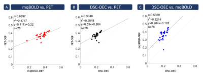 |
Quantitative cerebral oxygenation mapping by MRI with whole brain coverage compared to PET
Jan Kufer1, Christine Preibisch1,2, Samira Epp1, Jens Goettler1,3, Kilian Weiss4, Mikkel Bo Hansen5, Claus Zimmer1, Kim Mouridsen5, Fahmeed Hyder3, and Stephan Kaczmarz1,3
1Department of Neuroradiology, School of Medicine, Technical University of Munich (TUM), Munich, Germany, 2Department of Neurology, School of Medicine, Technical University of Munich (TUM), Munich, Germany, 3Department of Radiology & Biomedical Imaging (MRRC), Yale University, New Haven, CT, United States, 4Philips Healthcare, Hamburg, Germany, 5Department of Clinical Medicine, Aarhus University, Aarhus, Denmark
Clinical imaging of the oxygen extraction fraction (OEF) is highly promising to improve stratification of patients with various neurological diseases. Measurement of OEF by multiparametric quantitative blood oxygen level dependent (mqBOLD)-MRI could greatly increase clinical applicability compared to the current gold standard PET. Furthermore, oxygen extraction capacity (OEC) has recently emerged as another MRI-based biomarker of cerebral oxygenation. However, studies comparing both MRI techniques to PET reference data are still lacking. Here, we present data from an MRI study in young healthy volunteers, demonstrating good agreement of both, MRI-based OEF and OEC, with PET data from a similar subject group.
|
|||
1306.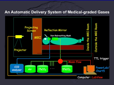 |
Carbogen-based Cerebrovascular Reserve Using BOLD-based fMRI of Human Brain : tissue, territorial and cortical specificity
Tzu-chen Yeh1,2, Chou-ming Cheng3, and Chi-che Chou3
1Department of Radiology, Taipei Veterans General Hospital, Taipei, Taiwan, 2Institue of Brain Science, National Yang-Ming University, Taipei, Taiwan, 3Department of Medical Research, Taipei Veterans General Hospital, Taipei, Taiwan
Carbogen-based cerebrovascular reserve (CO2-CVR), derived from 90 normal subjects, was constructed for data-based approaches or analysis. Tissue, territorial and cortical specificities of CO2-CVR were evaluated by regions of interest (ROI) approaches. Global ratio of GM/WM CO2-CVR was about 1.57. Territorial and cortical specificities of CO2-CVR demonstrated (1) limited CO2-CVR in territories of posterior circulation which supported the pathophysiology of posterior reversible encephalopathy syndrome and (2) vital function in distal territories of anterior cerebral artery as verified by cortical parcellation with the highest of CO2-CVR at ventral portion of Brodmann area 23 (co-localized with posterior cingulate cortices of default mode network).
|
|||
1307.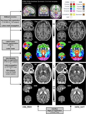 |
Clinical Whole-Brain R2* and Quantitative Susceptibility Maps at 3T – Reproducibility and Parameter Optimization Towards Millimetric 5min Scan
Thomas Troalen1, Arnaud Le Troteur2, Sylviane Confort-Gouny2, Patrick Vioux2, Claire Costes2, Lauriane Pini2, Jean-Philippe Ranjeva2, Maxime Guye2, and Ludovic de Rochefort2
1Siemens Healthcare SAS, Saint-Denis, France, 2CRMBM UMR7339 CNRS Aix-Marseille Université, Marseille, France
Advanced quantitative susceptibility mapping techniques allow to jointly estimate R2* and QSM in the brain. This work aims at comparing several multi gradient-recalled echo sequences in terms of coil setup and acceleration factors using available product sequences and modern hardware. In addition, an automatic and standardized post-processing pipeline is proposed for clinical studies. We show that quantitative values do not differ from the head coil and acceleration factor that were used, thus allowing for a millimetric whole brain coverage within 5 minutes scan time.
|
|||
1308.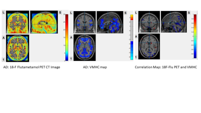 |
Interhemispheric Functional Connectivity and 18F-fluatmetamol PET differentiate AD, amyloid and non-amyloid Mild cognitive decline
Eva YW Cheung1, Patrick KC Chiu2, YF Shea2, Joseph SK Kwan3, and Henry KF Mak1
1The University of Hong Kong, Hong Kong, Hong Kong, 2Queen Mary Hospital, Hong Kong, Hong Kong, 3Imperial College London, London, United Kingdom
Interhemispheric functional connectivity and 18F-flutametamol PET
|
|||
1309.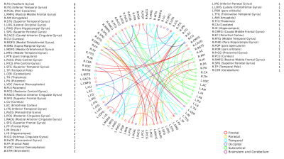 |
Associations of Musical Aptitude with High Angular Resolution Diffusion Imaging (HARDI) derived structural connectivity
Archith Rajan1, Apurva Shah1, Madhura Ingalhalikar1, and Nandini C Singh2
1Symbiosis Centre for Medical Image Analysis, Symbiosis International(Deemed) University, Pune, India, 2Language,Literacy and Music Lab, National Brain Research Centre, Gurgaon, India
Pre-existing structural connectivity could well explain predisposition to musical aptitude. The study aimed to associate Individual differences in High Angular Resolution Diffusion Imaging (HARDI) derived structural connectivity to music perception abilities as assessed by the performance in a music perception test. An increased whole brain connectivity was found to be associated with increased performance especially in the sequential music perception measures that comprised of Standard Rhythm, Embedded Rhythm, Accent and Melody. A prevalence of interhemispheric connectivity over intrahemispheric connectivity was also observed. Distinct structural connectivity patterns could thus be a determinant of sequential processing aptitude in music.
|
|||
1310.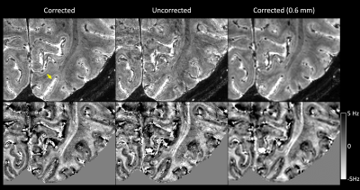 |
Imaging intracortical structure using navigator-based, motion and B0-corrected T2*-weighted MRI at 7 T
Jiaen Liu1, Peter van Gelderen1, Jacco A. de Zwart1, and Jeff H. Duyn1
1AMRI, LFMI, NINDS, National Institutes of Health, Bethesda, MD, United States
High-resolution T2*-weighted 7 T MRI can be used to delineate intracortical structure owning to its high signal-to-noise and contrast-to-noise ratio. However, subject motion remains a major challenge to obtain reliable high-resolution data; the long scan time and increased sensitivity of T2*-weighted signal at 7 T to motion and motion-induced B0 fluctuation make high-resolution T2*-weighted MRI particularly motion sensitive. In this study, the performance of a previously developed navigator-based motion and B0 correction approach was evaluated for intracortical imaging.
|
|||
1311. |
Detecting normal Fetal brain development with T1Mapping Imaging Technique
Yan-Chao Liu1, Bo-Hao Zhang2, De-Sheng Xuan2, Xue-Yuan Wang2, Kai-Yu Wang3, Xin Zhao2, and Xiao-An Zhang2
1Department of Radiology, The Third Affiliated Hospital of Zhengzhou University, Zhengzhou, China, 2Department of Radiology, the Third Affiliated Hospital of Zhengzhou University, Zhengzhou, 450052, China, Zhengzhou, China, 3MR Research China, GE Healthcare, Beijing 100000, PR China, Beijing, China
Fetal brain development is an ongoing process, and it is necessary to find a sensitive monitoring tool to detect potential brain developmental abnormalities earlier. In this work, T1Mapping allowed quantitative assessment of fetal brain development.
|
The International Society for Magnetic Resonance in Medicine is accredited by the Accreditation Council for Continuing Medical Education to provide continuing medical education for physicians.