Digital Posters
Mapping Brain Volumes & Lesions
ISMRM & SMRT Annual Meeting • 15-20 May 2021

Digital Posters
Mapping Brain Volumes & Lesions
| Concurrent 6 | 15:00 - 16:00 |
2142.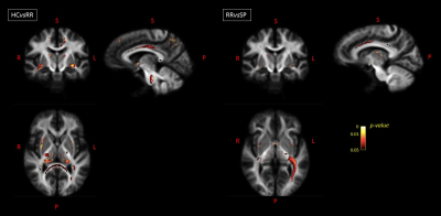 |
Assessment of White Matter Atrophy in Multiple Sclerosis Using Advanced Diffusion Weighted Imaging Models
Loredana Storelli1, Elisabetta Pagani1, Paolo Preziosa1,2, Massimo Filippi1,2,3,4,5, and Maria A. Rocca1,2,5
1Neuroimaging Research Unit, Division of Neuroscience, IRCCS San Raffaele Scientific Institute, Milan, Italy, 2Neurology Unit, IRCCS San Raffaele Scientific Institute, Milan, Italy, 3Neurorehabilitation Unit, IRCCS San Raffaele Scientific Institute, Milan, Italy, 4Neurophysiology Service, IRCCS San Raffaele Scientific Institute, Milan, Italy, 5Vita-Salute San Raffaele University, Milan, Italy
When investigating white matter (WM), its complex microstructure should be considered. Neurite Orientation Dispersion and Density Imaging (NODDI) and the constrained spherical deconvolution (CSD) are diffusion weighted imaging models that account for this complexity, compared to the commonly used MRI techniques. In this study, we applied volumetric, diffusion tensor, NODDI and CSD models to 86 patients with multiple sclerosis and 55 healthy controls at baseline and after 1-year of follow-up. The comparison of these techniques both globally and voxel-based showed that the CSD model was able to identify WM atrophy offering greater anatomical specificity and biological interpretability.
|
|||
2143.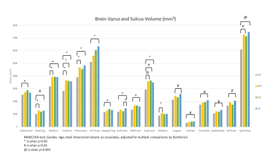 |
Association between interhemispheric functional connectivity (IFC) and brain structural volume in patients with AD, MCI and VD
Eva YW Cheung1, Patrick KC Chiu2, YF Shea2, Joseph SK Kwan3, and Henry KF Mak1
1The University of Hong Kong, Hong Kong, Hong Kong, 2Queen Mary Hospital, Hong Kong, Hong Kong, 3Imperial College London, London, United Kingdom
The findings demonstrated the interhemispheric functional connectivity correlates with brain regional volume.
|
|||
2144. |
Towards to quantitative mapping of grey matter deficits in children with treatment-naïve attention deficit/hyperactivity disorder
Shu Su1, Yingqian Chen1, Yan Dai1, Long Qian2, Hongyu Zhang1, and Zhiyun Yang1
1First Affiliated Hospital, Sun Yat-sen University, Guang Dong, China, 2MR Research, GE Healthcare, Beijing, China
As the most common childhood-onset neurodevelopmental disorder, the pathogenesis of attention deficit hyperactivity disorder (ADHD) is still unclear. The goal of current study aims at investigating the quantitative profiles of brain grey matter in pediatric treatment-naïve ADHD using Synthetic MRI (SyMRI).
|
|||
2145.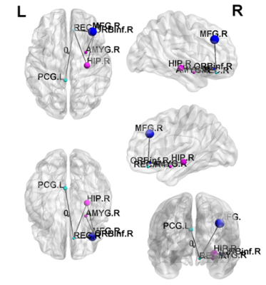 |
Mapping brain structure and network alterations in male chronic smokers using generalized q-sampling MRI
Jun-Cheng Weng1,2,3, Li-Bang Zheng1, Ming-Shih Lee4,5, and Ming-Chou Ho6,7
1Department of Medical Imaging and Radiological Sciences, and Bachelor Program in Artificial Intelligence, Chang Gung University, Taoyuan, Taiwan, 2Medical Imaging Research Center, Institute for Radiological Research, Chang Gung University and Chang Gung Memorial Hospital at Linkou, Taoyuan, Taiwan, 3Department of Psychiatry, Chang Gung Memorial Hospital, Chiayi, Taiwan, 4Department of Medical Laboratory and Biotechnology, Chung Shan Medical University, Taichung, Taiwan, 5Clinical Laboratory, Chung Shan Medical University Hospital, Taichung, Taiwan, 6Department of Psychology, Chung Shan Medical University, Taichung, Taiwan, 7Clinical Psychological Room, Chung Shan Medical University Hospital, Taichung, Taiwan
The World Health Organization suggests that cigarette smoking causes more than 7 million deaths every year. However, few studies have focused on the structural alternations of chronic smoking by using generalized q-sampling imaging (GQI). We aimed to use GQI to evaluate the impact of the neurological structure and network caused by chronic smoking. Our results provided further evidence indicating that chronic smoking may lead to brain structure and connectivity changes.
|
|||
2146.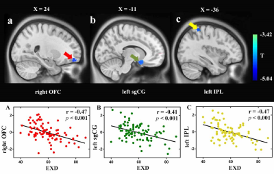 |
Regional Gray Matter Volume Associated with Exercise Dependence: A Voxel-Based Morphometry Study
Zhang Feifei1, Zhiyun Jia2, and Qiyong Gong2
1Sichuan University, Chengdu, China, 2Huaxi MR Research Center (HMRRC), Department of Radiology, West China Hospital of Sichuan University, Chengdu 610041, China, ChengDu, China
The present study provides initial evidence indicating that a higher risk of EXD is linked with a smaller GMV in the right OFC, left sgCG, and left IPL in a sample of regular exercisers, which sheds light on the neuroanatomical basis of EXD. Furthermore, our study found that the right OFC mediates the relationship between stress and EXD, revealing a potential neuropsychological mechanism for how stress affects EXD. Our findings might facilitate the diagnosis of EXD and the target selection for the corresponding intervention (e.g., behavioral or brain intervention ) to help individuals reduce EXD and improve their quality of life.
|
|||
2147.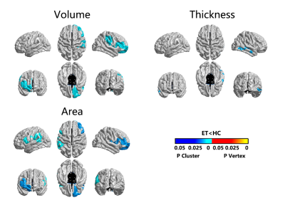 |
Essential tremor: Structural Characterization with 3-T MR Imaging
Jing Yang1, Du Lei2, Xueling Suo3, Jiaxin Peng4, Rong Peng4, and Qiyong Gong3
1Huaxi MR Research Center (HMRRC), West China Hospital of Sichuan University, sichuan, China, 2Department of Psychiatry and Behavioral Neuroscience, University of Cincinnati, Cincinnati, OH, United States, 3Huaxi MR Research Center (HMRRC), West China Hospital of Sichuan University, Chengdu, China, 4Department of Neurology, West China Hospital of Sichuan University, Chengdu, China
Present neuroradiological studies demonstrated significant alterations in surface-based morphology in patients with essential tremor (ET) relative to healthy controls (HC). The current study was achieved by exploring the abnormal brain morphological features of patients with ET by using three-dimensional T1-weighted imaging, as well as the relationship between demographic/clinical variables and affected structural cortical features. We found that ET patients showed significant structural changes both in the motor and non-motor brain regions and some abnormities were correlated with the clinical scales. Further, abnormalities attributed to ET can also be affected by various demographic and clinical variables.
|
|||
2148. |
Volume-based brain morphometry and T1 mapping: comparison between MP2RAGE accelerated by parallel imaging and compressed sensing at 3 Tesla
Blanche Bapst1,2, Aurélien Massire3, Tobias Kober4,5,6, Gian Franco Piredda4,5,6, Bénédicte Maréchal4,5,6, and Pierre Brugières1
1Department of Neuroradiology, Henri Mondor Universitary Hospital, AP-HP, Créteil, France, 2Faculty of Medicine, Université Paris Est Créteil, Créteil, France, 3Siemens Healthcare SAS, Saint-Denis, France, 4Advanced Clinical Imaging Technology, Siemens Healthcare AG, Lausanne, Switzerland, 5Department of Radiology, Lausanne University Hospital and University of Lausanne, Lausanne, Switzerland, 6Signal Processing Laboratory (LTS 5), École Polytechnique Fédérale de Lausanne (EPFL), Lausanne, Switzerland
MP2RAGE is 3D T1-weighted MR sequence used for high GM/WM contrast imaging, enabling morphometry, brain segmentation and T1 mapping. Despite these advantages, it suffers from long acquisition times, hindering broad clinical adoption. In this work, brain data obtained from 29 patients with various pathologies using the GRAPPAx3-accelerated reference (8:22 min) and a new compressed-sensing version (4:00 min) of the MP2RAGE were compared with respect to brain volumetry and T1 mapping results. Very high agreement was observed between the two versions. These results pave the way for a broader use of CS-MP2RAGE, eventually enabling its clinical use.
|
|||
2149. |
Comparison of cortical gray matter and subcortical gray matter using double diffusion encoding
Shimpei Kato1,2, Kouhei Kamiya3, Koji Kamagata1, Hiroshi Kusahara4, Masahiro Abe4, Shohei Fujita1,2, Toshiaki Akashi1, Katsuhiro Sano1, Akihiko Wada1, Masaaki Hori3, Osamu Abe2, and Shigeki Aoki1
1Department of Radiology, Juntendo University School of Medicine, Tokyo, Japan, 2Department of Radiology, Graduate School of Medicine, The University of Tokyo, Tokyo, Japan, 3Department of Radiology, Toho University Omori Medical Center, Tokyo, Japan, 4Advanced MRI development PJ Team, Canon Medical Systems corp., Kanagawa, Japan
Cortical and subcortical gray matter (GM) are histologically different and have different DTI values. In this study, we measured the microscopic fractional anisotropy (μFA) of cortical and subcortical GM in healthy subjects using double-diffusion encoding. We confirmed that the μFA and fractional anisotropyof subcortical GM were higher than those of cortical GM. Additionally, the μFA in the globus pallidus was high than in the striatum. Myelin was higher in subcortical GM than in cortical GM, and it was higher in globus pallidus than in the striatum; therefore, higher μFA may reflect these histological features.
|
|||
2150.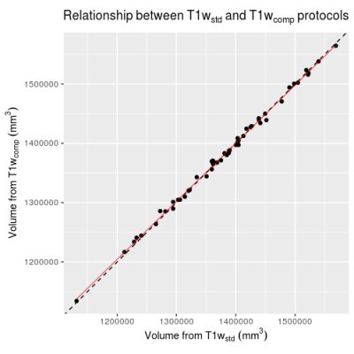 |
Structural 3DT1 scans with compressed sensing are suitable for cross-sectional brain volume measures in multiple sclerosis
Jonathan Stutters1, Marco Battiston1, Nevin John1, Thomas Williams1, Claudia Wheeler-Kingshott1,2,3, Frederik Barkhof1,4,5, Jeremy Chataway1, and Ferran Prados1,4,6
1NMR Research Unit, Queen Square MS Centre, Department of Neuroinflammation, UCL Queen Square Institute of Neurology, Faculty of Brain Sciences, University College London (UCL), London, United Kingdom, 2Department of Brain & Behavioural Sciences, University of Pavia, Pavia, Italy, 3Brain Connectivity Center Research Department, IRCCS Mondino Foundation, Pavia, Italy, 4Centre for Medical Image Computing (CMIC), Department of Medical Physics and Bioengineering, University College London (UCL), London, United Kingdom, 5Department of Radiology and Nuclear Medicine, VU University Medical Centre, Amsterdam, Netherlands, 6e-Health Centre, Universitat Oberta de Catalunya, Barcelona, Spain
Optimizations of a 3D T1 sequence to take advantage of recent improvements to the hardware and software of a Philips Achieva MRI scanner allowed an acquisition to be performed in one third of the original time. We compared a number of cross-sectional volume measures, often used in research studies or as outcome measures of clinical trials, computed from images obtained with this protocol and the more widely used non-accelerated one. We find that cross-sectional volume measures are highly correlated between the accelerated and non-accelerated protocols, warranting the adoption of the accelerated one in clinical studies and clinical trials.
|
|||
2151.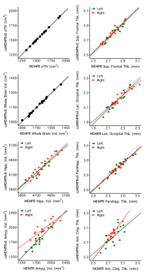 |
Exploration of highly accelerated multi-echo MPRAGE using compressed sensing for brain morphometry applications
Lindsay C Hanford1,2,3, Emily M Iannazzi1,2, Tom Hilbert4,5,6, Tobias Kober4,5,6, Randy L Buckner1,2,3, and Ross W Mair1,3
1Center for Brain Science, Harvard University, Cambridge, MA, United States, 2Department of Psychology, Harvard University, Cambridge, MA, United States, 3Martinos Center for Biomedical Imaging, Massachusetts General Hospital, Charlestown, MA, United States, 4Advanced Clinical Imaging Technology, Siemens Healthcare, Lausanne, Switzerland, 5Department of Radiology, Lausanne University Hospital and University of Lausanne, Lausanne, Switzerland, 6LTS5, École Polytechnique Fédérale de Lausanne, Lausanne, Switzerland
Compressed sensing (cs) has the potential to shorten scan acquisition time. This time savings may help to reduce patient burden, MR artifacts due to motion, and cost for repeat acquisitions. A key open question is whether time-saving cs T1w structural images will yield quantitative morphometric estimates comparable to traditional longer sequences. To test this, images were acquired for numerous cs T1w image variations across two independent datasets. Structural estimates were compared within- and between-subjects to assess scan stability and the effect of acceleration. x6 cs acceleration (~86s scan) was sufficient to yield comparable morphometrics for most measures in typical applications.
|
|||
2152. |
Are rapid structural scans viable for automated lesion assessment in MS patients ?
Mathilde Carrière1, Bénédicte Maréchal2,3,4, Jérémy Deverdun1, Thomas Troalen5, Tobias Kober2,3,4, Ricardo Corredor-Jerez2,3,4, and Emmanuelle Le Bars1
1I2FH, Neuroradiology, CHU Montpellier, University of Montpellier, Montpellier, France, 2Advanced Clinical Imaging Technology, Siemens Healthcare AG, Lausanne, Switzerland, 3Department of Radiology, Lausanne University Hospital and University of Lausanne, Département de radiologie, Hôpital Universitaire de Lausanne et Université de Lausanne (UNIL), Lausanne, Switzerland, 4LTS 5, École Polytechnique Fédérale de Lausanne (EPFL), Lausanne, Switzerland, 5Siemens SAS Healthcare, Saint-Denis, France
Current MR protocol guidelines for multiple sclerosis (MS) diagnosis and follow-up recommend the acquisition of 3D sequences as high isotropic resolution improves lesion conspicuity; however, this prolongs clinical scan protocols. We present a qualitative and quantitative comparison of lesion assessment using a standard 3D FLAIR and an optimized CAIPIRINHA version in conjunction with compressed sensing MPRAGE in 44 MS patients. We compared the automated lesion segmentation results between protocols, with and without additional manual corrections of the lesion masks. Volumes using the optimized protocol highly correlated with the results from the conventional protocol, while reducing the acquisition time by 47%.
|
|||
2153.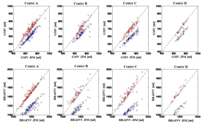 |
Atrophy Quantification in Multiple Sclerosis: Application to the Multicenter INNI Dataset
Loredana Storelli1, Elisabetta Pagani1, Patrizia Pantano2,3, Nikolaos Petsas2, Gioacchino Tedeschi4, Antonio Gallo4, Nicola De Stefano5, Marco Battaglini5, Paola Zaratin6, Maria A. Rocca1,7,8, and Massimo Filippi1,7,8,9,10
1Neuroimaging Research Unit, Division of Neuroscience, IRCCS San Raffaele Scientific Institute, Milan, Italy, 2Department of Human Neurosciences, "La Sapienza" Rome University, Rome, Italy, 3Department of Radiology, IRCCS Neuromed, Pozzilli, Italy, 4Department of Advanced Medical and Surgical Sciences, and 3T MRI Center, University of Campania “Luigi Vanvitelli”, Naples, Italy, 5Department of Medicine, Surgery and Neuroscience, University of Siena, Siena, Italy, 6Fondazione Italiana Sclerosi Multipla, Genoa, Italy, 7Neurology Unit, IRCCS San Raffaele Scientific Institute, Milan, Italy, 8Vita-Salute San Raffaele University, Milan, Italy, 9Neurorehabilitation Unit, IRCCS San Raffaele Scientific Institute, Milan, Italy, 10Neurophysiology Service, IRCCS San Raffaele Scientific Institute, Milan, Italy
Using the multicenter dataset available within the Italian Neuroimaging Network Initiative, we compared the performance of different atrophy tools on brain MRI from 457 multiple sclerosis (MS) patients and 271 healthy controls, since monitoring neurodegeneration is one of the most important goals of therapeutic strategies in MS. We found an acceptable agreement and comparable performances among the software for GM and whole brain atrophy quantification. The free licence, speed and facility of integration in the clinical routine are also important aspects to consider for the selection of the atrophy pipeline. These results should be confirmed on large-scale longitudinal data.
|
|||
| 2154. | Cross-species homogeneity of brain structural covariance between humans and non-human primates based on a primate brain parcellation
Ge Zhang1, Zheng Wang2, and Meiyun Wang3
1Radiology, Henan Provincial People's Hospital, Zhengzhou, China, 2CAS, Shanghai, China, 3Henan Provincial People's Hospital, Zhengzhou, China
To understand the structural consistency across primate brains is very important to perform neuroimaging computation, especially when it comes to study human brains through monkey-based experiments. Here we investigate the brain alignment by comparing structural covariance network calculated with the Regional Map parcellation templates. The unimodal areas and part of frontal areas exhibit consistency in cortical thickness and structural covariance. But M1 regions and lateral part of PFC were not similar in brain morphology. The result provide reference in cross-species brain imaging calculation based on Regional Map.
|
|||
2155.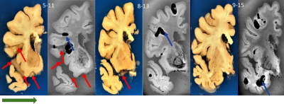 |
Reusable 3D printed enclosure with integrated cutting guides for the alignment of ex-vivo MRI with ex-vivo gross brain photographs
Nadim Farhat1, Julia Kofler2,3, Jacob Berardinelli1, Mark Stauffer4, Tales Santini1, Neilesh Vinjamuri1, Andrea Sajewski 1, Salem Alkhateeb1, Tiago Martins1, Noah Schweitzer 1, Milos Ikonomovic3,5, Howard J. Aizenstein1,6, and Tamer S Ibrahim1,6,7
1Bioengineering, University of Pittsburgh, Pittsburgh, PA, United States, 2Pathology, UPMC, Pittsburgh, PA, United States, 3Alzheimer's Disease Research Center, University of Pittsburgh, Pittsburgh, PA, United States, 4Pathology, University of Pittsburgh, Pittsburgh, PA, United States, 5Neurology, University of Pittsburgh, Pittsburgh, PA, United States, 6Psychiatry, University of Pittsburgh, Pittsburgh, PA, United States, 7Radiology, University of Pittsburgh, Pittsburgh, PA, United States In this project, we implemented a novel approach to align ex-vivo MRI brain images and gross neuropathology photographs with minimal image processing. The approach included the design and implementation of a reusable 3D printed enclosure with integrated cutting guides . Our results show a good alignment between ex-vivo high field MRI and gross brain image photographs. |
|||
2156.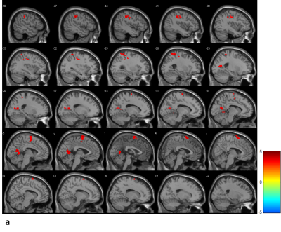 |
Differential cognitive impairment of lateral ventricle small infarct: Brain atrophy and blood oxygen activity under the cortical layer
Xuchen Yu1,2, Jiawei Han 3, Hui Zhang1,2, Min He4, Weibo Chen5, Jing Ding4, and He Wang1,2,3
1Institute of science and technology for Brain-Inspired Intelligence, Shanghai, China, 2Key Laboratory of Computational Neuroscience and Brain-Inspired Intelligence (Fudan University), Ministry of Education, Shanghai, China, 3Human Phenome Institute, Fudan University, Shanghai, China, 4Department of Neurology, The Affiliated Zhongshan Hospital of Fudan University, Shanghai, China, 5Philips Healthcare, Shanghai, China
Most of the lacunar infarcts are small and asymptomatic, while the location and accumulation of multiple lacunar infarctions could cause significant physical and cognitive disabilities. In this study, we explore the association between cognitive impairment and brain atrophy in patients with lateral ventricle infarction and whether there were differences in neuro-radiological features.
|
|||
2157.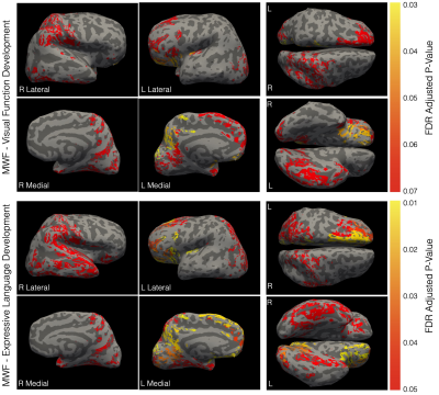 |
Changing Cortical Myeloarchitecture and Not Morphology Are Associated with Early Cognitive Development
Sean Deoni1, Lexie Volpe2, Jennifer Beauchemin2, and Viren D'Sa3
1Bill & Melinda Gates Foundation, Seattle, WA, United States, 2Advanced Baby Imaging Lab, Rhode Island Hospital, Providence, RI, United States, 3Pediatrics, Rhode Island Hospital, Providence, RI, United States
Early brain development is punctuated by rapid changes in the white and gray matter, including myelination and advancing microstructure, architecture, volume, and geometry. Here we explored the relationships between, and influence of, these changing neuroanatomical properties and advancing cognitive skill in healthy and typically developing toddlers and young children. Unlike past results in older children and adults, we find few significant associations between cortical morphometry measures and cognition. Instead, we find strong associations between intra-cortical myelin content and measures of language and visual reception function.
|
|||
2158.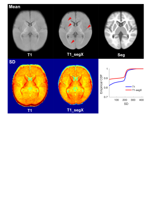 |
Mapping Infant Anatomical Brain Images Between Birth and Six Months Using Segmented Brain Images
Longchuan Li1, Pui-Yee Wong1, Warren Jones1, Ami Klin1, and Sarah Shultz1
1Department of Pediatrics, Marcus Autism Center, Children's Healthcare of Atlanta, Emory University, Atlanta, GA, United States
Image registration is a critical step for robust and accurate atlas-based analyses on brain volumetric changes. This is especially a challenge in brain growth studies in infants between birth and 6 months of life, when both T1w and T2w magnetic resonance images experience time-varying contrast changes. In this study, we compared image registration outcomes via T1w and tissue segmented images derived using a deep learning method. Our results demonstrated superior registration accuracy based on segmentation images, probably due to clear boundaries between tissues types, as well as their time-constant contrasts over development.
|
|||
2159.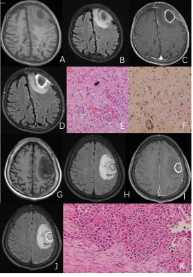 |
The value of contrast enhanced T2 FLAIR in the differential diagnosis of intracranial ring-enhanced lesions
Meng-Yao Su1, Zhi-Peng Zhou1, Mei Long1, Wen Su1, Xiao-Wei Lu1, and Long Qian2
1Affiliated Hospital of Guilin Medical University, Guilin, China, 2MR Research, GE Healthcar, Beijing, China
The goal of current study is to validate whether the contrast enhanced T2 FLAIR (CE-T2-FLAIR) could provide additional value for clinical diagnosis beyond the regular sequence. A retrospective analysis of 52 cases of intracranial ring-enhanced lesions validated by menstrual surgery, pathology or clinical follow-up observation from January 2019 to June 2020 was performed in current study. Our results demonstrated that the CE-T2-FLAIR "edge enhancement sign" was more closely related to inflammatory lesions, and indicated a high possibility of that disease.
|
|||
2160.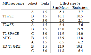 |
Sensitivity of structural brain MRI in clinical cross-sectional studies
Leighton Barnden1, Sonya Marshall-Gradisnik1, Donald Staines1, Ben Crouch2, and Zack Shan3
1Griffith University, Southport, QLD, Australia, 2Nuclear Medicine, Royal Adelaide Hospital, Adelaide, SA, Australia, 3University of Sunshine Coast, Sunshine Coast, QLD, Australia
The sensitivity of different structural MRI sequences for the detection of clinical abnormalities in cross-sectional studies was evaluated using actual population studies. We studied 3 patient cohorts on two MRI scanners (1.5T and 3T), using T1wSE, T2wSE, T1GRE, T2SPACE and MTC sequences for clinical group comparisons. Novel insights into their clinical potential of different structural MRI scans indicated T1wSE and T2wSE offer advanced sensitivity and should be standard in clinical cross-sectional studies. . Although our sensitivity metric was computed for separate voxels, we expect inter-subject variance will have a similar effect on the sensitivity for cluster detection.
|
|||
2161.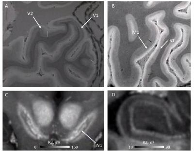 |
Ultra-high resolution quantitative multi-parameter mapping (MPM) for post-mortem whole brain microstructure imaging
Evgeniya Kirilina1,2, Ilona Lipp1, Kerrin Pine1, Luke Edwards1, Carsten Jäger1,3, Kirsten Garus4, Markus Cremer4, Katrin Amunts4,5, and Nikolaus Weiskopf1,6
1Neurophysics, Max Planck Institute for Human Cognitive and Brain Sciences, Leipzig, Germany, 2Center for Cognitive Neuroscience Berlin, Freie Universität Berlin, Berlin, Germany, 3Paul Flechsig Institute of Brain Research, University of Leipzig, Leipzig, Germany, 4INM-1, Structural and Functional Organisation of the Brain, Forschungszentrum Juelich GmbH, Juelich, Germany, 5Cecile und Oskar Vogt Institute for Brain Research, Heinrich Heine University Duesseldorf, Duesseldorf, Germany, 6Felix Bloch Institute for Solid State Physics, Faculty of Physics and Earth Sciences, Leipzig University, Leipzig, Germany
Reference neuroanatomical brain data are required to validate qMRI microstructural metrics and link them to histological measures. The BigBrain dataset provides unique cytoarchitectonic 3D atlas based on several post-mortem human brains. We aim to complement the BigBrain atlas by ultrahigh-resolution quantitative multi-parameter maps. Here, we present ultra-high resolution quantitative multi-parameter maps (MPM) acquired at 3T and 7T on a post-mortem brain. We demonstrate high data quality and feasibility of cortical layer and subcortical nuclei analysis. The brain will be processed following the BigBrain cytoarchitectonic atlasing procedure and will reveal specific qMRI fingerprints of cortical areas with distinct cytoarchitecture.
|
The International Society for Magnetic Resonance in Medicine is accredited by the Accreditation Council for Continuing Medical Education to provide continuing medical education for physicians.