Digital Posters
MR Fingerprinting: Artifacts, Optimisation & Applications in the Body
ISMRM & SMRT Annual Meeting • 15-20 May 2021

Digital Posters
MR Fingerprinting: Artifacts, Optimisation & Applications in the Body
| Concurrent 1 | 19:00 - 20:00 |
1551.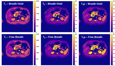 |
T1ρ Magnetic Resonance Fingerprinting of Chronic Pancreatitis
Cory R. Wyatt1, Kaveh R. Sharzehi2, Erin R. Gilbert3, Brett R. Sheppard3, and Alexander R. Guimaraes1
1Diagnostic Radiology, Oregon Health and Science University, Portland, OR, United States, 2Gastroenterology and Hepatology, Oregon Health and Science University, Portland, OR, United States, 3Surgery, Oregon Health and Science University, Portland, OR, United States
T1 relaxation mapping has been shown to demonstrate significant differences in the pancreas of patients with chronic pancreatitis. However, T2 and T1ρ relaxation times have been largely unexplored in pancreas, due to difficulty acquiring these values in the abdomen. In this study, magnetic resonance fingerprinting (MRF) techniques are applied to simultaneously acquire T1, T2, and T1ρ relaxation times in the pancreas of healthy volunteers and patients with clinically diagnosed chronic pancreatitis (CP). A significant increase in T1 relaxation was found with near significant increases in T2 and T1ρ relaxation times in CP patients.
|
|||
1552.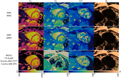 |
Myocardial T1, T2, T2* and Fat Fraction Quantification via Low-Rank Motion-Corrected Cardiac MRF
Gastao Cruz1, Carlos Velasco1, Olivier Jaubert1, Haikun Qi1, René M. Botnar1, and Claudia Prieto1
1School of Biomedical Engineering and Imaging Sciences, King's College London, London, United Kingdom
Cardiac Magnetic Resonance Fingerprinting (MRF) has been proposed for simultaneous myocardial T1, T2 and fat fraction quantification using ECG-triggering, mid-diastolic acquisition window and a 3-echo gradient echo sequence. Here we extend this framework to further enable T2* quantification. This is achieved with an 8-echo sequence with increased acquisition window (to acquire sufficient data within a breath-hold) and a low-rank motion correction reconstruction to correct for cardiac motion within this increased window. The proposed approach enables simultaneous mapping of T1, T2, T2* and fat fraction within a single breath-hold with similar quality to conventional (sequential) single parameter approaches.
|
|||
1553.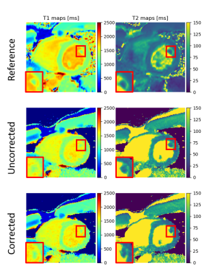 |
Cardiac motion-corrected image reconstruction for Cardiac Magnetic Resonance Fingerprinting
Constance G.F. Gatefait1, Kirsten M. Kerkering1, Sebastian Schmitter1, and Christoph Kolbitsch1
1Physikalisch-Technische Bundesanstalt (PTB), Braunschweig and Berlin, Germany
Cardiac magnetic resonance fingerprinting (cMRF) is a promising framework for quantitative assessment of various cardiomyopathies. One major challenge is cardiac motion. The majority of cMRF methods use ECG triggering and gating to select only certain cardiac phases to reconstruct cMRF data. In this study, we propose an iterative motion-correction approach utilizing the entire cardiac MRF acquisition. Obtained results show an improvement in obtained maps and consistent quantifications of T1 and T2.
|
|||
1554.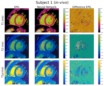 |
A Neural Network for Rapid Generation of T1, T2, T1ρ Dictionaries for Cardiac MR Fingerprinting
Thomas James Fletcher1, Carlos Velasco1, Talent Fong1, Gastão Cruz1, René Michael Botnar1, and Claudia Prieto1
1School of Biomedical Engineering and Imaging Sciences, King's College London, London, United Kingdom
Dictionary generation for multi-parametric cardiac Magnetic Resonance Fingerprinting (MRF) is a significant bottleneck as subject-specific dictionaries must be created accounting for the subject’s heart rate variability and dictionaries grow exponentially with the number of parameters considered. Here we propose a feedforward neural network to generate cardiac MRF dictionaries for T1, T2 and T1ρ. The proposed approach was tested on simulations and in-vivo data, generating dictionaries in 3 seconds. The proposed method achieved a good match to dictionaries generated with Extended Phase Graph (EPG) simulations with mean relative errors for myocardium T1, T2 and T1ρ ranging from 1.7% to 5.1%.
|
|||
1555.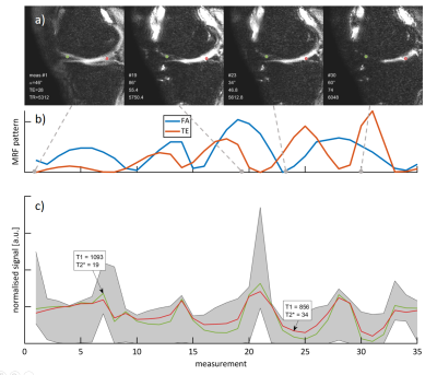 |
Whole-knee quantification of the articular cartilage: magnetic resonance fingerprinting for joint T1 and T2* mapping of 16 slices in 3 minutes
Telly Ploem1, Jaap Boon1, Ingo Hermann1,2, Cole S. Simpson3, Joe F Juffermans4, Tom M. Piscaer5, Hildo J Lamb4, Nazli Tümer6, Joao Tourais1, and Sebastian Weingärtner1
1Magnetic Resonance Systems Lab, Department of Imaging Physics, Delft University of Technology, Delft, Netherlands, 2Computer Assisted Clinical Medicine, Medical Faculty Mannheim, Heidelberg University, Mannheim, Germany, 3Department of Mechanical Engineering, Stanford University, Stanford, CA, United States, 4Department of Radiology, Leiden University Medical Center, Leiden, Netherlands, 5Orthopaedic Surgery, Erasmus University Medical Centre, Rotterdam, Netherlands, 6Department of Biomechanical Engineering, Delft University of Technology, Delft, Netherlands
Quantitative tissue characterization of the articular cartilage is a promising method for early assessment of degeneration. However, current techniques have limited spatial coverage and are at risk of missing localized alterations. In this work, we implemented and validated an MRF-EPI sequence for the simultaneous T1 and T2* quantification with whole knee coverage across 16 slices in 3 minutes. Initial evaluation in phantom, ex vivo animal tissue and in one healthy subject show promising results compared with conventional methods.
|
|||
1556. |
Golden-angle radial MR fingerprinting for high-resolution quantitative prostate MRI
Victoria YuiWen Yu1, Ergys Subashi1, Can Wu1, Peter Koken2, Mariya Doneva2, Ricardo Otazo1, and Ouri Cohen1
1Medical Physics, Memorial Sloan Kettering Cancer Center, New York, NY, United States, 2Philips Healthcare, Hamburg, Germany
We demonstrated the feasibility of golden-angle radial MR fingerprinting for high-resolution quantitative multi-parametic prostate MRI. The variation in T1 and T2 values for different scan times within the normal prostate and the required number of spokes per temporal time frame required for robust quantitation parameter mapping were examined. A decrease in average T1 and T2 values was observed as scan time increased. Radial spokes of 2 or more per temporal frame achieved parameter maps that are in good agreement with reported normal prostate transition zone values.
|
|||
1557.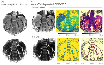 |
Abdominal Water/Fat Separated MR Fingerprinting on a Lower-Field 0.75T MRI
Christian Guenthner1, Peter Koken2, Peter Boernert2,3, and Sebastian Kozerke1
1University and ETH Zurich, Zurich, Switzerland, 2Philips Research, Hamburg, Germany, 3Leiden University Medical Center, Leiden, Netherlands
We have investigated the feasibility of concurrent water/fat separation and T1/T2 mapping using spoiled FISP-MR Fingerprinting on a lower-field 0.75T MRI. Water/fat separation is performed in k-space and combined with seven-peak fat-spectrum deblurring and B0-deblurring using multi-frequency interpolation. Matching is performed for water and fat separately and takes B1+ inhomogeneities into account. At 0.75T, T1 was 491ms (liver), 911ms (spleen), 958ms (kidney), 744ms (muscle), and 195ms (fat); and T2 was 77ms (liver), 91ms (spleen), 111ms (kidney), 50ms (muscle), and 105ms (fat).
|
|||
1558.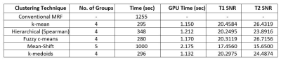 |
GPU Accelerated Grouped Magnetic Resonance Fingerprinting using Clustering Techniques
Abdul Moiz Hassan1, Rana Muhammad Saad1, Irfan Ullah1, and Hammad Omer1
1Medical Image Processing Research Group (MIPRG), Department of Electrical and Computer Engineering, COMSATS University, Islamabad, Pakistan
Magnetic Resonance Fingerprinting (MRF) has a limited use in clinics due to a considerable reconstruction time and large memory requirements. This paper utilizes clustering in MRF Dictionary to reduce the reconstruction time and memory requirements for MRF image reconstruction. The proposed method is further optimized for parallel processing to significantly reduce the pattern matching time with minimum memory usage by incorporating a multi-core GPU framework. As an outcome, the MRF reconstruction time is accelerated, keeping the SNR of the resulting images in a clinically acceptable range.
|
|||
1559.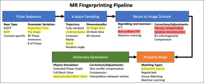 |
Uncertainty analysis framework for quantifying error propagation in MR Fingerprinting
Megan E Poorman1, Zydrunas Gimbutas2, Dan Ma3, Andrew Dienstfrey2, and Kathryn E Keenan1
1Physical Measurement Laboratory, National Institute of Standards & Technology, Boulder, CO, United States, 2Information Technology Laboratory, National Institute of Standards & Technology, Boulder, CO, United States, 3Department of Biomedical Engineering, Case Western Reserve University, Cleveland, OH, United States
This study demonstrated functionality of an error analysis framework applied to understand the impacts of variations in the MR Fingerprinting pipeline. Our preliminary analysis using both simulation and experimental methods showed small differences in the accuracy and precision of the reconstructed property maps with choice of k-space to image space reconstruction method. We demonstrated the need for a better understanding of error propagation within the pipeline, improved quantitative metrics of error, and future work will include a full Monte Carlo analysis using this framework.
|
|||
1560.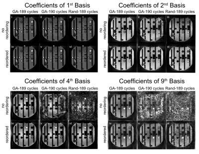 |
Minimization of Eddy Current Related Artefacts in Hybrid-State Sequences
Sebastian Flassbeck1,2 and Jakob Assländer1,2
1Dept. of Radiology, Center for Biomedical Imaging, New York, NY, United States, 2Center for Advanced Imaging Innovation and Research, New York, NY, United States In this work, we investigate the influence of eddy currents on hybrid state free precession (HSFP) sequences with 3D radial (koosh-ball) readout trajectories and propose a simulated annealing-based algorithm to minimize their impact. The proposed approach of reordering the temporal succession of radial spokes successfully minimized the influence of eddy currents on HSFP experiments. Although these results were shown in the context of a HSFP experiment, we believe that this approach could find a broader application in multi-shot bSSFP sequences. |
|||
1561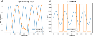 |
Sequence Optimisation for Multi-Component Analysis in Magnetic Resonance Fingerprinting Video Permission Withheld
David Heesterbeek1,2, Frans Vos1,3, Martin van Gijzen2, and Martijn Nagtegaal1
1Department of Imaging Physics, Delft University of Technology, Delft, Netherlands, 2Department of Numerical Analysis, Delft University of Technology, Delft, Netherlands, 3Department of Radiology and Nuclear Medicine, Erasmus MC, Rotterdam, Netherlands
Quantitative MRI and especially MR Fingerprinting make use of complicated acquisition schemes and signal models to measure tissues parameters. The sequence choice is crucial for the noise robustness for parameter estimations of different tissues. Multi-component (MC) signal models for MRF are of importance to estimate partial volume effects or myelin water fractions for example. We propose to use the Cramér-Rao bound to assess and optimise the multi-component parameter estimations for MRF. The optimised flip angle and TR patterns for MC-MRF were highly structured which was also observed for the optimisation based on the single-component model, but structural differences were noticed.
|
|||
1562.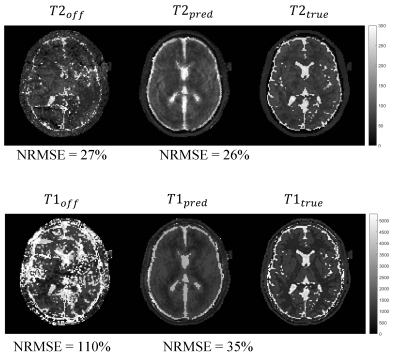 |
Magnetic Resonance Fingerprinting GAN-Transformer: removing off-resonance artifacts
Ronal Manuel Coronado1,2, Gabriel Manuel della Maggiora1,2, Carlos Manuel Castillo-Passi1,2, Gastão Cruz 3, Sergio Manuel Uribe1,2, Cristian Manuel Tejos1,2, Claudia Prieto2,3,4, and Pablo Manuel Irarrazaval2,4
1Centro de Imagenes Biomedicas-Universidad Catolica de Chile, Santiago, Chile, 2Millennium Nucleus for Cardiovascular Magnetic Resonance, Santiago, Chile, 3School of Biomedical Engineering and Imaging Sciences, King's College London, London, United Kingdom, 4Centro de Imagenes Biomedicas- Pontificia Universidad Catolica de Chile, Santiago, Chile
Magnetic Resonance Fingerprinting (MRF) acquisitions with balanced Steady State Free Precession (bSSFP) and spiral trajectories are prone to off-resonance artifacts. Thus, those artifacts hinder the reconstruction of the tissue maps (T1 and T2). In this work, we propose a model based on Generative Adversarial Networks (GANs) mixed with transformer blocks to decrease these artifacts. Our method improved the NMSE for both quantitative maps T1 and T2. Heavily reducing the effects of the off-resonance in comparison to classical bSSFP-MRF.
|
|||
1563.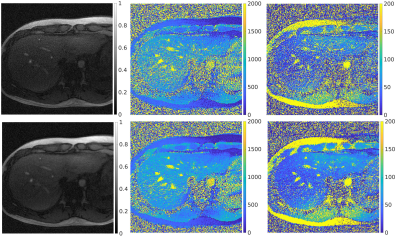 |
Motion Robust Free-Breathing MR-Fingerprinting
Ergys Subashi1, Victoria Yu1, Can Wu1, Peter Koken2, Mariya Doneva2, Ricardo Otazo1, and Ouri Cohen1
1Memorial Sloan Kettering Cancer Center, New York, NY, United States, 2Philips Research, Hamburg, Germany
The acquisition of quantitative MRI biomarkers with sufficiently high spatiotemporal resolution and coverage remains challenging. This is particularly difficult in organs of the abdomen, where respiratory, pulsative, and peristaltic motion may introduce biases in the expected evolution of signal intensity. This work describes an implementation of free-breathing MR-fingerprinting (MRF) for quantitative imaging of the abdomen. The method relies on golden-angle radial sampling combined with compressed sensing and parallel imaging. We compare MRF-derived parametric maps in vivo as a function of reconstruction algorithm and in free-breathing (FB) and breath-hold (BH) acquisitions.
|
|||
1564.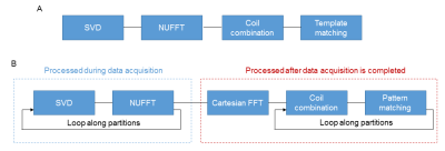 |
Rapid 3D MR Fingerprinting reconstruction using a GPU-based framework
Yong Chen1, Wei-Ching Lo2,3, Andrew Dupuis2, Rasim Boyacioglu1, Michael Hansen4, and Mark Griswold1
1Radiology, Case Western Reserve University, Cleveland, OH, United States, 2Biomedical Engineering, Case Western Reserve University, Cleveland, OH, United States, 3Siemens Medical Solutions, Boston, MA, United States, 4Health Futures, Microsoft Research, Seattle, WA, United States
In this study, we proposed an efficient online map reconstruction framework for 3D MRF using a GPU-based reconstruction. Both SVD compression and 2D NUFFT were performed mostly during data acquisition and GPU computation was further implemented to accelerate coil combination and pattern matching. Our simulation results demonstrated that 1) accurate T1 and T2 mapping was obtained with the proposed method and 2) rapid MRF tissue mapping can be achieved in ~ 0.7 sec per slice, which enables rapid volumetric quantitative imaging using MRF.
|
|||
1565.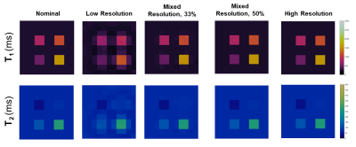 |
Multi-Resolution MR Fingerprinting: High-Resolution Maps from a Combination of High- and Low-Resolution Data
Kathleen Ropella-Panagis1, Jesse Hamilton1, and Nicole Seiberlich1
1Department of Radiology, University of Michigan, Ann Arbor, MI, United States
Increasing the spatial resolution of MR Fingerprinting can result in longer acquisition times due to the need to sample a larger extent of k-space. In this work, a novel data sampling scheme consisting of interleaved high- and low-resolution spiral trajectories is introduced to achieve high-resolution maps during a shortened acquisition time, which could have advantages for sequences that require a breath-hold.
|
|||
1566.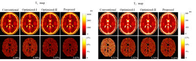 |
An Efficient Approach to Optimal Design of MR Fingerprinting Experiments with B-Splines
Evan Scope Crafts1 and Bo Zhao1,2
1Oden Institute for Computational Engineering and Sciences, The University of Texas at Austin, Austin, TX, United States, 2Department of Biomedical Engineering, The University of Texas at Austin, Austin, TX, United States
Optimal design of acquisition parameters utilizing the Cramer-Rao bound provides improved SNR efficiency for MR fingerprinting experiments. The early work demonstrates that smooth magnetization evolutions resulting from constraining the flip angle variation lead to improved estimation performance. Here we introduce a new formulation, in which we constrain the sequence of acquisition parameters in the low-dimensional spline space. The proposed formulation enforces smooth magnetization evolutions with significantly reduced degrees of freedom. Compared to the state-of-the-art experiment design approach, it improves the computational efficiency by two orders of magnitude, while achieving a similar or slightly better SNR efficiency of the imaging experiments.
|
|||
1567.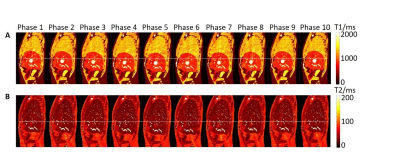 |
Investigation of Different Acquisition Schemes for Four-dimensional Magnetic Resonance Fingerprinting
Tian Li1, Di Cui2, Ge Ren1, Edward S. Hui3, and Jing Cai1
1Department of Health Technology and Informatics, The Hong Kong Polytechnic University, Hong Kong, Hong Kong, 2Department of Diagnostic Radiology, The University of Hong Kong, Hong Kong, Hong Kong, 3Department of Rehabilitation Science, The Hong Kong Polytechnic University, Hong Kong, Hong Kong
Magnetic resonance fingerprinting (MRF) is an imaging technique that effectively samples the transient state signal to achieve the goal of fast multi-parameter measurement. However, the applications of MRF only focus on static images, mostly brain image. Therefore, this study aims to investigate the feasibility of different acquisition schemes of four-dimensional magnetic resonance fingerprinting (4D-MRF) by computer simulation.
|
|||
1568.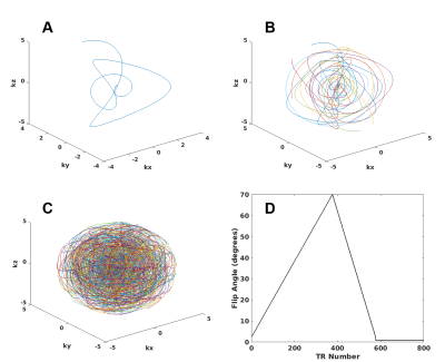 |
3D Magnetic Resonance Fingerprinting Using Seiffert Spirals
Cory R. Wyatt1 and Alexander R. Guimaraes2
1Oregon Health and Science University, Portland, OR, United States, 2Diagnostic Radiology, Oregon Health and Science University, Portland, OR, United States
3D magnetic resonance fingerprinting (MRF) techniques have been developed to efficiently acquire 3D volumes of quantitative parameters. Most techniques are based on applications of 2D trajectories rotated or stacked in 3D k-space. By acquiring data across all of 3D k-space each TR, we believe that efficient imaging and quantification can be obtained. In this study, seiffert spirals are used to acquire 3D k-space in an MRF acquisition of an isotropic 3D volume for the quantification of T1 and T2 relaxation times. High resolution T1 and T2 maps of a human brain were acquired in less than 3 minutes.
|
|||
1569.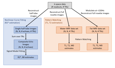 |
Rosette MRF for simultaneous T1, T2, and R2* mapping
Evan Cummings1,2, Yuchi Liu2, Kathleen Ropella-Panagis2, Jesse Hamilton1,2, and Nicole Seiberlich1,2
1Biomedical Engineering, University of Michigan, Ann Arbor, MI, United States, 2Radiology, University of Michigan, Ann Arbor, MI, United States
This abstract proposes a method for estimating T1, T2, and R2* values from rosette MRF data. R2* and B0 can be extracted from a series of images generated by subdividing the rosette data into partial readouts, and T1 and T2 are determined using the standard MRF pattern matching pipeline. This method was tested on the ISMRM/NIST MRI system phantom to assess the accuracy of the quantitative relaxation measurements.
|
|||
1570.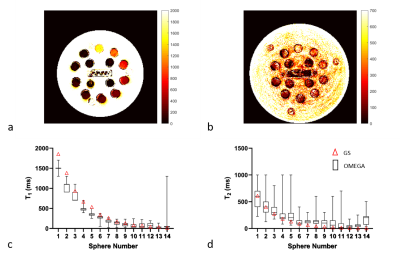 |
Open source Magnetic rEsonance finGerprinting pAckage (OMEGA)
Enlin Qian1 and Sairam Geethanath1
1Columbia Magnetic Resonance Research Center, New York, NY, United States
This work develops an end to end, open source, vendor neutral package that allows for fast prototyping of magnetic resonance fingerprinting using Pulseq. In this work, a previously published sequence was implemented in Pulseq, and data were acquired on ISMRM/NIST phantom. The raw data were reconstructed and matched with an extended phase graph simulated dictionary. Automated region of interest analysis was performed to extract T1 and T2 estimation for each sphere of the phantom. The results show that T1 and T2 estimations for 17 spheres of T1 and T2 arrays are within 20% of gold standard measurements.
|
The International Society for Magnetic Resonance in Medicine is accredited by the Accreditation Council for Continuing Medical Education to provide continuing medical education for physicians.