Digital Posters
Relaxometry in the Body: Thorax & Abdomen
ISMRM & SMRT Annual Meeting • 15-20 May 2021

Digital Posters
Relaxometry in the Body: Thorax & Abdomen
| Concurrent 1 | 19:00 - 20:00 |
3285.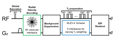 |
A method to rapidly quantify whole-organ metabolic rate of O2 with interleaved background-suppressed T2-oximetry and blood flow measurement
Rajiv S Deshpande1,2, Michael C Langham2, and Felix W Wehrli2
1Dept. of Bioengineering, University of Pennsylvania, Philadelphia, PA, United States, 2Dept. of Radiology, University of Pennsylvania, Philadelphia, PA, United States
A T2-based oximetry pulse sequence has been developed by interleaving a phase-contrast module preceding a background-suppressed T2-prepared echoplanar imaging readout. The method enables rapid simultaneous measurement of blood flow velocity and T2 of blood water protons from a single anatomic location in 18 seconds scan time. The pulse sequence improves on T2-based oximetry methods, including T2-relaxation-under-spin-tagging (TRUST), by incorporating background-suppression to eliminate the need for “control” and “label” images. The pulse sequence also interleaves phase-contrast MRI so that a separate measurement of blood flow velocity is not required to quantify whole-organ metabolic rate of oxygen.
|
|||
3286.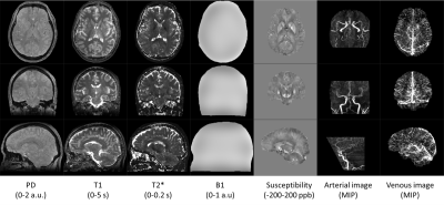 |
Simultaneous arterial and venous imaging and 3D quantitative parameter mapping with RF-spoiled gradient echo
Tomoki Amemiya1, Suguru Yokosawa1, Yo Taniguchi1, Ryota Sato1, Hisaaki Ochi1, and Toru Shirai1
1Healthcare Business Unit, Hitachi, Ltd., Tokyo, Japan
We proposed a method to acquire venous and arterial image in addition to maps of multiple MR parameters (T1, T2*, proton density, and susceptibility) at the same time. The method applies venous extraction to susceptibility map obtained in previously developed multiple parameter mapping method using RF-spoiled gradient echo. The venous image of the proposed method were similar to those of conventional method. The result suggests that the proposed method enables simultaneous acquisition of arterial and venous images with quantitative MR parameter maps, and it may contribute to more efficient MR examination.
|
|||
3287. |
Accelerated 3-Tesla Cardiac T2-Mapping at End-Systole for Improved Transmural Map Consistency and Accuracy
Ronald J Beyers1, Adil Bashir1, and Thomas S Denney1
1MRI Research Center, Auburn University, Auburn University, AL, United States
CMR T2-mapping is needed to assess myocardial edema after such events as acute myocardial infarction, myocarditis and tako-tsubo cardiomyopathy. Many T2mapping sequences have been developed, but most suffer from design shortcuts that degrade their accuracy, consistency and prognostic value. T2maps from four-point T2 curve-fits at end-systole (ES) would have maximum wall thickness and fewest artifacts for better prognostic value. We present a new sequence that produces accurate four-point T2maps at ES within a 24-second breathhold at 3T. This sequence was tested on phantoms and healthy human volunteers to present superior results.
|
|||
3288. |
Optimization of Spoiled GRE-based IR Acquisition Scheme for 3D Cardiac T1 Mapping at 3T
Paul Han1, Thibault Marin1, Vanessa Landes2, Yanis Djebra1,3, Georges El Fakhri1, and Chao Ma1
1Gordon Center for Medical Imaging, Department of Radiology, Massachusetts General Hospital and Harvard Medical School, Boston, MA, United States, 2GE Healthcare, Boston, MA, United States, 3LTCI, Télécom Paris, Institut Polytechnique de Paris, Paris, France
Spoiled gradient echo (GRE) is an attractive alternative to balanced steady-state free precession (bSSFP) for ECG-gated recovery (IR)-based volumetric myocardial T1 mapping at 3T. However, the robustness of T1 estimation from spoiled GRE-based ECG-gated IR acquisitions has not been thoroughly investigated under different schemes and in the presence of B1 inhomogeneity. This work investigated effects of B1 inhomogeneity in the context of T1 estimation, considering B1 inhomogeneity in the model for T1 estimation, and characterized effects of flip angle and heart rate to optimize a spoiled GRE-based ECG-gated IR acquisition scheme for 3D cardiac T1 mapping.
|
|||
3289.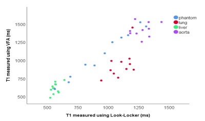 |
Comparison of lung T1 mapping using variable flip angle and Look-Locker techniques
Laura Saunders1, Paul J. C. Hughes1, James Eaden1, Andy J Swift1, Stephen Bianchi2, and Jim M Wild1
1Infection Immunity and Cardiovascular Disease, University of Sheffield, Sheffield, United Kingdom, 2Sheffield Teaching Hospitals NHS, Sheffield, United Kingdom
Lung T1 is sensitive to lung pathology, tissue and perfusion, and can be used to the calculation of quantitative DCE perfusion metrics. However, no comparisons have been made of common MRI T1 mapping sequences in the human lung. The aim of this work was to compare Look-Locker and variable flip angle T1 mapping sequences in phantoms and in vivo, in the lung, liver and blood. T1 measured in the phantom, blood and liver was significantly lower when measured using a Look-Locker acquisition than VFA, whereas lung T1 was significantly higher when measured using Look-Locker acquisition than when measured using VFA.
|
|||
3290.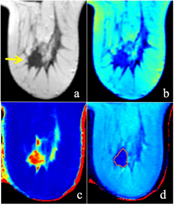 |
Synthetic MRI with quantitative mappings as biomarkers for prediction of prognostic factors and molecular subtypes of breast cancer
Weibo Gao1, Quanxin Yang1, Xin Chen1, and Xiaocheng Wei2
1Radiology, Second Affiliated Hospital of Xi’an Jiaotong University, Xi’an, Shaanxi, China, 2MR Research, GE Healthcare, Beijing, China
In this study, we aim to investigate whether different parameters of synthetic MRI can be used for the prediction of prognostic factors and molecular subtypes of breast cancer. It was concluded that SD of PD-Pre, SD of PD-Gd, T2-Pre, T2-Gd, T1-Pre and PD-Pre can be used as quantitative imaging biomarkers for the different receptor status of breast cancer. Among them, SD of PD-Pre, SD of PD-Gd, T2-Pre and T2-Gd were significantly different molecular subtypes, which worth further study.
|
|||
3291.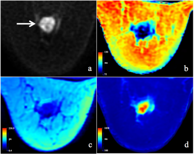 |
Synthetic relaxometry and diffusion measures in the differentiation of breast lesions: a contrast-free alternative to BI-RADS?
Weibo Gao1, Xin Chen1, Quanxin Yang1, and Xiaocheng Wei2
1Radiology, Second Affiliated Hospital of Xi’an Jiaotong University, Xi’an, Shaanxi, China, 2MR Research, GE Healthcare, Beijing, China
In this study, we aim to investigate the performance of quantitative measurements from synthetic MRI and DWI as well as their combinations in differentiating malignant from benign breast lesions and to compare with BI-RADS. Quantitative T2, PD and ADC values were significant lower in malignant than that of benign breast lesions. Quantitative multi-parameters of T2+PD+ADC had the best performance compared to all the other quantitative plans and BI-RADS. The approach, without contrast agents that combined synthetic MRI and DWI outperformed BI-RADS and may serve as an alternative and effective strategy for the improvement of breast lesion differentiation.
|
|||
3292.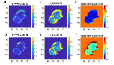 |
Why You Should Fit Signal Intensity, Not Relaxivity, for Quantitative DCE-MRI
Julie Camille DiCarlo1,2, Anum S Kazerouni3, and Thomas E Yankeelov1,2,4,5,6
1Oden Institute for Computational Engineering and Sciences, The University of Texas at Austin, Austin, TX, United States, 2Livestrong Cancer Institutes, The University of Texas at Austin, Austin, TX, United States, 3Department of Radiology, University of Washington, Seattle, WA, United States, 4Department of Biomedical Engineering, The University of Texas at Austin, Austin, TX, United States, 5Department of Diagnostic Medicine, The University of Texas at Austin, Austin, TX, United States, 6Department of Oncology, The University of Texas at Austin, Austin, TX, United States
Pharmacokinetic modeling of DCE-MRI is based on fitting perfusion parameters to contrast agent concentration or relaxivity curves computed using the nonlinear spoiled gradient-echo (SPGR) signal equation, T1 mapping values, and the linear relationship between T1 and contrast agent relaxivity. The nonlinear term of the SPGR equation has implications for how image noise scales in the concentration. By simulating image noise at different levels for ideal curves of different parameter values, we show why it’s advantageous to fit signal intensity curves rather than relaxivity curves.
|
|||
3293.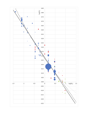 |
Survey of water proton longitudinal relaxation in liver in vivo.
John Charles Waterton1,2
1Centre for Imaging Sciences, University of Manchester, MANCHESTER, United Kingdom, 2Bioxydyn Ltd, MANCHESTER, United Kingdom
Liver R1 was surveyed in vivo in 3464 subjects at 16 field strengths from 106 publications. R1 was plotted against B0 and fitted both to a biophysical model, and to a log-log heuristic. An investigator who wishes to compare their own findings with prior literature, and who finds their average liver R1 to be within 9% of our fit, with between-subject CoV <8%, can conclude that their study is consistent with the majority of the literature.
|
|||
3294.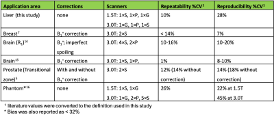 |
Bias, repeatability and reproducibility of liver T1 mapping with variable flip angles
Sirisha Tadimalla1,2, Daniel J Wilson3, Margaret A Saysell4, Martin John Graves5, Iosif A Mendichovszky6,7, Geoffrey JM Parker8, and Steven Sourbron2,9
1Institute of Medical Physics, The University of Sydney, Sydney, Australia, 2Department of Biomedical Imaging Sciences, The University of Leeds, Leeds, United Kingdom, 3Department of Medical Physics and Engineering, Leeds Teaching Hospital NHS Trust, Leeds, United Kingdom, 4Cardiac MRI, Leeds Teaching Hospitals NHS Trust, Leeds, United Kingdom, 5MR Physics and Radiology, Cambridge University Hospitals NHS Foundation Trust, Cambridge, United Kingdom, 6Department of Radiology, University of Cambridge, Cambridge, United Kingdom, 7Department of Nuclear Medicine, Cambridge University Hospitals NHS Foundation Trust, Cambridge, United Kingdom, 8Bioxydyn, Manchester, United Kingdom, 9Department of Infection, Immunity and Cardiovascular Disease, University of Sheffield, Sheffield, United Kingdom
A multi-centre, multi-vendor study in 8 travelling healthy volunteers was conducted for technical validation of variable flip angle (VFA) T1 mapping in the liver across 6 scanners (3 vendors and 2 field strengths). The 95% CI was 28±8% for the bias in VFA liver T1, 10±3% for the intra-scanner repeatability CV and 28±6% for the inter-scanner reproducibility CV. These values are comparable to B1+ corrected VFA T1 in prostate, brain, and breast. Any proposed refinement of the VFA method in the liver should demonstrate a significant improvement on those benchmarks before it can be recommended as a future standard.
|
|||
3295.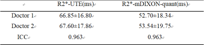 |
Correlation between multi-echo ultrashort TE and mDIXON-quant imaging for R2* mapping in liver cirrhosis
Qiang Wei1, Nan Wang1, Ailian Liu1, Qingwei Song1, Renwang Pu1, Lihua Chen1, Jiazheng Wang2, and Liangjie Lin2
1The First Affiliated Hospital of Dalian Medical University, Dalian, China, 2Philips Healthcare, Dalian, China
Ultrashort echo time (UTE) imaging is a valuable technique for imaging short T2 and T2* samples. While the mDIXON-quant sequence enables reliable separation of water and fat signals, and measurement of fat fraction. With introduction of multiple echo acquisition, both the UTE and mDIXON-quant sequence can be used for measurement of the quantitative R2* values. This study investigated the correlation between R2* values measured by UTE and mDIXON-quant sequences. Results show that a moderate correlation between R2 values by the two methods, and R2* values measured by UTE were slightly higher than those by mDIXON-quant.
|
|||
3296.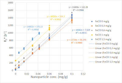 |
Transversal Relaxometry of a Mixture of Iron Compounds at Different Concentrations
Arthur Peter Wunderlich1,2, Eva Leithner1, Richard Fiedler3, Meinrad Beer1, Stefan Andreas Schmidt1, and Mika Lindén3
1Dept. of Diagnostic Radiology, Ulm University, Medical Center, Ulm, Germany, 2Section for Experimental Radiology, Ulm University, Medical Center, Ulm, Germany, 3Institute for Inorganic Chemistry II, Ulm University, Ulm, Germany
Liver R2* determined with a breath hold GRE sequence is commonly used to address liver iron content in vivo. However, iron occurs in the body bound to various chemical substances. To study influence of chemical composition on R2* on a clinical MRI scanner, we scanned mixtures of two iron compounds, polymer coated nanoparticles on the one hand and diluted Fe3+ ions on the other, at different concentrations of both compounds. We found that the relaxivity of one compound depended on the concentration of the other, suggesting that the interactions between compounds are more complex than previously assumed.
|
|||
3297.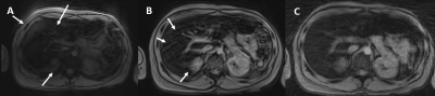 |
Hepatic iron quantification using a Free-breathing 3D Radial Dixon technique and validation with a 2D GRE biopsy calibration
Shawyon Chase Rohani1,2, Cara Morin1, Xiaodong Zhong3, Stephan Kannengiesser4, Joseph Holtrop1, Ayaz Khan1, Ralf Loeffler1,5, Claudia Hillenbrand1,5, Jane Hankins6, and Aaryani Tipirneni Sajja1,2
1Department of Diagnostic Imaging, St. Jude Children's Research Hospital, Memphis, TN, United States, 2Department of Biomedical Engineering, The University of Memphis, Memphis, TN, United States, 3Siemens Medical Solutions USA, Inc., Los Angeles, CA, United States, 4MR Application Development, Siemens Healthcare, Erlangen, Germany, 5University of New South Wales, Sydney, Australia, 6Department of Hematology, St. Jude Children's Research Hospital, Memphis, TN, United States
Radial acquisitions are less motion sensitive and a viable alternative in patients unable to breath-hold. This study investigates a free-breathing 3D Radial Dixon technique to assess HIC by validating with 3D Cartesian Dixon and biopsy-calibrated 2D GRE method. All three acquisitions showed excellent correlation with each other. The free-breathing 3D Radial Dixon produced sharper images whereas the 2D GRE and 3D VIBE Cartesian-based techniques exhibited motion artifacts and a slight underestimation in R2* values compared to 3D Radial VIBE likely due to motion artifacts.
|
|||
3298.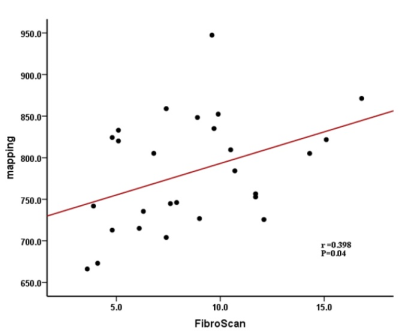 |
The application of B1 corrected VFA T1-mapping in staging of liver fibrosis
YANLI JIANG1, Pin Yang1, FengXian Fan1, Wanjun Hu1, Jing Zhang1, and Shaoyu Wang2
1Department of Magnetic Resonance, LanZhou University Second Hospital, LanZhou, China, 2MR Scientific Marketing, Siemens Healthineers, Shanghai, China
The aim of this study was using B1 corrected native T1 value to assess their diagnostic accuracy for staging of liver fibrosis. We found that native T1 values increased with the level of liver fibrosis, and a positive correlation was found between liver FibroScan results and the native T1 values. In conclusion, the B1 corrected native T1 values might be useful for staging of liver fibrosis.
|
|||
3299.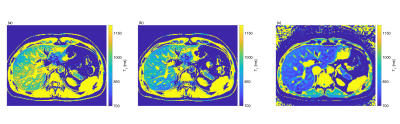 |
A comparison of two B1+ mapping methods for 3D VFA T1 mapping in the liver at 3T
Gabriela Belsley1, Damian J. Tyler1, Matthew D. Robson1,2, and Elizabeth M. Tunnicliffe1
1Oxford Centre for Clinical Magnetic Resonance Research, Radcliffe Department of Medicine, University of Oxford, Oxford, United Kingdom, 2Perspectum, Oxford, United Kingdom
Correcting B1+ inhomogeneities is imperative in T1 mapping using a VFA 3D SPGR sequence. We compared B1+ mapping using a preconditioning RF (preRF) pulse and TurboFLASH readout against B1+ mapping using a 2D double angle method (DAM) with EPI readout. The preRF method had a residual B1+-related variation in the T1 maps of 143ms, 95% CI [135, 150] and 170ms, 95% CI [158, 181] for 2 healthy volunteers while the DAM reduced these, with B1+-related variations of -27ms, 95% CI [-33, -20] and -8ms, 95% CI [-18, 2].
|
|||
3300.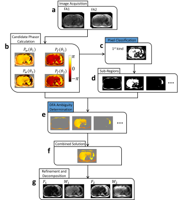 |
Resolving the fat-water ambiguity based on T1 difference
Hao Peng1,2, Liwen Wan1, Qian Wan1, Jianxun Lv1, Chuanli Cheng1, Yi Wang3, Wenzhong Liu2, Xin Liu1, Hairong Zheng1, and Chao Zou1
1Paul C. Lauterbur Research Center for Biomedical Imaging, Shenzhen Institute of Advanced Technology,Chinese Academy of Sciences, Shenzhen, China, 2Key Laboratory of Imaging Processing and Intelligence Control, School of Artificial Intelligence and Automation, Huazhong University of Science & Technology, Wuhan, China, 3Department of Radiology, Peking University Peoples Hospital, Beijing, China
Fat-water ambiguity is an intrinsic problem from chemical shift encoded imaging model. When this problem is not well solved throughout the whole image, fat-water swaps appear and result in inaccurate fat quantification. The success of solving the ambiguity relies on pre-assumptions, e.g. the magnetic field continuity or the prior knowledge of the adipose/aqueous tissue distribution etc. In our work, we proposed to incorporate the T1 difference of fat and water to help solve the ambiguity problem. Proposed method can obtain PDFF and accurate T1 mapping simultaneously with B1+ correction. The sequence could cover the whole liver within a single breath-hold.
|
|||
3301.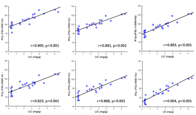 |
Ultrashort echo time R1ρ for detection of rat liver iron overload at 11.7T MRI
Qianfeng Wang1, Hong Xiao2, He Wang1,3, and Fuhua Yan2
1Institute of Science and Technology for Brain-Inspired Intelligence, Fudan University, Shanghai, China, 2Department of Radiology, Ruijin Hospital, Shanghai Jiao Tong University School of Medicine, Shanghai, China, 3Human Phenome Institute, Fudan University, Shanghai, China, Shanghai, China
In this study, R1ρ images of 24 rat liver samples were acquired with 2D UTE-R1ρ pulse sequences. The results showed that mean R1ρ values displayed dispersion, with decrease in R1ρ at higher FSLs. Spearman’s correlation analysis (two-tailed) indicated that the R1ρ values were significantly associated with liver iron concentration at all 6 spin-lock frequencies (all r > 0.8 and all P < 0.001).
|
|||
3302.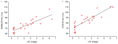 |
Comparison of T1ρ imaging between rapid acquisition with relaxation enhancement (RARE) and ultrashort TE (UTE) sequence of rat liver at 11.7T MRI
Qianfeng Wang1, Hong Xiao2, Xuchen Yu1, He Wang1,3, and Fuhua Yan2
1Institute of Science and Technology for Brain-Inspired Intelligence, Fudan University, Shanghai, China, 2Department of Radiology, Ruijin Hospital, Shanghai Jiao Tong University School of Medicine, Shanghai, China, 3Human Phenome Institute, Fudan University, Shanghai, China, Shanghai, China
In this study, T1ρ images of 24 rat liver samples were acquired with two types of pulse sequences: RARE and UTE. The mean T1ρ value of the rat liver with RARE was significantly higher compared to UTE (p<0.001). This study shows that the quantitative UTE-R1ρ has a better correlation with LIC than RARE-R1ρ.
|
|||
3303.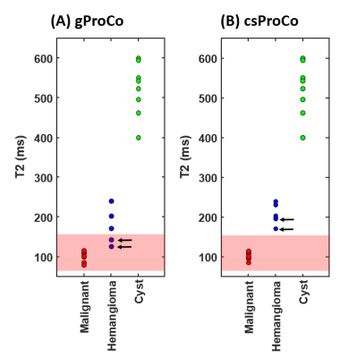 |
Multi-component T2 Modeling for Improved Characterization of Abdominal Neoplasms
Mahesh Bharath Keerthivasan1,2, Jean-Philippe Galons2, Diego Martin3, Ali Bilgin2,3,4, and Maria Altbach2
1Siemens Medical Solutions USA Inc, New York, NY, United States, 2Medical Imaging, University of Arizona, Tucson, AZ, United States, 3Biomedical Engineering, University of Arizona, Tucson, AZ, United States, 4Electrical and Computer Engineering, University of Arizona, Tucson, AZ, United States
Turbo spin-echo based T2 quantification of neoplasms is affected by partial volume effects resulting in underestimation of T2 values leading to lesion misclassification. Bi-exponential models proposed for two-component T2 estimation do not account for flip angle variations across the excited slice due to RF imperfections. This work presents a two-component model for accurate T2 estimation in the presence of partial volume which considers the slice profile of the excitation and refocusing pulses. The proposed model has <4% relative error in slices affected by partial volume and allows good separation between benign and malignant lesions in the abdomen.
|
|||
 |
3304.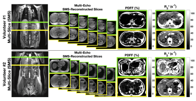 |
Accelerating 2D Chemical Shift Encoded MRI with Simultaneous Multislice Imaging
Nathan Tibbitts Roberts1,2, Ruvini Navaratna1,3, Diego Hernando1,3, and Scott B Reeder1,3,4,5,6
1Radiology, University of Wisconsin - Madison, Madison, WI, United States, 2Electrical and Computer Engineering, University of Wisconsin - Madison, Madison, WI, United States, 3Medical Physics, University of Wisconsin - Madison, Madison, WI, United States, 4Biomedical Engineering, University of Wisconsin - Madison, Madison, WI, United States, 5Medicine, University of Wisconsin - Madison, Madison, WI, United States, 6Emergency Medicine, University of Wisconsin - Madison, Madison, WI, United States
Chemical shift-encoded (CSE) MRI allows for measurement of important tissue parameters with established and emerging quantitative MRI applications, including proton density fat fraction (PDFF) and R2*. Respiratory and bulk patient movement can affect the accuracy and precision of CSE-MRI measurements, which makes rapid and motion robust imaging of high clinical value. This work demonstrates the combination of simultaneous multislice (SMS) imaging to accelerate 2D motion robust CSE-MRI acquisitions. Results with computer simulations and in vivo experiments confirm the feasibility of combining these two techniques to achieve rapid, whole-liver, motion robust quantitative tissue characterization.
|
The International Society for Magnetic Resonance in Medicine is accredited by the Accreditation Council for Continuing Medical Education to provide continuing medical education for physicians.