Digital Posters
Relaxometry in the Brain: Accuracy, Robustness & Analysis
ISMRM & SMRT Annual Meeting • 15-20 May 2021

Digital Posters
Relaxometry in the Brain: Accuracy, Robustness & Analysis
| Concurrent 1 | 17:00 - 18:00 |
3065.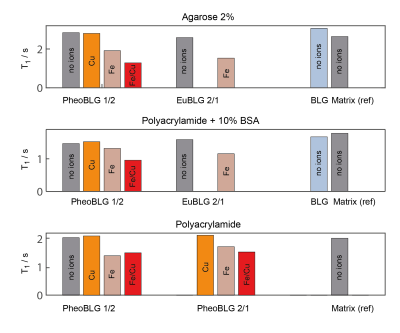 |
Neuromelanin-Related Proton Relaxation of Water: Influence from Iron and Copper
Niklas Wallstein1, André Pampel 1, Andrea Capucciati2, Carsten Jäger1, Fabio A. Zucca3, Luigi Casella2, Luigi Zecca3, and Harald E. Möller1
1Max Planck Institute for Human Cognitive and Brain Sciences, Leipzig, Germany, 2Department of Chemistry, University of Pavia, Pavia, Italy, 3Institute of Biomedical Technologies, National Research Council of Italy, Segrate, Milan, Italy
Neuromelanin (NM) MRI has received increasing interest in applications to diagnosis of Parkinson’s disease. A reduction of T1 associated with paramagnetic melanin-iron complexes is assumed to be the primary contrast mechanism. NM is able to chelate large quantities of iron but also, to a lesser extent, other transition metals, in particular copper. Here, we investigated the effect of different copper/iron concentrations bound to NM on water T1 and T2. Our results corroborate previous studies suggesting a concentration-dependent decrease of T1 for iron-loaded melanin. Additionally, we found a synergetic effect if both metals are simultaneously present leading to a pronounced T1-shortening.
|
|||
3066.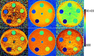 |
FAST T1 MAPPING: ACCURACY AND REPRODUCIBILITY OF VOLUMETRIC SEQUENCES FOR BRAIN RELAXOMETRY
Stefano Tambalo1,2, Alberto Finora2, Diego Cavalli2, and Jorge Jovicich1
1CIMeC, University of Trento, Trento, Italy, 2Department of Radiology, G.B. Rossi Hospital, University of Verona, Verona, Italy
The gold standard T1 mapping method is the Inversion Recovery (IR). Volumetric sequences such as MP2RAGE without or with compressed sensing (csMP2RAGE) allow faster T1-mapping, but potential biases are not well characterized. Here we investigated T1-mapping and test-retest reproducibility effects of sequence (IR, MP2RAGE, csMP2RAGE) and head RF coil (64 and 20-channel), both in-vitro and in-vivo. The 64-channel coil results are more accurate and reliable, showing good agreement with the gold standard and a beneficial reduction of scan time.
|
|||
3067.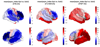 |
Asymmetries in the distribution of quantitative MRI parameters in the brain
Jonas Kielmann*1, Ana-Maria Oros-Peusquens*1, Nora Bittner2, Svenja Caspers2, and N. Jon Shah1,3,4,5
1INM-4, Research Centre Juelich, Juelich, Germany, 2INM-1, Research Centre Juelich, Juelich, Germany, 3Faculty of Medicine, JARA, RWTH Aachen University, Aachen, Germany, 4INM-11, JARA, Research Centre Juelich, Juelich, Germany, 5Department of Neurology, RWTH Aachen University, Aachen, Germany
Multicontrast quantitative MRI is used to characterize left-right and sulcal-gyral asymmetries of the brain in 26 volunteers. Several regions with significant asymmetries are found in different parameters (H2O, fbound, R2*, R1, kex, QSM). Good correlation between sulcal-gyral H2O-defined asymmetry and local gyrification index is found.
|
|||
3068.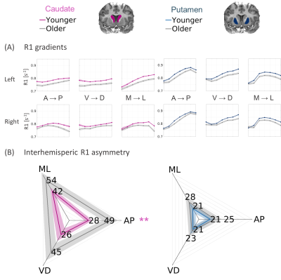 |
Aging-Related Spatial Changes in the Microstructure of the Human Striatum Detected Through Quantitative MRI in vivo
Elior Drori1, Shir Filo1, and Aviv Mezer1
1The Edmond and Lily Safra Center for Brain Sciences, The Hebrew University of Jerusalem, Jerusalem, Israel
The striatum is a heterogeneous brain structure with microstructural gradients along its main axes. Changes in its organization are associated with normal aging and disease. Yet the spatial variability in the human striatum is not well characterized and is mostly limited to postmortem studies. We propose a robust non-invasive method for detection and quantification of microstructural gradients along axes of the striatum in individuals in vivo, using qMRI. We show distinct profiles of spatial and aging-related changes in the striatum, associated with different biophysical sources such as tissue density and iron content, estimated in vivo.
|
|||
3069.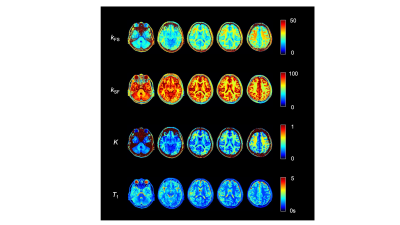 |
Derivation of Water Exchange Constants between Components using Quantitative Parameter Mapping (QPM).
Naoki Maeda1, Yuki Kanazawa2, Masafumi Harada2, Yo Taniguchi3, Yuki Matsumoto2, Hiroaki Hayashi4, Kosuke Ito3, Yoshitaka Bito3, and Akihiro Haga2
1Graduate of Health Science, Tokushima University, Tokushima, Japan, 2Graduate School of Biomedical Sciences, Tokushima University, Tokushima, Japan, 3Healthcare Business Unit, Hitachi, Ltd., Tokyo, Japan, 4College of Medical, Pharmaceutical and Health Sciences, Kanazawa University, Kanazawa, Japan
We developed a method for deriving water exchange rate based on mcDESPOT approach between two structures using quantitative parameters calculated from QPM-MRI. Our method was performed with human study for ten healthy volunteers and one patient with brain tumor. There was a significant negative correlation between kFS and kSF derived from QPM in each region, e.g., white matter, gray matter, and cerebral spinal fluid of healthy volunteers. (R = -0.75, P < 0.001). In conclusion, water exchange rates derived from QPM make it possible to obtain more detailed information of the brain associated with proton diffusion between structures, i.e., cross-relaxation.
|
|||
3070.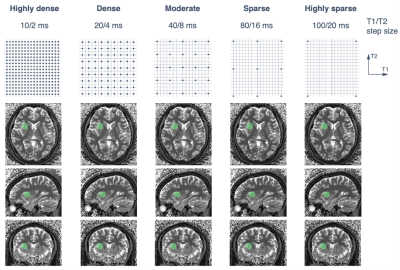 |
Radiomics with 3D MR fingerprinting: Influence of dictionary design on radiomic features and a potential mitigation strategy
Shohei Fujita1,2, Akifumi Hagiwara1, Koichiro Yasaka3, Hiroyuki Akai3, Akira Kunimatsu3, Shigeru Kiryu4, Issei Fukunaga1, Shimpei Kato1,2, Toshiaki Akashi1, Koji Kamagata1, Akihiko Wada1, Osamu Abe2, and Shigeki Aoki1
1Department of Radiology, Juntendo University, Tokyo, Japan, 2Department of Radiology, The University of Tokyo, Tokyo, Japan, 3Department of Radiology, The Institute of Medical Science, The University of Tokyo, Tokyo, Japan, 4Department of Radiological Sciences, International University of Health and Welfare, Narita, Japan
Understanding the dependencies of radiomic features on the dictionary step size in MR fingerprinting is crucial in the clinical implementation of MR fingerprinting-based radiomics. We investigated the influence of dictionary design in the radiomic analysis of MR fingerprinting and observed that radiomic features vary significantly for different designs. Moreover, special attention is required when using datasets containing maps generated from different dictionaries. We also demonstrated that an inverse quantization process can mitigate the influence of dictionary design on the radiomic features obtained with MR fingerprinting.
|
|||
3071.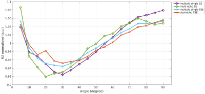 |
Diffusion sensitivity of spin echo sequences affects white matter R2 fiber orientation dependency
Christoph Birkl1, Christian Kremser2, Alexander Rauscher3, and Elke Ruth Gizewski1
1Department of Neuroradiology, Medical University of Innsbruck, Innsbruck, Austria, 2Department of Radiology, Medical University of Innsbruck, Innsbruck, Austria, 3UBC MRI research Centre, University of British Columbia, Vancouver, BC, Canada
We investigated how the sensitivity of spin-echo and turbo spin echo MRI sequences to diffusion processes affects the estimation of fiber orientation dependent R2 in white matter. Our results indicate that the strongest R2 orientation dependency is observed when R2 is estimated using multiple single spin echo sequences. The lowest R2 orientation dependency was observed for the multi-echo spin echo sequence.
|
|||
3072.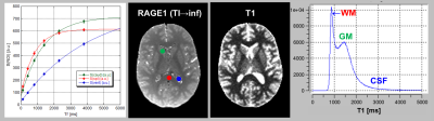 |
Isotropic T1-mapping of the whole brain by MP-RAGE at different inversion times
Gunther Helms1 and Hampus Olsson1
1Medical Radiation Physics, Clinical Sciences Lund, Lund University, Lund, Sweden
Fitting the inversion recovery (IR) at multiple TI provides T1 estimates of high accuracy if performed in a single slice with full relaxation. We present an isotropic 3D variant based on MP-RAGE, where a T1-weighted driven equilibrium is prepared prior to inversion by a second RAGE readout. This abolishes the need for long recovery times and provides volumes of similar contrast for co-registration. The method was tested at 3T on a small cohort and compared to 3D IR-TSE and 3D variable flip angle mapping.
|
|||
3073.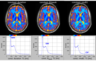 |
A simple approach to control rms(B1) by pulse length in variable flip angle (VFA) T1 mapping of human brain
Gunther Helms1
1Medical Radiation Physics, Clinical Sciences Lund, Lund University, Lund, Sweden
Controlling the saturation of bound magnetization in variable flip angle (VFA) experiments improves the derived parameter maps. Instead of using special CSMT RF pulses, we kept the rmsB1 constant simply by increasing the RF pulse duration with the square of the flip angle. At 3T, VFA T1 mapping of human brain thus showed better compliance to the Ernst equation than at constant pulse duration or B1 amplitude, but yielded shorter T1.
|
|||
3074. |
T1 Relaxation of White Matter Following Adiabatic Inversion
Luke A Reynolds1, Sarah R Morris1,2, Irene M Vavasour2,3, Laura Barlow3, Alex L MacKay1,2,3, and Carl A Michal1
1Physics & Astronomy, University of British Columbia, Vancouver, BC, Canada, 2Radiology, University of British Columbia, Vancouver, BC, Canada, 3UBC MRI Research Centre, Vancouver, BC, Canada
Adiabatic pulses have advantageous properties, such as an independently scalable bandwidth, which makes them more clinically versatile compared to standard soft pulses. However, their application to white matter has produced some unvalidated assumptions about the subsequent relaxation mechanisms. We examined the form of the longitudinal relaxation following adiabatic inversion in-vivo with simple inversion recovery experiments, which we found to be biexponential in character. This arises from cross-relaxation/exchange between aqueous and non-aqueous tissue components prepared in different magnetization states. We suggest a straightforward and accessible scheme for reproducible measurements of monoexponential T1 using a specific saturation recovery method.
|
|||
3075.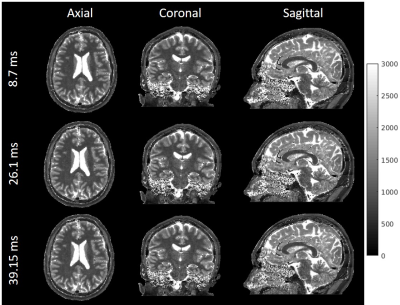 |
High spatial and temporal resolution rapid 3D IR-radFLASH pulse sequence for T1 mapping in the brain
Zhitao Li1,2, Zhiyang Fu3, and Maria I Altbach4
1Department of Radiology, Stanford University, Palo Alto, CA, United States, 2Electrical Engineering, Stanford University, Palo Alto, CA, United States, 3Electrical and Computer Engineering, The University of Arizona, Tucson, AZ, United States, 4Department of Medical Imaging, The University of Arizona, Tucson, AZ, United States
A 3D IR-radFLASH pulse sequence and a corresponding model-based reconstruction algorithm is presented for in vivo brain T1 mapping. The proposed pulse sequence can acquire data for high SNR 1.0mm isotropic resolution T1 mapping within clinical time constraints. The proposed model-based reconstruction algorithm can reconstruct the acquired data with high temporal resolution, which can be used to map spin species with relatively low T1 values. When combined with a slab-selective inversion pulse, the proposed technique can achieve whole brain coverage under 5 minutes.
|
|||
3076.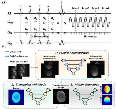 |
Single-shot T2 mapping in severe head motion with multiple overlapping-echo detachment planar imaging
Qinqin Yang1, Jiechao Wang1, Qizhi Yang1, Shuhui Cai1, Hongjian He2, Congbo Cai1, Zhong Chen1, and Jianhui Zhong2,3
1Department of Electronic Science, Xiamen University, Xiamen, China, 2The Center for Brain Imaging Science and Technology, the Collaborative Innovation Center for Diagnosis, and the Treatment of Infectious Diseases, Zhejiang University, Hangzhou, China, 3Department of Imaging Sciences, University of Rochester, Rochester, NY, United States
Subject motion often leads to serious quality degradation of MR images, especially in quantitative MRI. In this study, a novel reconstruction method was proposed for single-shot T2 mapping based on overlapping-echo detachment planar imaging and synthetic-data-driven learning (SDDL). This method was made robust to severe motion by improved parallel reconstruction and motion correction methods without extra scan. The proposed method was evaluated with human brain experiments. The results show that SDDL-based method can significantly reduce ghosting and motion artifacts in T2 maps in the presence of randomly and continuously subject movement.
|
|||
3077.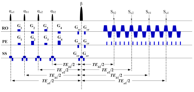 |
Single-shot Simultaneous Double-slice T2 Mapping based on Overlapping Echo Detachment Planar Imaging
Simin Li1, Jian Wu1, Shuhui Cai1, and Congbo Cai1
1Department of Electronic Science, Xiamen University, Xiamen, China
Most quantitative magnetic resonance imaging (qMRI) techniques remain time-consuming and sensitive to motion, especially when large volume imaging is needed. Simultaneous multi-slice (SMS), which is not restricted by single-slice evolution time, is an acceleration method for MRI. Here we propose a flexible SMS T2 mapping method based on overlapping echo detachment (OLED) planar imaging. Experimental results demonstrate the superior performance of our method. Reliable multi-slice T2 maps can be obtained in a single shot within milliseconds for the first time.
|
|||
3078.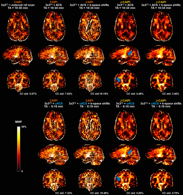 |
Joint-CAIPI reconstruction of multi-echo GRASE data for fast, high-resolution myelin water imaging
Gian Franco Piredda1,2,3, Tom Hilbert1,2,3, Berkin Bilgic4,5,6, Erick Jorge Canales-Rodríguez3, Marco Pizzolato3,7, Reto Meuli2, Jean-Philippe Thiran2,3, and Tobias Kober1,2,3
1Advanced Clinical Imaging Technology, Siemens Healthcare AG, Lausanne, Switzerland, 2Department of Radiology, Lausanne University Hospital and University of Lausanne, Lausanne, Switzerland, 3LTS5, École Polytechnique Fédérale de Lausanne (EPFL), Lausanne, Switzerland, 4Athinoula A. Martinos Center for Biomedical Imaging, Charlestown, MA, United States, 5Department of Radiology, Harvard Medical School, Boston, MA, United States, 6Harvard‐MIT Health Sciences and Technology, MIT, Cambridge, MA, United States, 7Department of Applied Mathematics and Computer Science, Technical University of Denmark, Kongens Lyngby, Denmark
In previous work, CAIPIRINHA was implemented in multi-echo gradient and spin echo (GRASE) acquisitions for 3D myelin water imaging, achieving significant acceleration. However, residual undersampling artifacts prevented further acquisition time reduction. In this work, we propose a joint-CAIPI reconstruction across echoes of GRASE k-space data to remove aliasing artifacts and further accelerate the acquisition. Exploiting the redundant anatomical information across the different GRASE echoes helped mitigate aliasing artifacts in myelin water fraction maps and provided an additional ~40% reduction in scan time to 6:18$$$\,$$$minutes for a whole-brain acquisition at 1.6$$$\,$$$mm3 isotropic resolution.
|
|||
3079.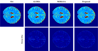 |
Fast T2 Mapping with Improved Accuracy Using Memory-Efficient Low-Rank Hankel Matrix Reconstruction
Xinlin Zhang1, Hengfa Lu2, Zi Wang1, Xi Peng3, Feng Huang4, Qin Xu4, Di Guo5, and Xiaobo Qu1
1Department of Electronic Science, School of Electronic Science and Engineering (National Model Microelectronics College), National Institute for Data Science in Health and Medicine, Xiamen University, Xiamen, China, 2College of Optical Science and Engineering, Zhejiang University, Hangzhou, China, 3Department of Radiology, Mayo Clinic, Rochester, MN, United States, 4Neusoft Medical System, Shanghai, China, 5School of Computer and Information Engineering, Fujian Provincial University Key Laboratory of Internet of Things Application Technology, Xiamen University of Technology, Xiamen, China
Being the state-of-the-art parallel magnetic resonance imaging methods other than the deep learning approaches, the low-rank Hankel approaches embrace the advantage of holding low reconstruction errors. However, they demand intensive computations and high memory consumptions, thereby result in long reconstruction time. We proposed a new strategy for exploiting the low rankness and applied it to accelerate 2D imaging and T2 mapping. It is shown that the proposed method outperforms the state-of-the-art approaches in terms of lower reconstruction errors and more accurate mapping estimations. Besides, the proposed method required much less computation and memory consumption.
|
|||
3080.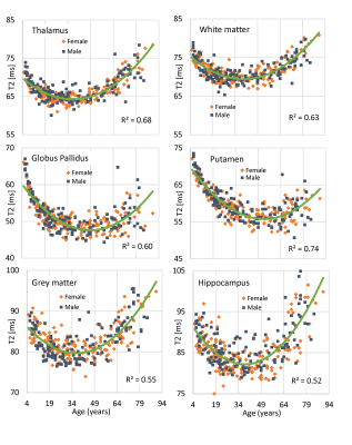 |
Robust T2 Mapping from Standard Brain Images: Repeatability and Lifespan Changes in Healthy Participants
Peter Seres1, Kelly C McPhee1,2, Emily Stolz1, Julia Rickard1, Jeff Snyder1, Christian Beaulieu1, and Alan H Wilman1
1Biomedical Engineering, University of Alberta, Edmonton, AB, Canada, 2CancerCare Manitoba, Winnipeg, MB, Canada
Transverse relaxation (T2) mapping from standard images is investigated in human brain using only a proton density and T2-weighted dual-echo turbo spin echo acquisition and applying Bloch fitting approach with transmit field map. In 24 subjects, repeatability of T2 maps was excellent with typically coefficient of variation (CoV) ~1% on the same scanner. In grey and white matter overall, CoV was 0.4%. The method is then applied to study T2 changes across the lifespan from 5-90 years old in 333 healthy participants, providing a normative population of true T2 values independent of refocusing flip angle effects.
|
|||
3081.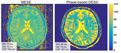 |
Phase-based T2 mapping using RF phase-modulated dual echo steady-state (DESS) imaging
Daiki Tamada1 and Scott B. Reeder1,2,3,4,5
1Radiology, University of Wisconsin-Madison, Madison, WI, United States, 2Medical Physics, University of Wisconsin-Madison, Madison, WI, United States, 3Biomedical Engineering, University of Wisconsin-Madison, Madison, WI, United States, 4Medicine, University of Wisconsin-Madison, Madison, WI, United States, 5Emergency Medicine, University of Wisconsin-Madison, Madison, WI, United States
A novel T2 mapping method is proposed using RF-phase modulated dual echo steady-state sequence (DESS). T2 information is encoded into the phase of the DESS signals by using a small RF phase increment. T2 value is estimated from the phase difference between the FISP and PSIF signal of the DESS acquisition. Phantom studies demonstrated that estimated T2 using the proposed method agrees closely with spin-echo-based T2 mapping, and volunteer studies demonstrate the feasibility of the proposed method in vivo. These results suggested that the proposed method enables fast and three-dimensional T2 mapping with a single acquisition.
|
|||
3082.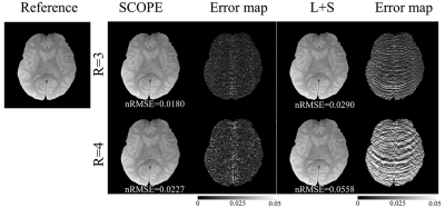 |
Accelerating spin-lock imaging using signal compensated low-rank plus sparse matrix decomposition
Yuanyuan Liu1, Yanjie Zhu1, Yuxin Yang2, Xin Liu1, Hairong Zheng1, and Dong Liang1
1Shenzhen Institutes of Advanced Technology, Shenzhen, China, 2Chongqing University of Technology, Chongqing, China, Chongqing, China
Recent studies show that variations in the rate of $$$T_{1\rho} (R_{1\rho})$$$ reflect changes in the spectral density of the local dipolar fields experienced by protons due to slow molecular motions. The quantitative data for $$$R_{1\rho}$$$ dispersion requires multiple $$$T_{1\rho}$$$-weighted images with different spin lock times (TSLs) and different locking fields, which makes the acquisition time very long. In this work, a signal compensation strategy with low-rank plus sparse model (SCOPE) was used to reconstruct $$$T_{1\rho}$$$-weighted images from highly undersampled data. We provide the reconstructed images, the estimated $$$R_{1\rho}$$$ maps, and the results of $$$R_{1\rho}$$$ dispersion curves at different acceleration factors.
|
|||
3083.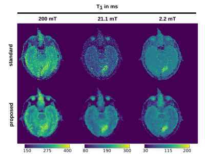 |
T1 quantification in fast field-cycling MRI using model-based reconstruction
Oliver Maier1, Markus Bödenler1,2, Rudolf Stollberger1,3, Mary-Joan MacLeod4, Lionel M Broche5, and Hermann Scharfetter1
1Graz University of Technology, Graz, Austria, 2Institute of eHealth, University of Applied Sciences FH JOANNEUM, Graz, Austria, 3BioTechMed Graz, Graz, Austria, 4Acute Stroke Unit, Aberdeen Royal Infirmary, Aberdeen, United Kingdom, 5Aberdeen Biomedical Imaging Centre, Univeresity of Aberdeen, Aberdeen, United Kingdom
T1 maps from fast field-cycling (FFC) MRI can provide insights to structural information and dynamics of molecular system not accessible by traditional MRI. The low SNR associated with the lower field strength in FFC imaging (0.2 T - 50 µT) leads to long acquisition times, impairing its clinical applicability. Hence, we propose a model-based reconstruction strategy to reduce acquisition time and improve image quality of multi-field T1 maps. The method is compared to standard fitting on numerical phantoms and a in vivo stroke patient. Model-based reconstruction clearly outperformed standard fitting, reducing noise and revealing previously unseen details in low-field T1 maps.
|
|||
3084.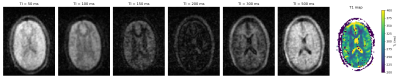 |
High resolution in-vivo relaxation time mapping at 50 mT.
Thomas O'Reilly1 and Andrew Webb1
1C.J. Gorter Center for High Field MRI, Leiden University Medical Center, Leiden, Netherlands
Recent years have seen a renewal in interest in low field MRI systems due to its lower cost, increased portability and robustness to medical implants, with the obvious disadvantage of lower SNR compared to clinical high field systems. In this work we present T1 and T2 relaxation maps of the brain and lower leg to facilitate the optimization of sequence parameters. Generally the T1 times are shorter and T2 times are slightly longer than at clinical field strengths.
|
The International Society for Magnetic Resonance in Medicine is accredited by the Accreditation Council for Continuing Medical Education to provide continuing medical education for physicians.