Oral
Digestive, Diabetes & Pancreas
ISMRM & SMRT Annual Meeting • 15-20 May 2021

| Concurrent 5 | 12:00 - 14:00 | Moderators: Tanya Chawla |
0251.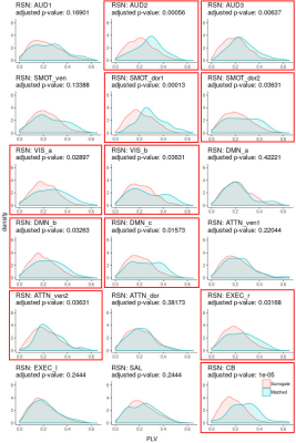 |
Phase synchronization of resting-state brain networks with the intrinsic electrical rhythm of the stomach
Ann S Choe1,2,3, Bohao Tang4, Kimberly R. Smith5, Hamed Honari6, Martin A. Lindquist4, Brian S. Caffo4, and James J. Pekar1,3
1F.M. Kirby Research Center for Functional Brain Imaging, Kennedy Krieger Institute, Baltimore, MD, United States, 2International Center for Spinal Cord Injury, Kennedy Krieger Institute, Baltimore, MD, United States, 3Department of Radiology, Johns Hopkins Medicine, Baltimore, MD, United States, 4Department of Biostatistics, Johns Hopkins Bloomberg School of Public Health, Baltimore, MD, United States, 5Department of Psychiatry and Behavioral Sciences, Johns Hopkins Medicine, Baltimore, MD, United States, 6Department of Electrical and Computer Engineering, Johns Hopkins University, Baltimore, MD, United States
Combining concurrent cutaneous electrogastrography (EGG) with resting-state fMRI (rsfMRI), we estimated resting-state brain networks from the rsfMRI data using independent component analysis, then used the amplitude-weighted Phase-Locking Value to assess whether those networks were synchronized with the intrinsic gastric rhythm estimated from the EGG data. We found 18 resting-state brain networks, of which 11 were found to be partially but significantly phase synchronized with the gastric rhythm. Disruptions to the gut-brain axis are thought to be involved in various disorders; manifestation of the infra-slow rhythm of the stomach in brain rsfMRI data could be useful for studies in clinical populations.
|
||
 |
0252.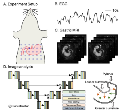 |
Deep learning based fully automatic analysis of gastric motility from contrasty-enhanced MRI
Xiaokai Wang1, Jiayue Cao1, Minkyu Choi2, and Zhongming Liu3
1Biomedical Engineering, University of Michigan, Ann Arbor, MI, United States, 2Electrical Engineering and Computer Science, University of Michigan, Ann Arbor, MI, United States, 3Biomedical Engineering, Electrical Engineering and Computer Science, University of Michigan, Ann Arbor, MI, United States
Contrast-enhanced gastrointestinal MRI serves as a non-invasive tool for studying and assessing gastric functions. Previous studies generally have their home-brewed semi-automatic algorithm for assessing gastric motility. This process is time-consuming and susceptible to errors. Here, we used deep learning to establish a fully automatic pipeline for assessing gastric motility with contrast-enhanced gastrointestinal MRI in rats. We cross-validated our analysis against simultaneously recorded electrogastrogram indicating gastric myoelectrical activity. Results from this analysis are consistent with electrogastrogram in terms of time, frequency, and power, and in addition, shed light on more detailed spatial characteristics of gastric motility.
|
|
0253.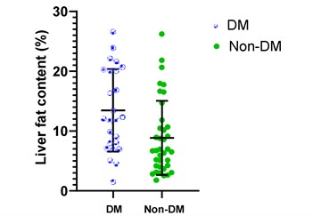 |
To quantify liver and pancreatic fat content in subjects with and without type 2 diabetes as measured by MRI-PDFF-observational study
Sonal Krishan1, Aparajita Pradhan2, and Shafi Kuchay3
1Radiology, Medanta Hospital, Gurgaon, India, 2Medanta Hospital, Gurgaon, India, 3Endocrinology, Medanta Hospital, Gurgaon, India
Aim of this study was to quantify liver (LFC) and pancreatic fat content (PFC) in subjects with and without type 2 diabetes (DM) as measured by MRI-PDFF. 25 adult patients who had recently diagnosed DM were compared with control group of 37 without DM. All underwent MRI PDFF for evaluation of LFC and PFC. The mean LFC in DM group was more: 12.1 % (8-20) vs Non-DM 6.7% (4.2-10.7).The overall prevalence of NAFLD in our study, was 72.6% (45/62 patients).In DM subgroup the NAFLD prevalence was more-88.0%. MRI PDFF can be a screening tool for NAFLD in patients with DM.
|
||
|
0254.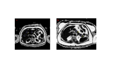 |
Quantitative MRI measurements of ectopic fat and body composition as predictors of T2D remission after bariatric surgery
Naomi S Sakai1, Kusuma Chaiyasoot1, Alan Bainbridge2, Margaret Hall-Craggs1, Rachel L Batterham1, and Stuart A Taylor1
1University College London, London, United Kingdom, 2University College London Hospitals, London, United Kingdom
Bariatric surgery can lead to rapid and substantial improvements in glycaemic control, often before significant weight loss has occurred. However, a proportion of patients do not achieve type 2 diabetes (T2D) remission after surgery despite significant post-surgical weight loss. Ectopic fat deposition and alterations in body composition are implicated in T2D pathoaetiology. Quantitative MRI (qMRI) can provide non-invasive measurements of ectopic organ fat and body composition which may have roles as predictors of T2D remission after bariatric surgery. This study aimed to assess the relationship between T2D remission status and qMRI-derived measurements of ectopic fat and body composition.
|
|
0255.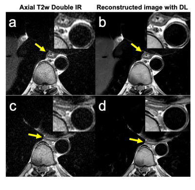 |
Optimizing T2-weighted MRI and DWI of the Esophagus
Jitka Starekova1, Lloyd Estkowski2, Ruiqi Geng1,3, Yuxin Zhang1,3, Diego Hernando1,3, and Scott B Reeder1,3,4,5,6
1Radiology, University of Wisconsin-Madison, Madison, WI, United States, 2Global MR Applications and Workflow GE Healthcare, Madison, WI, United States, 3Medical Physics, University of Wisconsin-Madison, Madison, WI, United States, 4Biomedical Engineering, University of Wisconsin-Madison, Madison, WI, United States, 5Medicine, University of Wisconsin-Madison, Madison, WI, United States, 6Emergency Medicine, University of Wisconsin-Madison, Madison, WI, United States
Esophageal MRI is emerging alternative to endoscopic ultrasound (EUS) and computed tomography (CT) for the detection and staging of cancer. Esophageal MRI has great potential for screening, active surveillance post-neodjuvant therapy and ruling out surgery for patients with higher stage cancer. The major challenges in esophageal imaging are the spatial resolution, as the esophageal wall is only approximately 3mm thick, and cardiac related motion artifacts. This prospective study aimed to address these challenges by developing a protocol for high-resolution T2-weighted and motion-robust diffusion-weighted imaging of the esophagus and periesophageal tissue.
|
||
0256.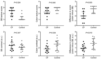 |
Using texture analysis to detect changes in intestinal contents in people with cystic fibrosis
Neele S Dellschaft1,2, Caroline Hoad1,2, Christabella Ng2,3, Luca Marciani2,4, Robin Spiller2,4, Alan Smyth2,3, Giles Major2,4, and Penny Gowland1,2
1Sir Peter Mansfield Imaging Centre, University of Nottingham, Nottingham, United Kingdom, 2Nottingham NIHR Biomedical Research Centre, University of Nottingham, Nottingham, United Kingdom, 3Division of Child Health, Obstetrics and Gynaecology, University of Nottingham, Nottingham, United Kingdom, 4Nottingham Digestive Diseases Centre, University of Nottingham, Nottingham, United Kingdom
Cystic Fibrosis (CF) is a genetic disease leading to sticky mucus, slower small bowel transit and maldigestion. We used MRI to characterize gastrointestinal function in CF. Changes in the image texture suggested increased bacterial load in the small bowel, which we have now quantified applying Haralick texture analysis. The difference in texture observed between small bowel and colon chyme in healthy subjects was less distinct in people with CF (Control median 2.11 a.u. [IQR 0.71, 3.30] v.CF 0.90 a.u. [0.38, 1.67], Wilcoxon P=0.010). This compared well to subjective analysis. These findings probably indicate overgrowth of colonic bacteria and maldigestion.
|
||
0257.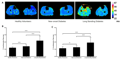 |
Noninvasive Assessment of Healthy People, New-onset and Long Standing Male T2DM Patients on Skeletal Muscle With T1ρMRI of Calf Muscle
Yufei Zhao1,2, Yang Jiang1,2, Jingyue Dai1,2, Honghong Wu1,2, Ying Cui1,2, Xinxiang Li1,2, and Xingui Peng1,2
1Jiangsu Key Laboratory of Molecular and Functional Imaging, Southeast University, Nanjing, China, 2Radiology, Zhongda Hospital Southeast University, Nanjing, China
T1ρ magnetic resonance can non-invasively assess the changes in the distribution of myofiber in male T2DM patients and this changes is gradually obvious as the course of the disease progresses.
|
||
 |
0258.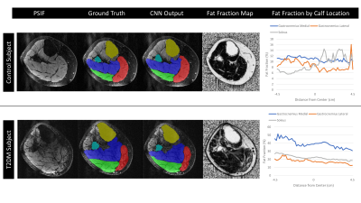 |
Deep Learning Based Segmentation and Fat Fraction Assessment of the Calf in Diabetic Subjects and Non-Diabetic Controls
Jill T Shah1, Katherine Medina2,3, Haresh R Rajamohan2,4, Justin Ho2,3, Cem M Deniz2,3, and Ryan Brown2,3
1New York University Grossman School of Medicine, New York, NY, United States, 2Center for Biomedical Imaging, Department of Radiology, New York University Grossman School of Medicine, New York, NY, United States, 3Center for Advanced Imaging Innovation and Research (CAI2R), Department of Radiology, New York University Grossman School of Medicine, New York, NY, United States, 4Center for Data Science, New York University, New York, NY, United States
Diabetes mellitus, muscular dystrophies, and other pathologies are characterized by metabolic impairment that can lead to lower extremity muscle degeneration. While MRI provides access to quantitative biomarkers to characterize muscle quality, analysis requires time-consuming manual image segmentation. To address this problem, we developed an automated segmentation algorithm based on a convolutional neural network that provided high dice similarity coefficient scores (>0.92) in the gastrocnemius medial, gastrocnemius lateral, and soleus muscles. We utilized the automatic segmentations to show volumetric fat fraction was elevated in individuals with diabetic peripheral neuropathy compared to controls in the soleus and gastrocnemius medial muscles (P<0.05).
|
|
0259.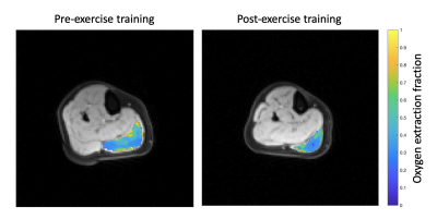 |
Evaluation of muscle oxygen extraction fraction in response to 15 weeks of exercise training: Comparison of people with and without type 2 diabetes
Erin K Englund1, Deirdre Rafferty2, Jie Zheng3, Hongyu An3, Judith G Regensteiner2,4, Alex J Barker1,5, and Jane EB Reusch2,4
1Department of Radiology, University of Colorado Anschutz Medical Campus, Aurora, CO, United States, 2Department of Medicine, University of Colorado Anschutz Medical Campus, Aurora, CO, United States, 3Department of Radiology, Washington University in St. Louis, St. Louis, MO, United States, 4Center for Women’s Health Research, University of Colorado Anschutz Medical Campus, Aurora, CO, United States, 5Department of Bioengineering, University of Colorado Anschutz Medical Campus, Aurora, CO, United States
Oxygen extraction fraction (OEF) provides insight into muscle oxygen consumption. As part of a larger exercise training study, 16 sedentary participants (12 controls, 4 with diabetes) underwent an MRI scan to determine OEF before and after 15 weeks of supervised exercise training. Pilot MRI data were acquired at rest with an asymmetric spin echo sequence and used to calculate OEF in the medial gastrocnemius muscle. Cardiovascular exercise capacity, measured as VO2Peak, was reduced in people with diabetes. MRI-measured OEF did not differ between groups, however a decrease in OEF was observed in all participants in response to exercise training.
|
||
0260.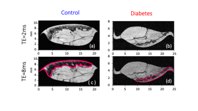 |
Dermal sodium space in controls and Type 2 Diabetes Mellitus patients characterised at histological length scales using 1H/23Na MRS and MRI at 9.4T
Galina E Pavlovskaya1,2, Christopher J Philp1, Thomas Meersmann1, Petra Hanson3,4, Harpal S Randeva4,5, Paul Paul O’Hare4,5, and Thomas Barber4,5
1SPMIC/Medicine, Univeristy of Nottingham, Nottingham, United Kingdom, 24Nottingham NIHR Biomedical Research Centre, Nottingham, United Kingdom, 3Warwick Medical School, University of Warwick, Warwick, United Kingdom, 42Warwickshire Institute for the Study of Diabetes Endocrinology and Metabolism, University Hospitals Coventry and Warwickshire, Coventry, United Kingdom, 5Warwick Medical School, University of Warwick, Coventry, United Kingdom
We reveal intriguing experimental results providing the evidence for dermal space storage for sodium, and how dynamics of the space changes in patients with Type2 Diabetes Mellitus.
|
The International Society for Magnetic Resonance in Medicine is accredited by the Accreditation Council for Continuing Medical Education to provide continuing medical education for physicians.